
Treatment of unruptured saccular intracranial aneurysms using stereotactic radiosurgery

a saccular and radiosurgery technology, applied in the field of intracranial aneurysm treatment, can solve the problems of high risk of rebleeding, high risk of ruptured aneurysms that are not treated, and difficult surgical repair of ruptured aneurysms in the posterior intracranial circulation, so as to prevent recurrence, facilitate clot formation, and promote neointimal proliferation and recruitment

Active Publication Date: 2020-09-15
LIPANI JOHN D
View PDF12 Cites 0 Cited by
  • Summary
  • Abstract
  • Description
  • Claims
  • Application Information

AI Technical Summary

Benefits of technology

[0033]Stereotactic radiosurgery that delivers ionizing radiation to the aneurysm would facilitate clot formation and prevent recurrence by the following mechanisms: (1) Lesions to the tunica intimata, to create a thrombotic surface along the inner wall of the aneurysm dome; to injure antithrombogenic endothelial cells that foster the activation of enzymes involved in clot removal; and to promote neointimal proliferation and recruitment of smooth muscle cells for progressive narrowing and occlusion of the aneurysm dome. (2) Lesions to the tunica adventitia: to activate the proliferation of cells at that location, which facilitates neointimal occlusion; and (3) Lesions to the wall (tunica intimata and tunica adventitia) of the aneurysm to induce radiation necrosis and fibrosis for permanent obliteration.

Problems solved by technology

When they do rupture, they are associated with significant morbidity and mortality.
Ruptured aneurysms that are not treated have a very high risk of rebleeding after the initial hemorrhage has occurred.
This leaves physicians with the therapeutic dilemma of either subjecting patients with borderline smaller aneurysms to a complex surgery having high morbidity and complications, or simply surveilling an aneurysm having a risk of rupture that might be acceptable.
Surgical repair of aneurysms in the posterior intracranial circulation, however, is extremely difficult due to technical access issues [POLEVAYA N V, Kalani M Y, Steinberg G K, Tse V C. The transition from hunterian ligation to intracranial aneurysm clips: a historical perspective.
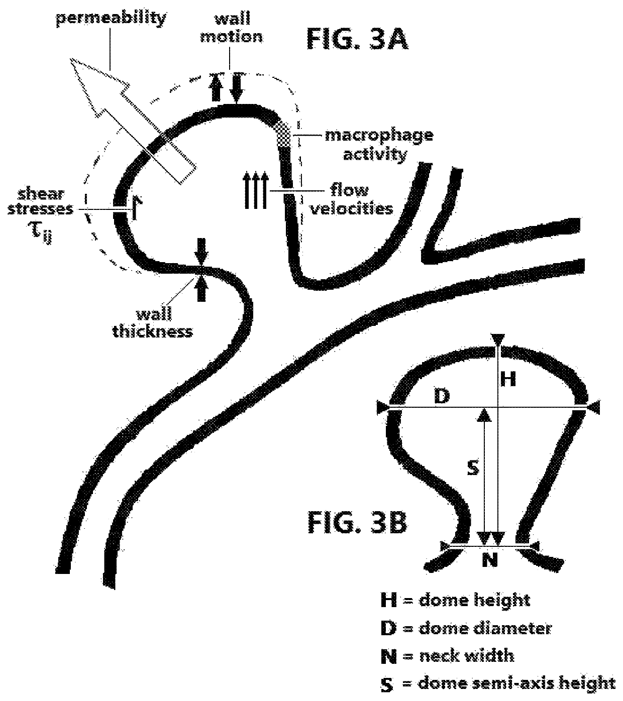
For example, a wide-neck saccular aneurysm has a neck width of at least 4 mm, or a neck at least twice as wide as the height of the aneurysm dome, and it may be difficult for the coils to remain in such an aneurysm without the use of a stent.
Sometimes, however, instead of remodeling the parent vessels and aneurysms, thrombus-associated autolysis of the aneurysm wall may also result in delayed rupture.
Also, a point source that emits photons or atomic particles in all directions cannot produce a beam of radiation (because omnidirectional rays are inherently not parallel with one another), unless the point source is accompanied by a waveguide or collimation-aperture device that minimizes ray divergence or eliminates rays of radiation that are not aligned substantially parallel to one another.
Another explanation for the disappearance of the vessel might be that there was excessive unintentional irradiation of the vessel itself, and the resulting damage to the vessel ultimately brought about its resorption, secondarily obliterating the aneurysm.
All except one died of a hemorrhage a few weeks to months after the Gamma Knife treatment.” Apparently, those additional 15 cases were never described in any publication, presumably because Forster was not inclined to publish negative results.
So, we have no way of knowing anything specific about the 15 cases, or what was different about the one patient who did not die soon after treatment.
How the patient recovered from the initial bleeding is still somewhat unknown and is a separate issue.
From a single case, it is impossible to determine if GKS made the aneurysm disappear or if it resolved spontaneously.
Or because tortuous vessels may make it difficult to access some aneurysms endovascularly, or if the aneurysm is located at a site that is dangerously close to vital anatomic regions, or if the patient is unable to tolerate conventional operative intervention, the aneurysms may instead be treated by stereotactic radiosurgery.
The resulting thickening of the wall will alter the geometry of the aneurysm, which in turn will alter the hemodynamics of the flow of blood into and out of the aneurysm, which will in turn alter (and possibly increase) the wall shear stress, leading to an interplay that causes to aneurysm to dynamically change its structure.
But if the destruction of the vessel wall, aided by any inflammatory response, weakens the wall more than the hyperplasia thickens and strengthens the wall, rupture will eventually occur when the wall stress exceeds the wall strength.

Method used


Image

  • Treatment of unruptured saccular intracranial aneurysms using stereotactic radiosurgery
  • Treatment of unruptured saccular intracranial aneurysms using stereotactic radiosurgery
  • Treatment of unruptured saccular intracranial aneurysms using stereotactic radiosurgery

Examples


Embodiment Construction

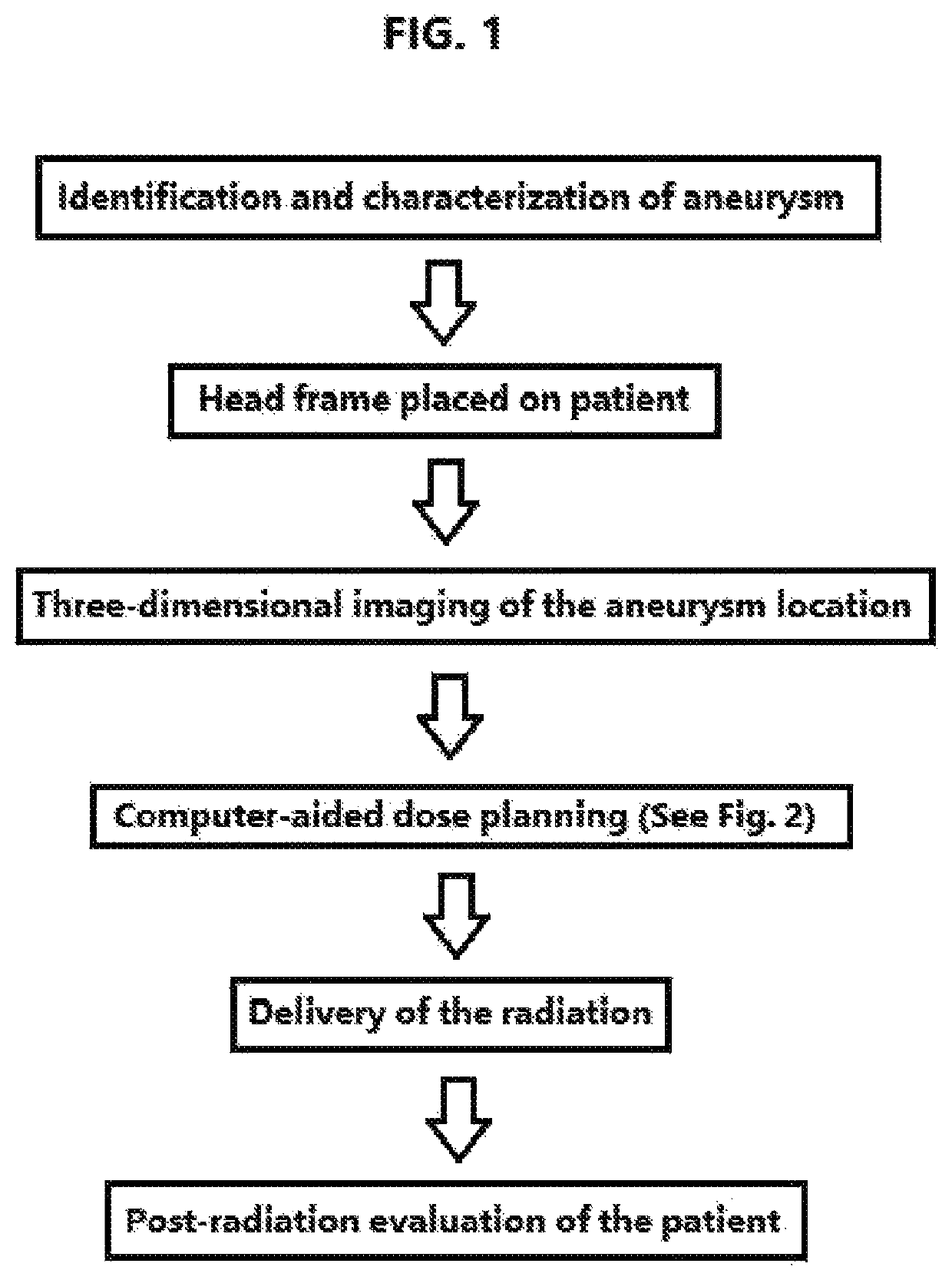
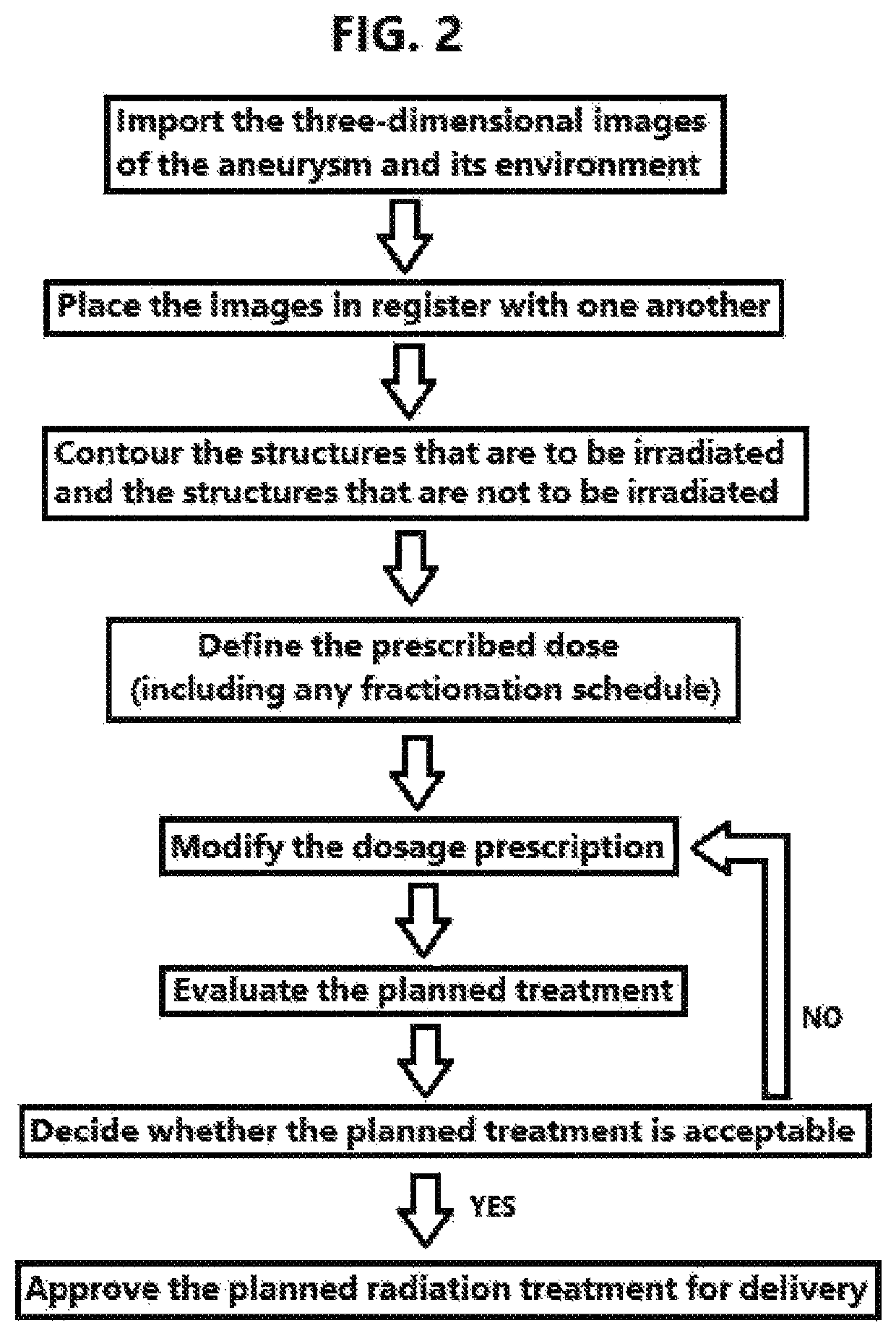
lass="d_n">[0041]Three types of radiosurgical devices have been used for stereotactic radiosurgery (SRS)—the Gamma Knife stereotactic radiosurgery apparatus (Elekta, Sweden); Linear Accelerator (LINAC) devices such as the CYBERKNIFE® (Accuray, Sunnyvale, Calif.), NOVALIS Tx™ (BrainLab, Germany), XKNIFE™ (Integra, New Jersey), and AXESSE™ (Elekta, Sweden) stereotactic radiosurgery apparatus, (among others); and proton beam devices. Because the Gamma Knife is the oldest and arguably best known such device, the description of the invention that follows presumes that it makes use of the Gamma Knife device, but without endorsing its use over other SRS devices. To give a specific embodiment of the invention, the specific radiosurgical equipment could comprise a Gamma Knife device, stereotactic frame, and planning software, respectively, as follows: Leksell Gamma Knife® Perfexion™ (Catalog No. 715000), Leksell® Coordinate Frame G (Catalog No. 1006442), and Leksell GammaPlan® for Perfexion™...


PUM


Abstract

Methods and devices are disclosed for treating a patient with an unruptured saccular aneurysm that is not associated with an arteriovenous malformation, wherein ionizing radiation is delivered to the brain of a patient through stereotactic radiosurgery. The radiosurgery may be performed with a Gamma Knife stereotactic radiosurgery apparatus, LINAC device such as CYBERKNIFE® stereotactic radiosurgery apparatus, or particle-beam device. The treatment impedes a natural progression of the aneurysm towards rupture and may actually obliterate the aneurysm. The treatment plan may be based in part on measured geometric properties of the aneurysm or on physical aneurysm properties such as permeability, wall thickness, wall motion, the presence of macrophages, shear stress, and flow velocities. The methods may be used in conjunction with endovascular coil embolization.

Description

BACKGROUND OF THE INVENTION[0001]The field of the present invention is the treatment of intracranial aneurysms using focused ionizing radiation. An intracranial aneurysm (brain aneurysm) is a disorder in which weakness in the wall of a cerebral artery causes a localized dilation or ballooning of the blood vessel. Intracranial aneurysms are classified into several basic types—the rounded saccular aneurysms; the spindle-shaped fusiform aneurysms; and Charcot-Bouchard aneurysms (also known as microaneurysms or miliary aneurysms). Charcot-Bouchard aneurysms are associated with hypertension and occur in blood vessels less than 300 microns in diameter, particularly in the lenticulostriate vessels of the basal ganglia. Blister-like aneurysms, traumatic and dissecting aneurysms, and infectious aneurysms have also been described. For each of these aneurysm types, all layers of the blood vessel wall are dilated or ballooned, such that a portion of the blood vessel lumen itself is enlarged. In...

Claims


Application Information

Patent Timeline
Patent Type & Authority Patents(United States)
IPC IPC(8): A61N5/10A61B90/10A61B17/00
CPCA61N5/1031A61N5/1084A61B90/10A61N5/1075A61N2005/1055A61N2005/1041A61N5/1049A61B2090/101A61N2005/1035A61B2017/00778A61N2005/1052A61N5/1039A61N2005/1087
Inventor LIPANI, JOHN D.
Owner LIPANI JOHN D
Learn More