Rapidly degrading GFP-fusion proteins and methods of use

a fusion protein and rapid degradation technology, applied in the field of biochemical assays and reagents, can solve the problems of inability to determine short-term or repetitive events, toxic to mammalian cells, prior art deficient in destabilized or short-lived gfp, etc., to avoid toxic levels of gfp, facilitate the development of stable cell lines, and accelerate the turnover of gfp

Inactive Publication Date: 2005-10-18
TAKARA BIO USA INC
View PDF1 Cites 7 Cited by
  • Summary
  • Abstract
  • Description
  • Claims
  • Application Information

AI Technical Summary

Benefits of technology

[0011]A rapid turnover or destabilized GFP can be used in research applications where prior art GFPs cannot. Such applications include using the destabilized GFP as a genetic reporter for analyzing transcriptional regulation and / or cis-acting regulatory elements, or as a tool for studying protein degradation. Further, a rapid turnover GFP permits easier development of stable cell lines which express the GFP gene, since toxic levels of GFP are avoided because the GFP protein is degraded quickly.

Problems solved by technology

GFP is a very stable protein which can accumulate and thus is often toxic to mammalian cells.
On the other hand, its stability makes it difficult to determine short-term or repetitive events.
The prior art is deficient in a destabilized or short-lived GFP.

Method used


Image

  • Rapidly degrading GFP-fusion proteins and methods of use
  • Rapidly degrading GFP-fusion proteins and methods of use
  • Rapidly degrading GFP-fusion proteins and methods of use

Examples


example 1

Construction of DNA Expression Vectors

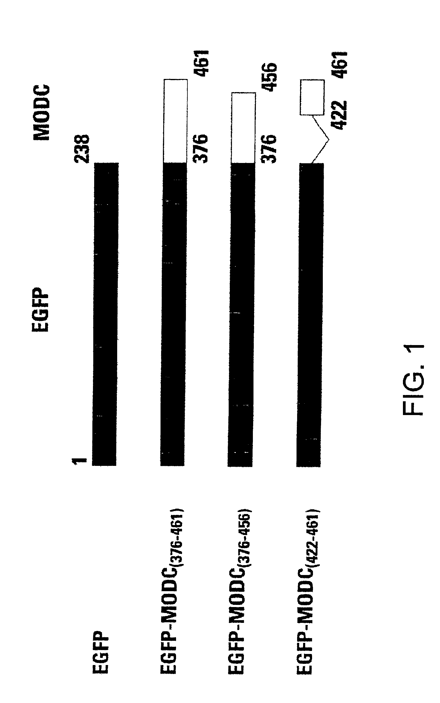
[0058]The cDNAs encoding EGFP and the C terminus of murine ODC (MODC) were amplified with pfu DNA polymerase (Stratagene, Inc., La Jolla, Calif.). EGFP was amplified with a pair of primers: 5′ incorporated with a SacII recognition sequence and 3′ with a Hind III sequence. The stop codon of EGFP was deleted from its C-terminus in order to make an open reading frame with the C terminus of murine ODC. The C terminus of murine ODC was also amplified with a pair of primers: 5′ incorporated with a Hind III recognition sequence and 3′ with an EcoRI sequence. Two amplified PCR products were ligated at the Hind III site and the fusion was cloned into pTRE expression vector, Tc-regulated expression system (Gossen M., and Bujard H. (1992) Proc. Natl. Acad. Sci. 89: 5547-5551).

[0059]Using these methods, fusion proteins of EGFP-MODC were constructed. The EGFP-MODC376-461 fusion protein included the complete C-terminus of murine ornithine decarboxylase. EGFP-...

example 2

Cell Transfection

[0060]The construct DNAs were purified and transfected into CHO K1-off cells for determination of protein degradation. CHO K1-off cells are CHO cells which were pre-transfected by a fusion protein of the tet-repressor and the herpes simplex virus VP16 gene (tTA). This pre-transfection allows expression of the gene coding for the fusion protein on a pTRE vector (Gossen and Bujard, ibid), which in turn initiates transcription by binding to a modified CMV promoter with tet-repressor binding elements. This binding can be blocked by tetracycline; hence, the expression can be controlled by tetracycline. The DNAs were introduced into these cells by CLONfectin (CLONTECH Laboratories, Inc., Palo Alto). After 24 hours, transfected cells were subject to functional analyses.

example 3

Fluorescence Analysis

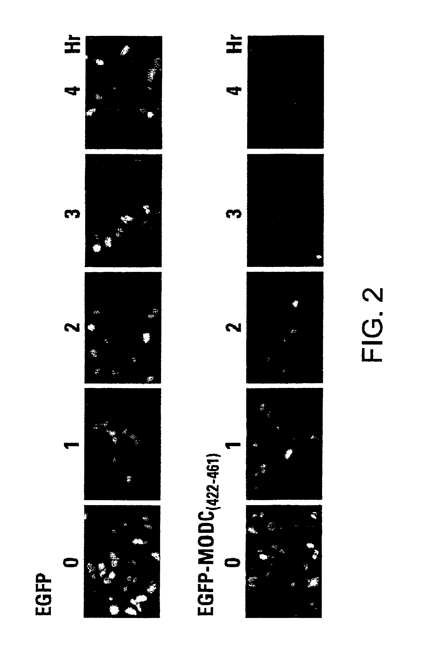
[0061]Cells were cultured on top of cover-slips to allow observation under a fluorescence microscope. After transfection, the cells were incubated at 37° C. for 24 hours on the cover-slips and then fixed with 4% paraformaldehyde for 30 minutes. The cover-slips were mounted on a glass slide for fluorescence examination with a Zeiss Axioskop Model 50 fluorescent microscope. To determine protein turnover, the cells were treated with cycloheximide at a final concentration of 100 μg / ml for variable times before paraformaldehyde fixation.

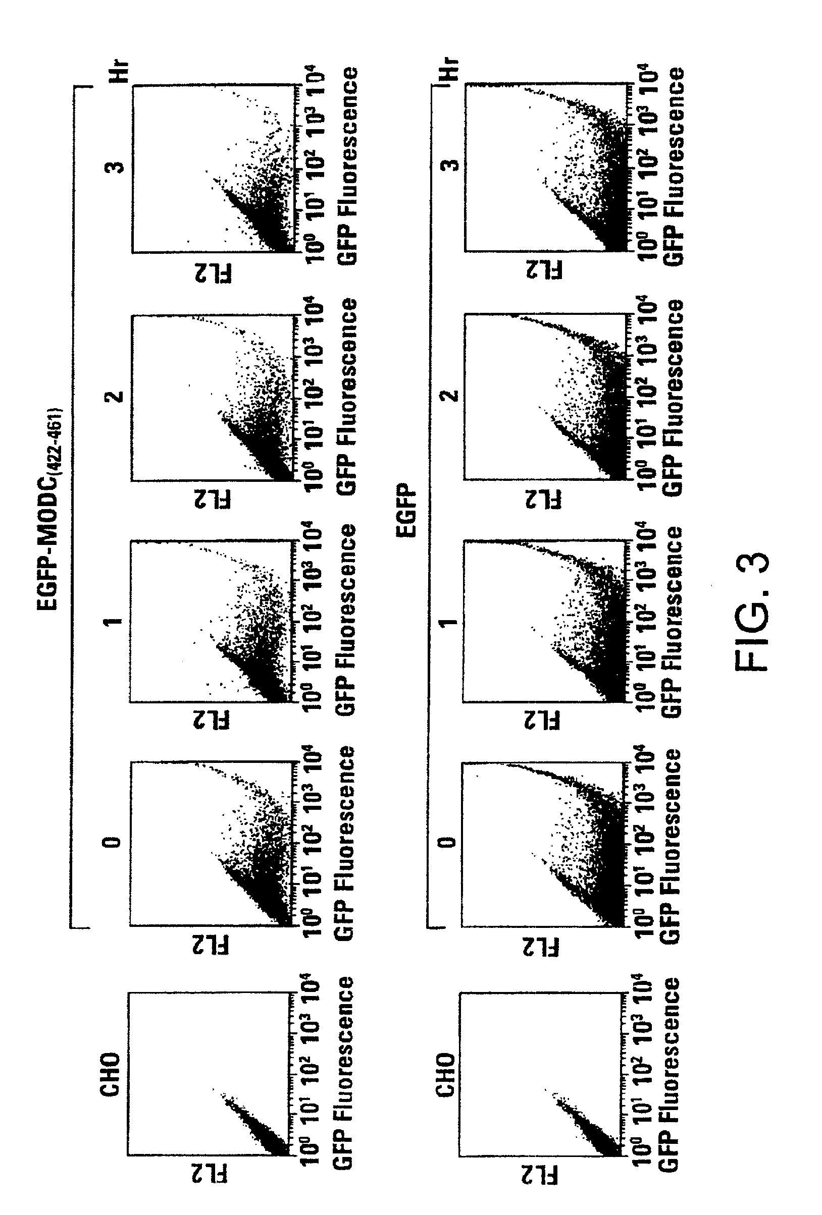
[0062]For FACS analysis, the transfected cells as well as cycloheximide-treated cells were collected by EDTA treatment and the cell pellets were resuspended in 0.5 ml of PBS. The cell suspensions were then analyzed for fluorescence intensity by FACS Calibur (Becton Dickson, Inc., San Jose, Calif.). EGFP was excited at 488 nm, and emission was detected using a 510 / 20 bandpass filter.


PUM

PropertyMeasurementUnit
half lifeaaaaaaaaaa
half lifeaaaaaaaaaa
half lifeaaaaaaaaaa

Abstract

Green fluorescent protein (GFP) is widely used as a reporter in determining gene expression and protein localization. The present invention provides fusion proteins with a half life of ten hours or less with several embodiments having half lives of 4 hours or less. Such proteins may be constructed by fusing C-terminal amino acids of the degradation domain of mouse ornithine decarboxylase (MODC), which contains a PEST sequence, to the C-terminal end of an enhanced variant of GFP (EGFP). Fluorescence intensity of the fusion protein in transfected cells is similar to that of EGFP, but the fusion protein, unlike EGFP, is unstable in the presence of cycloheximide. Specific mutations in the MODC region have resulted in mutants with varying half lives, useful for a variety of purposes.

Description

CROSS-REFERENCE TO RELATED APPLICATION[0001]This application is a continuation in part of U.S. patent application Ser. No. 09 / 062,102, filed Apr. 17, 1998 now U.S. Pat. No. 6,130,313.BACKGROUND OF THE INVENTION[0002]1. Field of the Invention[0003]This invention relates to the field of biochemical assays and reagents. More specifically, this invention relates to modified fluorescent proteins and to methods for their use.[0004]2. Description of the Related Art[0005]Because of its easily detectable green fluorescence, green fluorescent protein (GFP) from the jellyfish Aequorea Victoria has been used widely to study gene expression and protein localization. GFP fluorescence does not require a substrate or cofactor; hence, it is possible to use this reporter in numerous species and in a wide variety of cells. GFP is a very stable protein which can accumulate and thus is often toxic to mammalian cells.[0006]Recently, crystallographic structures of wild-type GFP and the mutant GFP S65T rev...

Claims


Application Information

Patent Timeline
Patent Type & Authority Patents(United States)
IPC IPC(8): C07K14/435C12N9/88C12N15/62C12P21/04
CPCC07K14/43595C12N9/88C12N15/62C07K2319/00C07K2319/60C07K2319/71C07K2319/80C07K2319/95
Inventor LI, XIANQIANGKAIN, STEVE
Owner TAKARA BIO USA INC
Why Eureka
Try Eureka