Method for Raman chemical imaging of endogenous chemicals to reveal tissue lesion boundaries in tissue

a chemical imaging and endogenous chemical technology, applied in the field of tissue evaluation, can solve the problems of significant cancer, relatively slow process, and loss of lives

Inactive Publication Date: 2005-11-15
CHEMIMAGE CORP
View PDF25 Cites 58 Cited by
  • Summary
  • Abstract
  • Description
  • Claims
  • Application Information

AI Technical Summary

Problems solved by technology

Cancer is significant, not only in lives lost, but also in the $107 billion cost to the United States economy in 2000 according to the National Institutes of Health.
Because of the tissue preparation required, this process is relatively slow.
However, in fine needle biopsies intact breast tissue morphology is disrupted often leaving only cellular structure for analysis which is often less revealing of disease state.
However, needle biopsies necessitate an outpatient surgical procedure and the resulting needle core sample must be embedded or frozen prior to analysis.
Despite years of research and development, two techniques that have not realized their potential are:(1) fluorescence optical biopsies, which fails due to the nonspecific nature of tissue autofluorescence; and(2) near-infrared optical diagnostics, in particular non-invasive glucose sensing, which fails due to interference from tissue major components, including predominantly water.
However, Raman spectroscopy has not been able to differentiate benign vs. malignant tissues due to the spectral similarities of these tissue types.
In addition, Raman spectroscopy of breast tissue samples requires large numbers of cell populations.

Method used


Image

  • Method for Raman chemical imaging of endogenous chemicals to reveal tissue lesion boundaries in tissue
  • Method for Raman chemical imaging of endogenous chemicals to reveal tissue lesion boundaries in tissue
  • Method for Raman chemical imaging of endogenous chemicals to reveal tissue lesion boundaries in tissue

Examples


Embodiment Construction

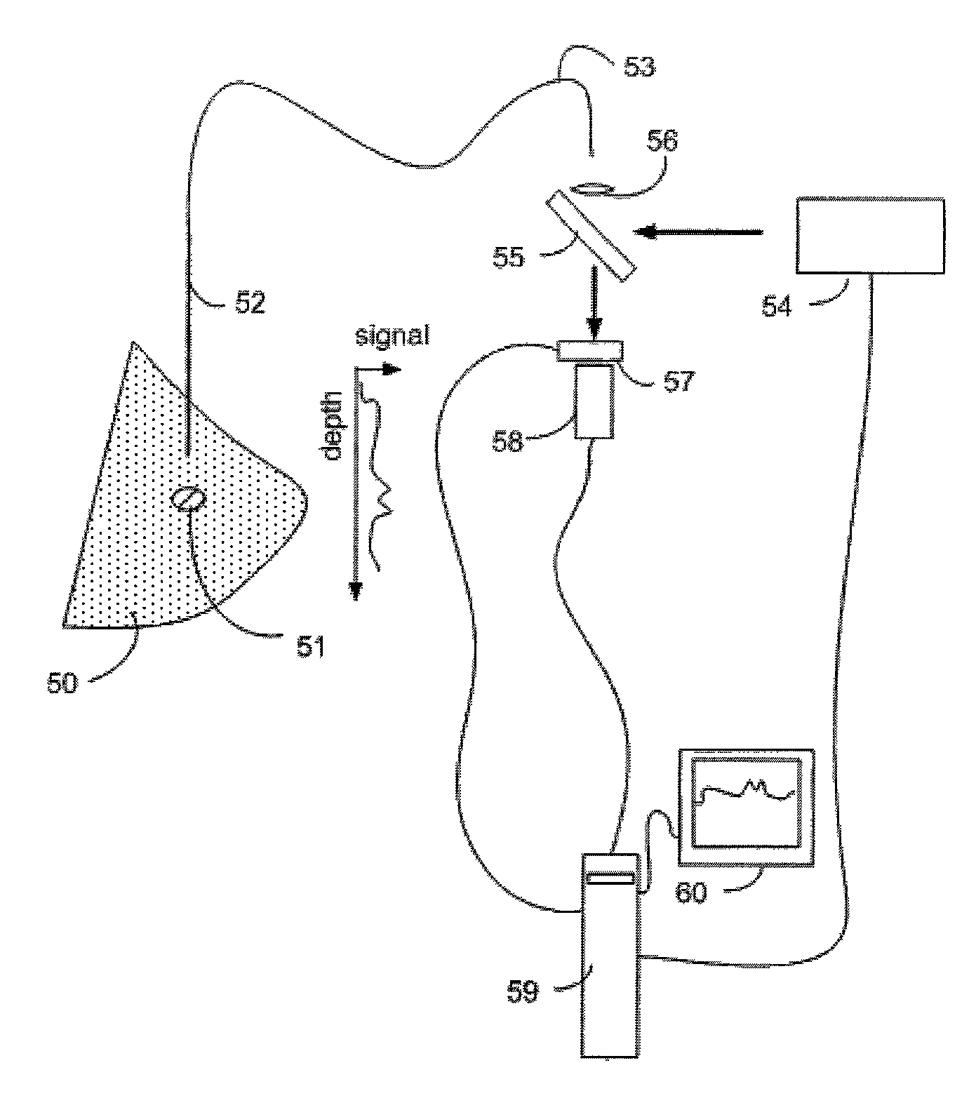
[0030]Raman Spectroscopy

[0031]When light interacts with matter, a portion of the incident photons are scattered in all directions. A small fraction of the scattered radiation differs in frequency (wavelength) from the illuminating light. If the incident light is monochromatic (single wavelength) as it is when using a laser source or other sufficiently monochromatic light source, the scattered light which differs in frequency may be distinguished from the light scattered which has the same frequency as the incident light. Furthermore, frequencies of the scattered light are unique to the molecular or crystal species present. This phenomenon is known as the Raman effect.

[0032]In Raman spectroscopy, energy levels of molecules are probed by monitoring the frequency shifts present in scattered light. A typical experiment consists of a monochromatic source (usually a laser) that is directed at a sample. Several phenomena then occur including Raman scattering which is monitored using instru...


PUM

PropertyMeasurementUnit
timeaaaaaaaaaa
thickaaaaaaaaaa
Raman chemical imagining spectrophotometeraaaaaaaaaa

Abstract

Apparatus and methods for spatially resolved Raman detection of molecules indicative of the borders of lesions with normal tissue are disclosed. A region of biological tissue was illuminated with monochromatic light. A Raman shifted light signal is detected from endogenous molecules in the region, the molecules being spatially organized in a localized first area of the region. These molecules are indicative of a border between normal tissue and a lesion. The Raman shifted light signal is then spatially resolved in at least one direction.

Description

CROSS REFERENCE TO RELATED APPLICATIONS[0001]This application claims priority pursuant to 35 U.S.C. 119(e) to U.S. Provisional Application No. 60 / 301,708 filed Jun. 28, 2001 which is incorporated herein by reference in its entirety including incorporated material.FIELD OF THE INVENTION[0002]The field of the invention is the field of tissue evaluation and, more particularly, the field of tissue evaluation using optical detection of light which has been Raman shifted in frequency.BACKGROUND OF THE INVENTION[0003]Cancer is the second leading cause of death in the United States and over 1.2 million people are diagnosed with this disease annually. Cancer is significant, not only in lives lost, but also in the $107 billion cost to the United States economy in 2000 according to the National Institutes of Health. It is widely recognized among the cancer research community, that there is a need to develop new tools to characterize normal, precancerous, cancerous, and metastatic cells and tis...

Claims


Application Information

Patent Timeline
Patent Type & Authority Patents(United States)
IPC IPC(8): A61B5/00G01N21/65G01N21/63
CPCA61B5/0091A61B5/4312G01N21/65
Inventor TREADO, PATRICK J.NELSON, MATTHEW P.
Owner CHEMIMAGE CORP
Why Eureka
Try Eureka