

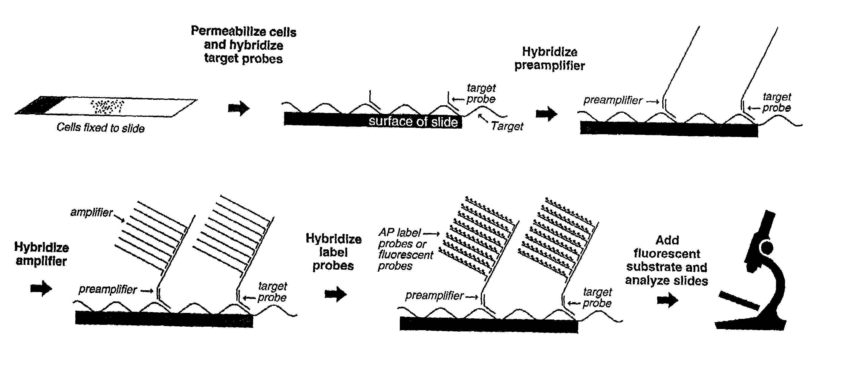
Highly sensitive gene detection and localization using in situ branched-DNA hybridization
a gene and in situ hybridization technology, applied in the field of nucleic acid chemistry and biochemical assays, can solve the problems of low sensitivity of the assay described in this study, no study suggests or discloses a bdna assay for the detection of nucleic acids in morphologically intact cells or tissues, and the number of challenges must be overcome. to achieve the effect of increasing sensitivity
- Summary
- Abstract
- Description
- Claims
- Application Information
AI Technical Summary
Benefits of technology
Problems solved by technology
Method used
Image

Examples
example 1
Detection of HPV DNA by bDNA ISH in Cells
A. Materials and Methods
[0100]i. Cell Culture
[0101]Because they contain different strains and amounts of human papilloma virus (HPV), the human cervical carcinoma cell lines of HeLa, CaSki and SiHa were used to evaluate the present invention. Table 1 describes each cell line.
[0102]
TABLE 1Cell Line*Viral strainDNA copies per cell 3CaSkiintegrated HPV-16 DNA400–600 copiesHeLaintegrated HPV-18 DNA10–50 copiesSiHaintegrated HPV-16 DNA1–2 copiesC33A (control)HPV negative0 copies*All cell lines were obtained from the American Type Cell Culture Collection (ATCC; Manassas, VA) and grown in flasks under conditions generally suggested by the ATCC. When required, cells were detached from the flasks using a mild trypsin treatment.
[0103]ii. Oligonucleotide Probes
[0104]The HPV-16-specific target probes consisted of a total of 26 DNA oligonucleotide probes covering approximately 90% of the E6 and E7 regions of the HPV genome. These probes were designed by f...
example 2
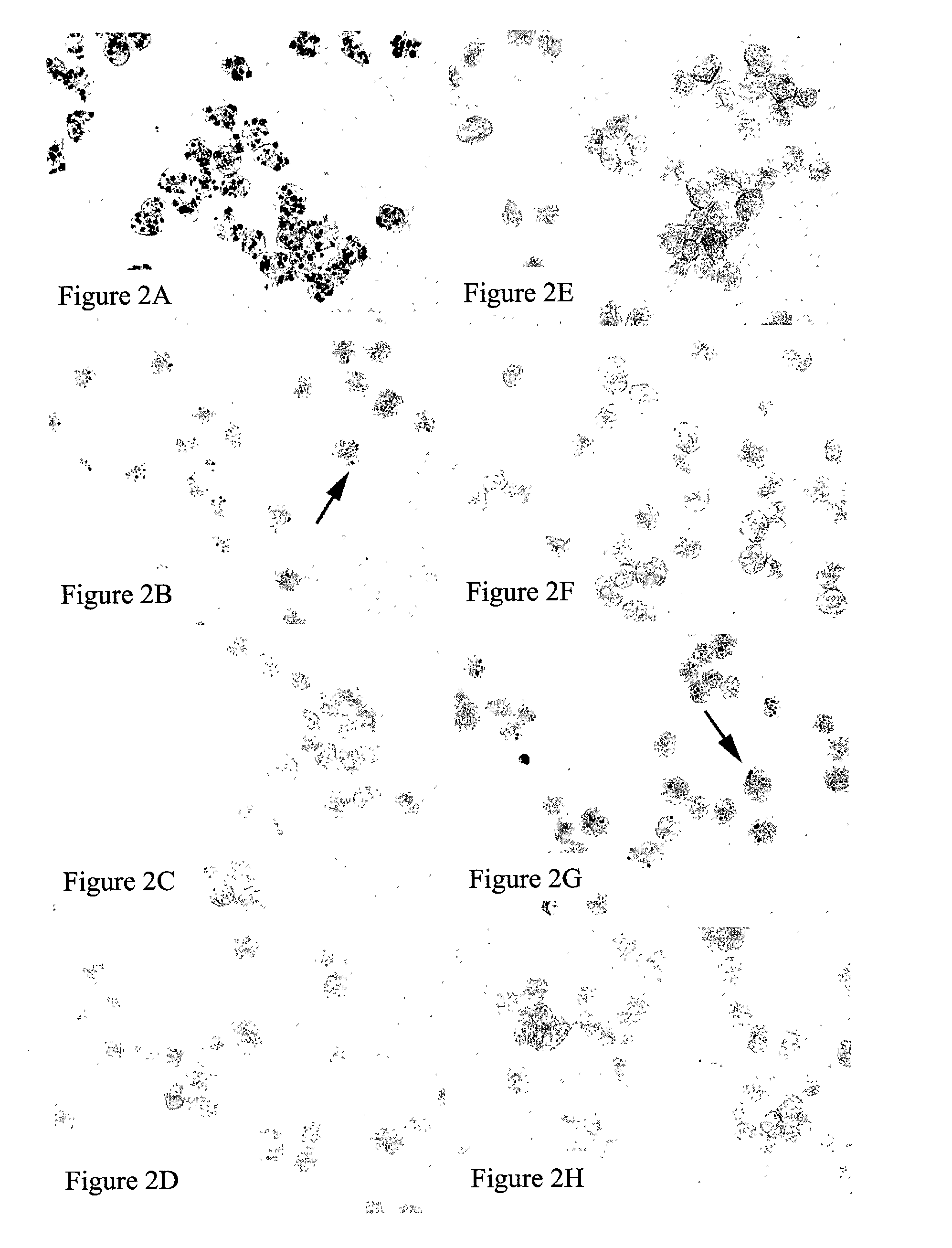
Assessing Specificity in Cells
[0112]Mixed cell populations were used to further assess the specificity of the present method for HPV DNA detection. Mixed cell samples were comprised of unlabeled CaSki cells (containing 400–600 DNA copies of HPV-16) and labeled HeLa cells (containing 10–50 DNA copies of HPV-18). The HeLa cells were labeled with the fluorescent cell tracer CFDA SE (Carboxyfluorescein diacetate, succinimidyl ester, Molecular Probes, Eugene, Oreg.) according to manufacturer's instructions. Both cell types were assayed according to the procedure of Example 1.
[0113]As shown in FIGS. 3A and 3C, hybridization with HPV-16 target probes yielded positive signal detection only in HPV-16-infected CaSki cells (arrow) and not in HeLa cells (arrow). Likewise, as shown in FIGS. 3B and 3D, hybridization with HPV-18 target probes yielded positive signal detection only in HPV-18-infected HeLa cells (arrow head) and not in CaSki cells (arrow head). As expected, greater signal intensity ...
example 3
Detection of HPV RNA and Localization of RNA / DNA within a Cell
[0115]For RNA detection, cells grown on chamber slides (#12-565-18, Fisher Scientific, Pittsburgh, Pa.) were fixed with 4% formaldehyde in PBS for 30 minutes at RT, treated with 10 μg / ml Proteinase K in PBS for 10 minutes at RT, and then washed twice for 5 minutes in PBS. Samples were incubated at 40° C. for 3 hours with 1 pmole HPV-specific target probes in a target probe buffer (6×SSC, 25% formamide, 0.2% BRIJ®-35, 0.2% casein), and then washed following the same decreasing series of SSC buffers as described in Example 1. Similarly, the pre-amplifier, amplifier, and AP-conjugated probe hybridization and wash conditions were the same as described in Example 1.
[0116]For subcellular localization of DNA targets, HeLa cells were grown overnight on poly-D-lysine-coated chamber slides. Poly-D-lysine is available from Sigma (product code P7280). The following day, the cell culture medium was removed; the cells were washed with ...
PUM
| Property | Measurement | Unit |
|---|---|---|
| concentration | aaaaa | aaaaa |
| temperature | aaaaa | aaaaa |
| concentration | aaaaa | aaaaa |
Abstract
Description
Claims
Application Information