Dynamic computed tomography imaging using positional state modeling

a computed tomography and dynamic technology, applied in the field of medical imaging arts, can solve the problems of inconsistent medical diagnosis of cardiac diseases, insufficient cardiac imaging modalities, and inability to fully satisfy existing cardiac imaging modalities, so as to improve the accuracy of diagnostic information, improve temporal and spatial resolution, optimally provide a selected angular coverage

Inactive Publication Date: 2006-06-06
KONINKLIJKE PHILIPS ELECTRONICS NV
View PDF19 Cites 140 Cited by
  • Summary
  • Abstract
  • Description
  • Claims
  • Application Information

AI Technical Summary

Benefits of technology

[0020]One advantage of the present invention is that it improves temporal and spatial resolution of images of moving anatomy.
[0021]Another advantage of the present invention is that it identifies a plurality of data acquisition windows in each cardiac cycle.
[0022]Yet another advantage of the present invention resides in improved accuracy of diagnostic information

Problems solved by technology

It is well known that cardiac diseases are a leading health problem in the United States at the present time.
In spite of this prevalence, however, medical diagnosis of cardiac diseases is inconsistent.
However, the existing cardiac imaging modalities, including cardiac angiography, ultrasound, magnetic resonance imaging (MRI), and various forms of computed tomography (CT), are not fully satisfactory.
Ultrasound suffers from poor image quality.
MRI typically does not detect calcification, and is subject to various artifacts.
However, cardiac CT imaging, including cone-beam CT imaging, suffers from motion blurring due to the relatively long CT image scan times needed to acquire projection data with at least 180° of angular coverage.
The motion of the cardiac muscle as data is acquired over this extended time causes motion-related image blurring.
The high pulse rates often characteristic of patients with coronary diseases further increases motion image blurring.
However, collecting imaging data over more cycles can introduce other artifacts.
However, the various physiological portions of the cardiac cycle, e.g. the systolic and diastolic portions and physiological events therein, do not linearly scale with changes in the cardiac cycle period, and so using a percentage temporal position does not fully compensate for changes in the pulse rate.
Third, image artifacts can be introduced in helical cone-beam CT due to an introduction of potential inconsistencies in the angular deviation in the Z-direction (i.e., the cone angle) at segment boundaries.
How

Method used


Image

  • Dynamic computed tomography imaging using positional state modeling
  • Dynamic computed tomography imaging using positional state modeling
  • Dynamic computed tomography imaging using positional state modeling

Examples


Embodiment Construction

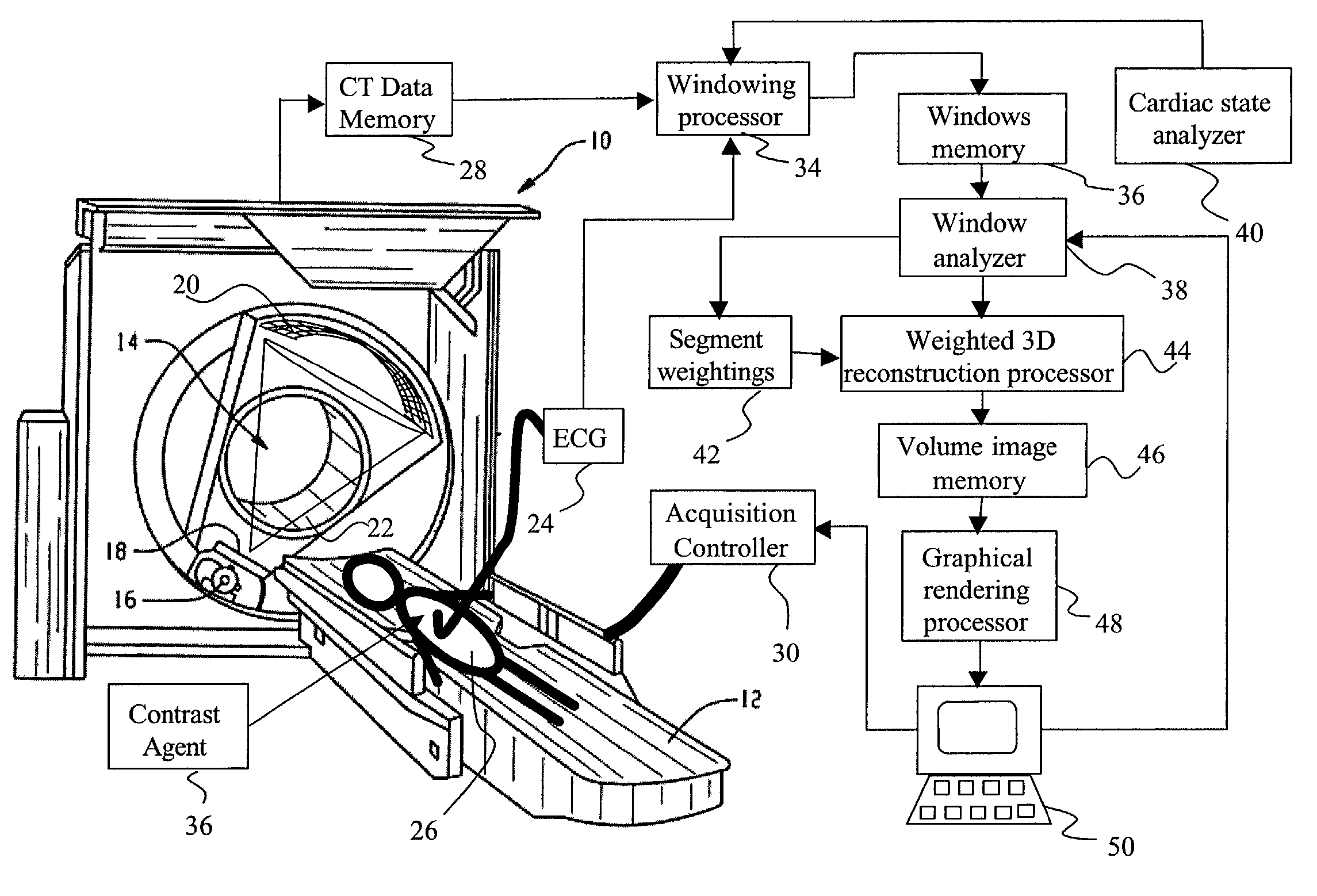
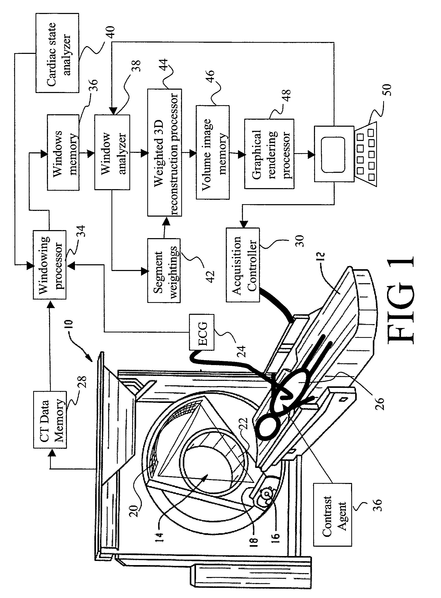
[0042]With reference to FIG. 1, an exemplary cone-beam computed tomography (CT) scanner 10 includes a subject support 12 such as a patient couch which is linearly movable inside an examination region 14. An x-ray tube assembly 16 mounted on a rotating gantry projects x-rays through the examination region 14. A collimator 18 collimates the radiation in two dimensions. In the exemplary CT scanner 10, a two-dimensional x-ray detector array 20 is disposed on the rotating gantry across the examination region from the x-ray tube. In an alternative embodiment (not shown), the detector array includes an array of two-dimensional detector rings mounted in a stationary fashion around the rotating gantry.

[0043]In the exemplary cone-beam CT scanner 10, the x-ray tube assembly 16 cooperates with the collimator 18 to produce a conical or wedge-shaped beam 22 which diverges conically as it passes through the examination region 14. The cone beam 22 substantially covers the detector array 20, which i...


PUM


Abstract

An apparatus for computed tomography (CT) imaging of a cyclically moving organ includes a positional state monitor (24, 40) that monitors a positional state of the cyclically moving organ such as the heart. A cone-beam CT scanner (10) acquires image data at least within a plurality of time windows. Each time window is centered about an occurrence of a selected positional state of the organ. A window analyzer (38) selects a data segment within each time window such that the data segments combine to form a complete data set covering a selected angular range. A reconstruction processor (44) reconstructs the selected data segments into an image representation.

Description

[0001]This application claims the benefit of U.S. Provisional Application No. 60 / 301,618, filed Jun. 28, 2001.BACKGROUND OF THE INVENTION[0002]The present invention relates to the medical imaging arts. It particularly relates to dynamic volumetric cardiac imaging using computed tomography (CT), and will be described with particular reference thereto. However, the invention will also find application in conjunction with the dynamic volumetric imaging of other non-stationary organs, such as the lungs, in conjunction with time evolution studies, and in conjunction with other imaging techniques and modalities.[0003]It is well known that cardiac diseases are a leading health problem in the United States at the present time. In spite of this prevalence, however, medical diagnosis of cardiac diseases is inconsistent. More than half of the individuals who die of a coronary attack do not exhibit previously recorded symptoms. Hence, there is a need for improved screening and accuracy in the d...

Claims


Application Information

Patent Timeline
IPC IPC(8): A61B5/05A61B5/352A61B6/00A61B6/03A61B8/00A61B8/08
CPCA61B6/032A61B6/4085A61B6/541A61B8/4416A61B6/027A61B6/5247A61B5/0456A61B5/7289A61B6/583A61B8/08A61B5/352
Inventor HEUSCHER, DOMINIC J.ZHAO, SHIYINGMATTHEWS, DAVID D.WANG, GE
Owner KONINKLIJKE PHILIPS ELECTRONICS NV
Why Eureka
Try Eureka