Method for spectrophotometric blood oxygenation monitoring

a spectrophotometric and blood oxygenation technology, applied in the field of noninvasive determination of biological tissue oxygenation, can solve the problems of inability to provide pulse oximetry with any information about venous blood, difficult to determine the absolute value of oxygen saturation within the biological tissue, and inability to provide a means for determining the absolute value of oxygen saturation

Active Publication Date: 2006-07-04
EDWARDS LIFESCIENCES CORP
View PDF67 Cites 271 Cited by
  • Summary
  • Abstract
  • Description
  • Claims
  • Application Information

AI Technical Summary

Benefits of technology

[0019]It is, therefore, an object of the present invention to provide a method for non-invasively determining the absolute oxygen saturation value within biological tissue.
[0020]It is a further object of the present invention to provide a method that provides calibration means to account for energy losses due to scattering as well as other background absorption from biological compounds.
[0021]It is a still further object of the present invention to provide a method that can non-invasively determine oxygen saturation within tissue at certain depth that limits the influence from the superficial tissues.
[0026]The calibrated sensor can be used subsequently to calibrate similar sensors without having to invasively produce a blood sample. Hence, the present method and apparatus enables a non-invasive determination of the blood oxygen saturation level within tissue. For example, an operator can create reference values by sensing a light signal or other reference medium using the calibrated sensor. The operator can then calibrate an uncalibrated sensor by sensing the same light signal or reference medium, and subsequently adjusting the uncalibrated sensor into agreement with the calibrated sensor. Hence, once a reference sensor is created, other similar sensors can be calibrated without the need for invasive procedure.
[0027]There are, therefore, several advantages provided by the present method and apparatus. Those advantages include: 1) a practical non-invasive method and apparatus for determining oxygen saturation within tissue that can be used to determine the total blood oxygen saturation within tissue as opposed to a change in blood oxygen saturation; 2) a calibration method that accounts for energy losses (e.g., light attenuation) due to light scattering within tissue, other background absorption losses from biological compounds, and other unknown losses including measuring apparatus variability; 3) a practical non-invasive method and apparatus for determining oxygen saturation within tissue that can distinguish between the contribution of oxygen saturation attributable to venous blood and that saturation attributable to arterial blood; and 4) a practical non-invasive method and apparatus for determining oxygen saturation within tissue at certain depth that limits the influence from the superficial tissues.

Problems solved by technology

Since venous blood is not pulsatile, pulse oximetry cannot provide any information about venous blood.
Absolute measurement of chromophore concentration (C) is very difficult because G is unknown or difficult to ascertain.
The above-described NIRS approach to determining oxygenation levels is useful, but it is limited in that it only provides information regarding a change in the level of oxygenation within the tissue.
It does not provide a means for determining the absolute value of oxygen saturation within the biological tissue.

Method used


Image

  • Method for spectrophotometric blood oxygenation monitoring
  • Method for spectrophotometric blood oxygenation monitoring
  • Method for spectrophotometric blood oxygenation monitoring

Examples


Embodiment Construction

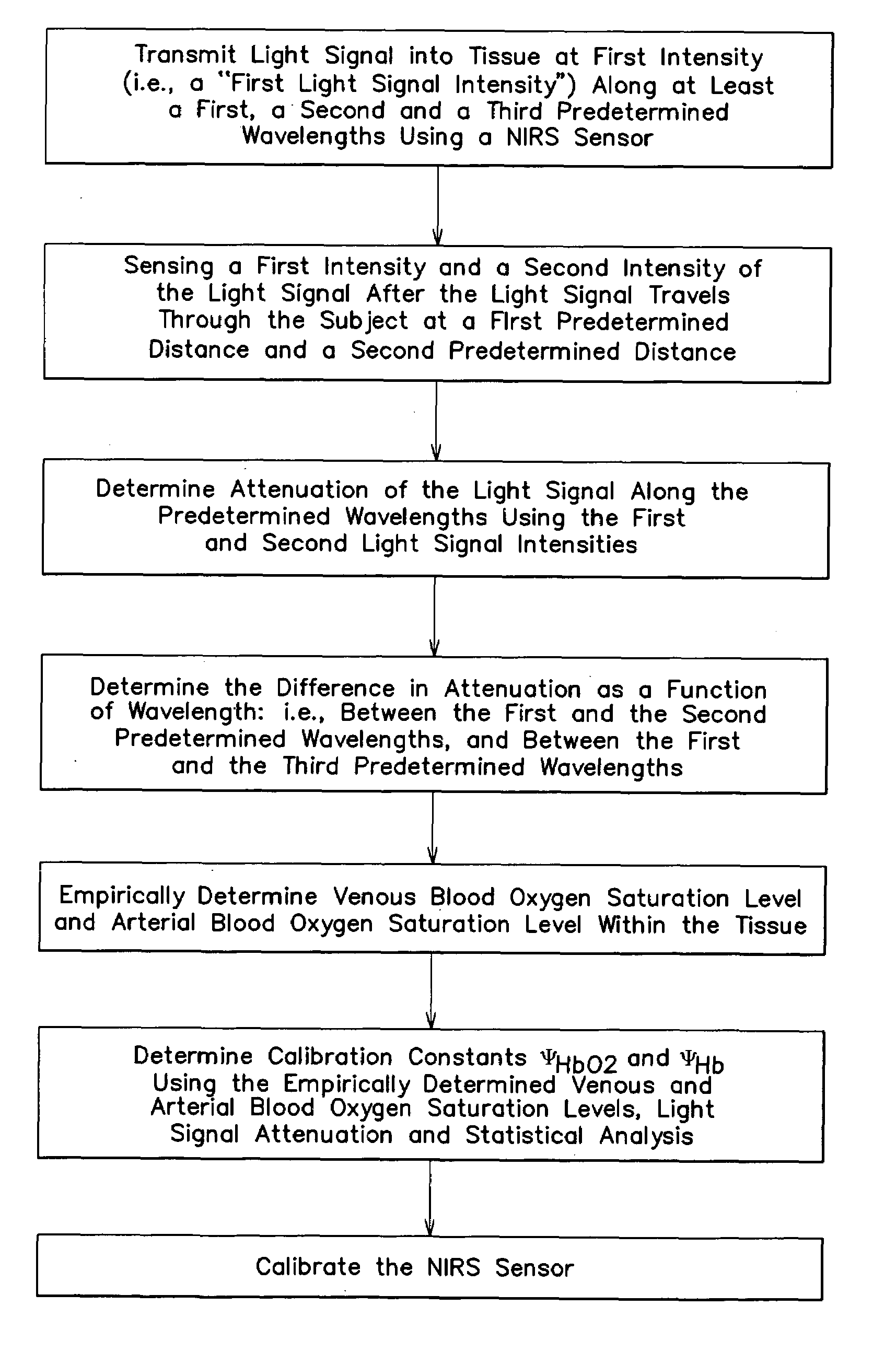
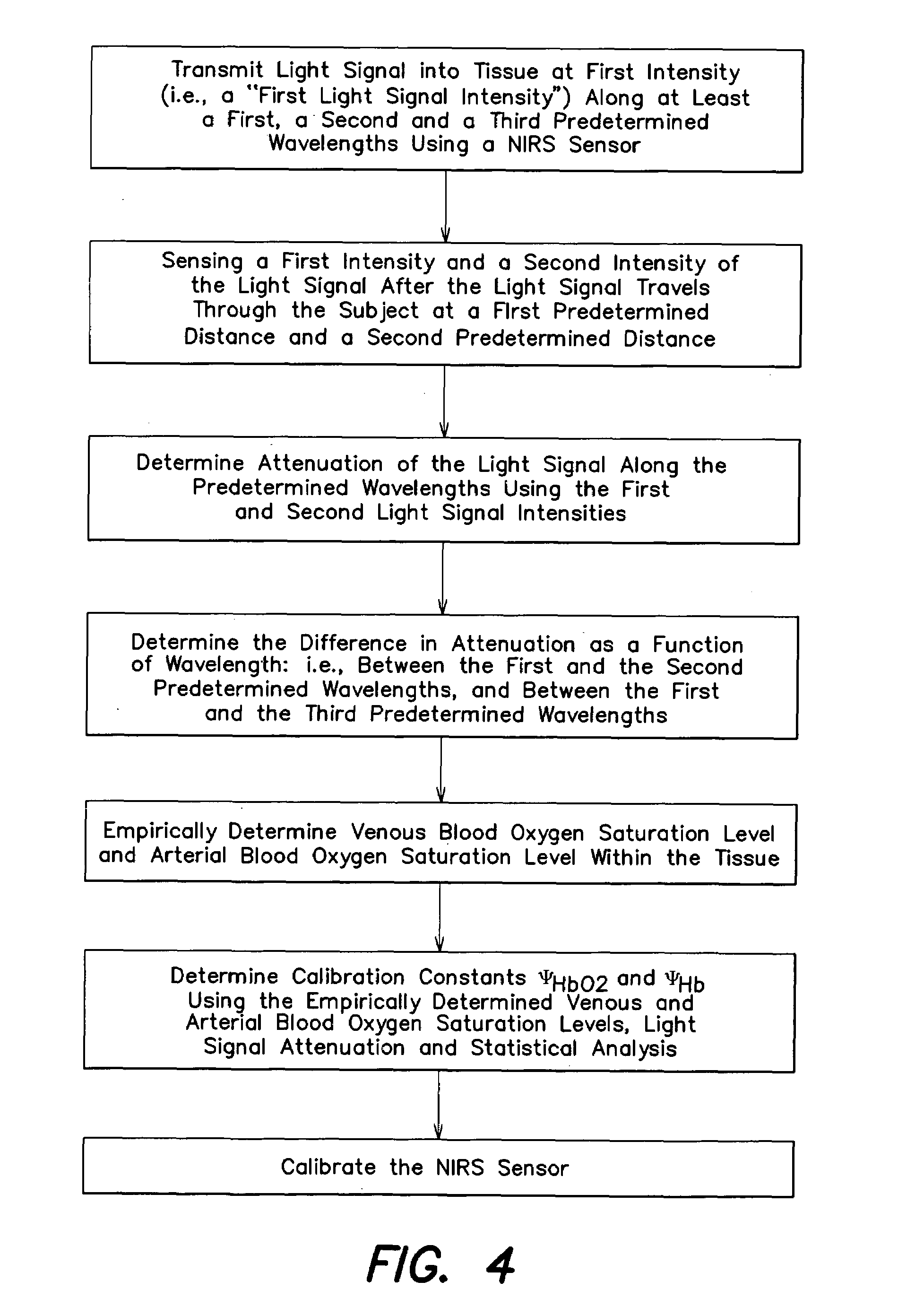
[0035]The present method of and apparatus for non-invasively determining the blood oxygen saturation level within a subject's tissue is provided that utilizes a near infrared spectrophotometric (NIRS) sensor that includes a transducer capable of transmitting a light signal into the tissue of a subject and sensing the light signal once it has passed through the tissue via transmittance or reflectance. The present method and apparatus can be used with a variety of NIRS sensors. The present method is not limited to use with this preferred NIRS sensor, however.

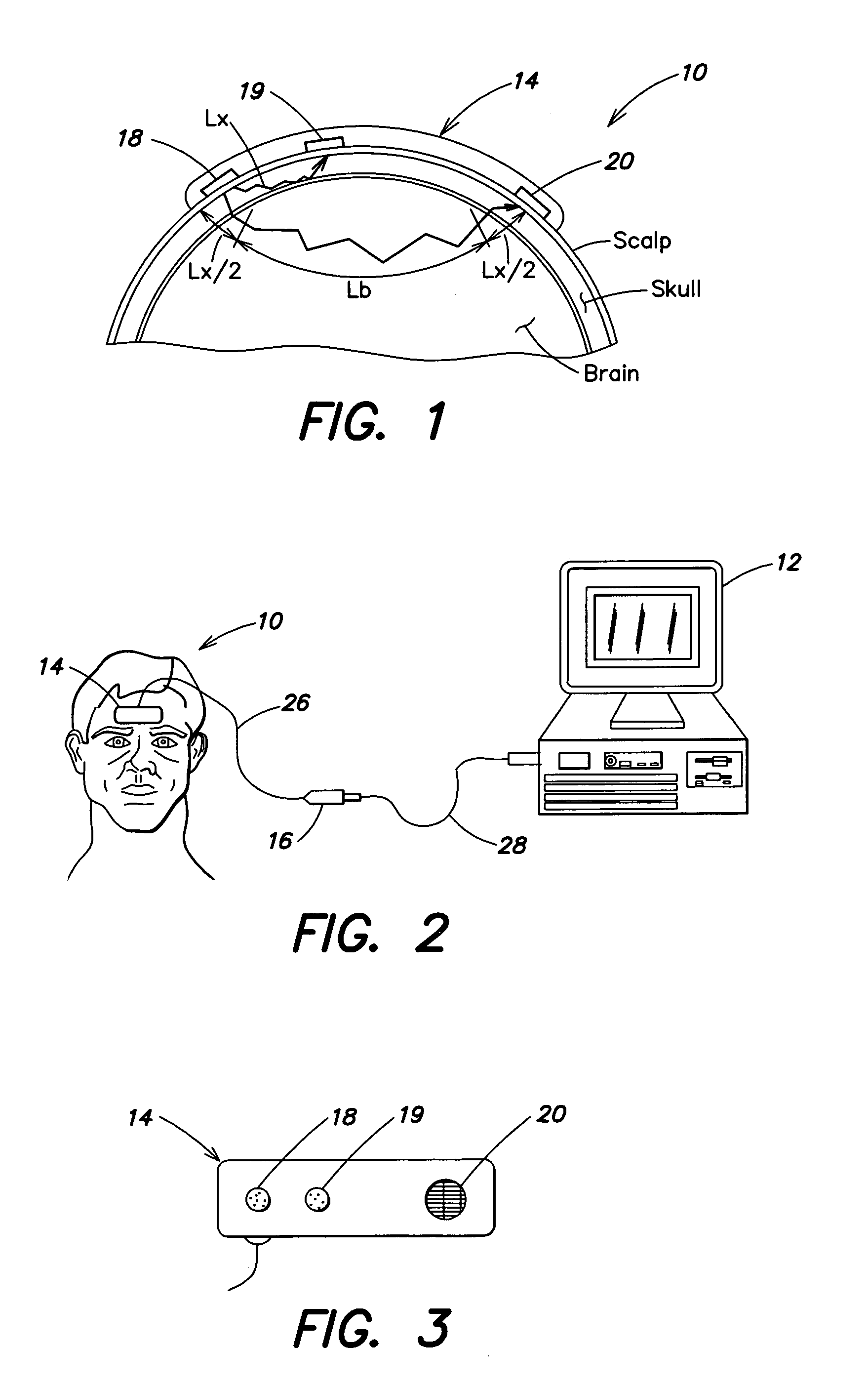
[0036]Referring to FIGS. 1–5, the preferred NIRS sensor includes a transducer portion 10 and processor portion 12. The transducer portion 10 includes an assembly housing 14 and a connector housing 16. The assembly housing 14, which is a flexible structure that can be attached directly to a subject's body, includes one or more light sources 18 and light detectors 19, 20. A disposable adhesive envelope or pad is used for mounting th...


PUM

PropertyMeasurementUnit
wavelength rangeaaaaaaaaaa
first wavelengthaaaaaaaaaa
second wavelengthaaaaaaaaaa

Abstract

A method and apparatus for non-invasively determining the blood oxygen saturation level within a subject's tissue is provided that utilizes a near infrared spectrophotometric (NIRS) sensor capable of transmitting a light signal into the tissue of a subject and sensing the light signal once it has passed through the tissue via transmittance or reflectance. The method includes the steps of: (1) transmitting a light signal into the subject's tissue, wherein the transmitted light signal includes a first wavelength, a second wavelength, and a third wavelength; (2) sensing a first intensity and a second intensity of the light signal, along the first, second, and third wavelengths after the light signal travels through the subject at a first and second predetermined distance; (3) determining an attenuation of the light signal for each of the first, second, and third wavelengths using the sensed first intensity and sensed second intensity of the first, second, and third wavelengths; (4) determining a difference in attenuation of the light signal between the first wavelength and the second wavelength, and between the first wavelength and the third wavelength; and (5) determining the blood oxygen saturation level within the subject's tissue using the difference in attenuation between the first wavelength and the second wavelength, and the difference in attenuation between the first wavelength and the third wavelength.

Description

[0001]This application claims the benefit of the filing date of U.S. Provisional Applications 60 / 398,937, filed Jul. 26, 2002, and 60 / 407,277 filed Aug. 30, 2002.[0002]This invention was made with Government support under Contract No. 1R43NS045488-01 awarded by the Department of Health & Human Services. The Government has certain rights in the invention.BACKGROUND OF THE INVENTION[0003]1. Technical Field[0004]This invention relates to methods for non-invasively determining biological tissue oxygenation in general, and to non-invasive methods utilizing near-infrared spectroscopy (NIRS) techniques in particular.[0005]2. Background Information.[0006]The molecule that carries the oxygen in the blood is hemoglobin. Oxygenated hemoglobin is called oxyhemoglobin (HbO2) and deoxygenated hemoglobin is deoxyhemoglobin (Hb). Total hemoglobin is the summation of the two states of hemoglobin (Total Hb=HbO2+Hb), and is proportional to relative blood volume changes, provided that the hematocrit or...

Claims


Application Information

Patent Timeline
Patent Type & Authority Patents(United States)
IPC IPC(8): A61B5/00G01N21/17A61BA61B5/145A61B5/1455A61B5/1495G01N21/35
CPCA61B5/14553
Inventor CHEN, BOBENNI, PAUL B.
Owner EDWARDS LIFESCIENCES CORP
Why Eureka
Try Eureka