Imaging apparatus and method with event sensitive photon detection

a photon detection and imaging apparatus technology, applied in the field of photon detection and imaging, can solve the problems of adversely affecting the image quality of a conventional ct, increasing the radiation exposure of patients, and deficient in providing detailed information regarding a small abnormal tissue, so as to improve the signal to noise ratio of an object examination system, accurate radiation detection, and accurate detection of generated particles

Inactive Publication Date: 2006-07-18
VARIAN MEDICAL SYSTEMS
View PDF10 Cites 60 Cited by
  • Summary
  • Abstract
  • Description
  • Claims
  • Application Information

AI Technical Summary

Benefits of technology

[0009]In accordance with another embodiment, a method for examining an object is provided. The method includes generating a particle in an object, detecting the particle, and generating a signal in response to the detected particle. By means of non-limiting examples, the particle may be generated by introducing a radioactive isotope into the object, or by a positron-electron annihilation process. Also by means of non-limiting examples, the particle may comprise a proton, a positron, a neutron, or an electron. The method also includes accessing the generated signal in an event sensitive mode, in which case, the signal is accessed as the signal is generated and before the signal integrates with another signal. Accessing the generated signals in an event sensitive mode allows an accurate detection of the generated particle, and improves a signal to noise ratio of an object examination system. In one embodiment, the object is at least a portion of a body of a patient who is undergoing a PET scanning or a single-photon emission computed tomography (SPECT) scanning. In another embodiment, the object includes a passenger, a luggage, or a cargo. In this case, the method may be used to determine whether the object contains contraband or explosive based on the generated data. In yet another embodiment, the object includes a mineral rock. In this case, the method may be used to determine whether the object contains a diamond.
[0010]In accordance with another embodiment, an imaging apparatus is provided. The imaging apparatus includes a conversion panel for generating a light photon in response to a radiation, and a photo detector array aligned with the conversion panel and configured to generate a signal in response to the light photon received from the conversion panel. The imaging apparatus also includes an access circuit coupled to the photo detector array. The access circuit operates in an event sensitive mode, and reads the signal as the signal is generated and before the signal integrates with another signal. Accessing the signals in an event sensitive mode allows an accurate detection of the radiation, and improves a signal to noise ratio of the imaging apparatus.

Problems solved by technology

A conventional CT is deficient in providing detailed information regarding a small abnormal tissue, e.g., a tumor, in the patient's body.
In addition, the intrinsic noise in the electronic circuits may adversely affect the image quality of a conventional CT.
Increasing the intensity of the X-ray radiation may improve the signal to noise ratio of the circuits, but will also increase radiation exposure of the patient.
Excessive increase in the radiation intensity may also adversely affect the resolution of the images.
The low spatial resolution of the photomultipliers may adversely affect the quality of PET and SPECT images.
Other constraints on the PET and SPECT image qualities include the temporal and spatial resolution and counting rate characteristics of the photomultipliers and associated circuitry.

Method used


Image

  • Imaging apparatus and method with event sensitive photon detection
  • Imaging apparatus and method with event sensitive photon detection
  • Imaging apparatus and method with event sensitive photon detection

Examples


Embodiment Construction

[0018]Various embodiments of the present invention are described hereinafter with reference to the figures. It should be noted that the figures are not drawn to scale and elements of similar structures or functions are represented by like reference numerals throughout the figures. It should also be noted that the figures are only intended to facilitate the description of specific embodiments of the invention. They are not intended as an exhaustive description of the invention or as a limitation on the scope of the invention. In addition, an aspect described in conjunction with a particular embodiment of the present invention is not necessarily limited to that embodiment and can be practiced in any other embodiments of the present invention.

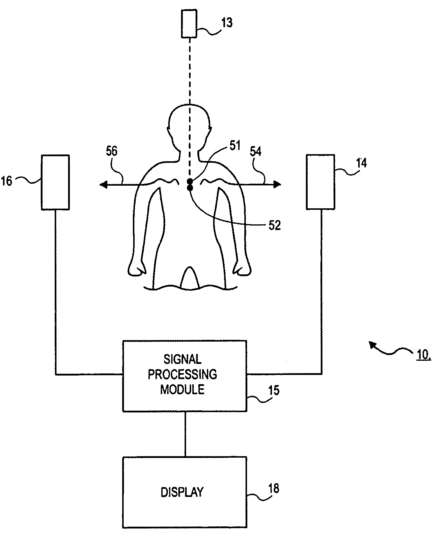
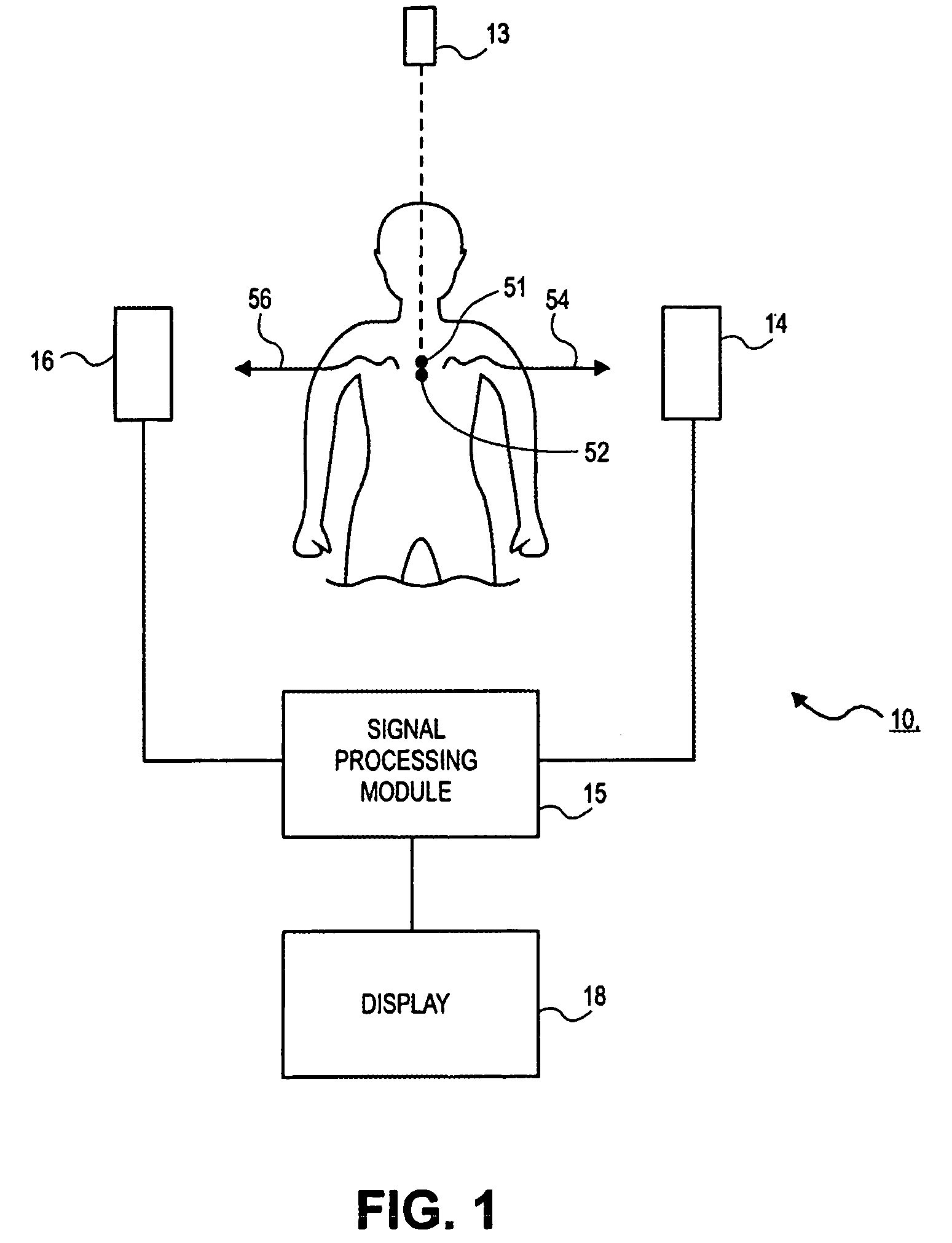
[0019]FIG. 1 is a functional block diagram illustrating a positron emission tomography (PET) apparatus 10 in accordance with an embodiment of the present invention. A PET process generates images of an object, such as tissues in a patient 11, by d...


PUM

PropertyMeasurementUnit
energy levelaaaaaaaaaa
massaaaaaaaaaa
energyaaaaaaaaaa

Abstract

An imaging apparatus (10) includes a photon detector (20) and an accessing circuit (44) coupled thereto. The photon detector (20) detects photons and generates signals in response thereto. The accessing circuit (44) reads out the signals from the photon detector (20) at a sufficiently high rate so that it operates in an event sensitive mode. The apparatus (10) also includes a signal processing module (15) for processing the signals and generating data regarding the images of the object. In accordance with various embodiments of the present invention, the signal processing module (15) may include a spatial resolution circuit (56), a photon energy resolution circuit (57), a temporal resolution circuit (58), or any combination thereof.

Description

RELATED APPLICATION DATA[0001]This application claims priority to U.S. Provisional Patent Application No. 60 / 416,021, filed on Oct. 4, 2002, the entire disclosure of which is expressly incorporated by reference herein.BACKGROUND OF THE INVENTION[0002]The present invention relates, in general, to photon detection and imaging and in one embodiment, specifically, to event sensitive photon detection in an imaging process and detection of photons produced through interactions between an incident beam and material in the object to be imaged.[0003]Computed tomography (CT) is widely used in medical diagnosis. In a typical CT process, a radiation source emits a thin X-ray beam while rotating around a patient. X-ray image detectors positioned at the opposite side of the patient from the radiation source pick up and record data regarding absorption of the X-ray beam by various tissues and bones in the patient. A computer processes the data and generates images that assimilate multiple X-ray im...

Claims


Application Information

Patent Timeline
Patent Type & Authority Patents(United States)
IPC IPC(8): G01N23/02A61B6/03G01NG01T1/164G01T1/29
CPCG01T1/2985A61B6/037G01V5/005A61B6/4258G01V5/0069
Inventor SEPPI, EDWARD J.
Owner VARIAN MEDICAL SYSTEMS
Why Eureka
Try Eureka