Materials and methods for treating oncological disease

a technology for oncological disease and materials, applied in the field of materials and methods for treating oncological disease, can solve the problems of mutagenic events, insufficient addition of normal genes, and inability to readily detect the potential of gene therapy for the treatment of malignancies

Inactive Publication Date: 2006-08-22
MORPHOGENESIS
View PDF9 Cites 16 Cited by
  • Summary
  • Abstract
  • Description
  • Claims
  • Application Information

AI Technical Summary

Problems solved by technology

While a place for somatic gene therapy in the treatment of inherited single gene disorders is no longer disputed, the potential of gene therapy for the treatment of malignancies may not be readily apparent.
Because most forms of cancer have been shown to be complex, multifactorial, and multigenic in nature, there are many conceptual and technical obstacles which must be overcome in order to approach this disease at the genetic level.
However, in other cases, the addition of a normal gene is not sufficient to repair the abnormality because this technique does not remove or correct the resident, nonfunctional mutant gene.
Furthermore, the random insertion of foreign sequences into nonspecific sites of the genome may result in mutagenic events such as insertional inactivation of genes necessary for the viability of that cell, or uncontrolled regulation of the transgene and / or flanking chromosomal sequences.
However, the vast majority of malignancies arise in immunocompetent hosts.
Although cellular activity is normally regulated by the various protein kinases, growth factors, growth factor receptors, and DNA binding proteins encoded by proto-oncogenes, together with genes that can suppress malignant transformation, such as the retinoblastoma and p53 genes, if one or more of these genes is or becomes defective, it can result in a clone with an abnormal pattern of growth control.
The fact that such a clone grows uncontrolled in an individual, indicates that the immune system has either failed to recognize tumor-specific antigens or has failed to effectively respond.
Despite the widespread use of chemotherapeutic agents for the treatment of solid tumors, efficacy has been restricted by their toxicity to normal cells.
However, the interaction of the superantigen with TCR is apparently of low avidity and is usually insufficient to generate a full response (Dohlsten et al., 1991, Herman et al., 1991, and Avery et al., 1994).

Method used


Image

  • Materials and methods for treating oncological disease
  • Materials and methods for treating oncological disease
  • Materials and methods for treating oncological disease

Examples


example 1

Tumorgenicity of Neuro-2a in A / J Syngeneic Mice

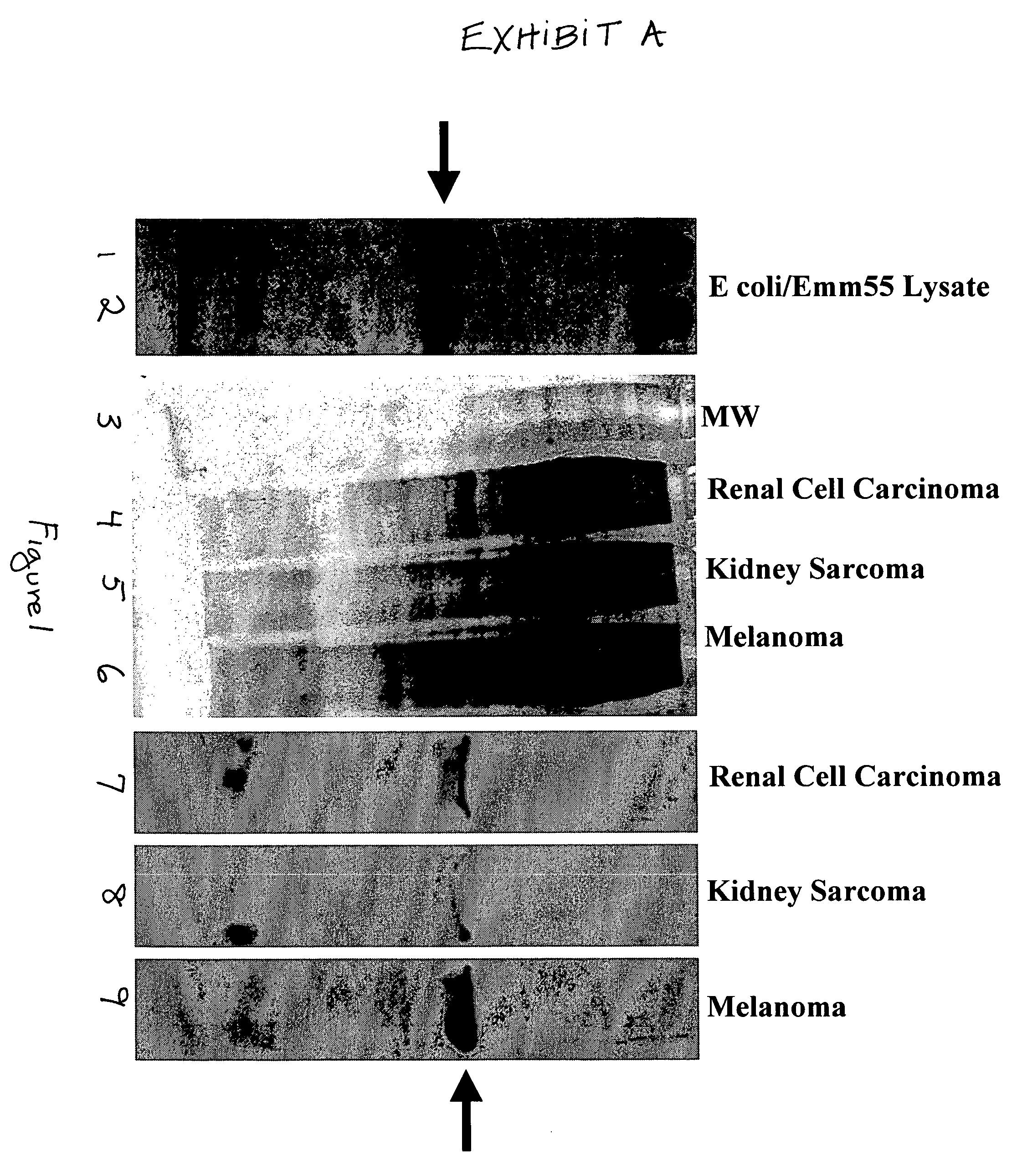
[0078]Neuroblastoma cells were highly tumorigenic in A / J syngeneic mice. Visible tumors appeared at the site of inoculation after a variable latency period. The duration of latency was dependent upon the number of cells injected (Table 1). Injection of 3×10⁶ and 1×10⁶ cells caused tumor formation in 100% of the mice tested, and in these cases, the latency period was 7–20 days. Eighty percent of the mice injected with 5×10⁵ and 1×10⁵ cells developed tumors within 10–28 days. Once the tumors appear, they grew rapidly and their size was not dependent on number of cells originally injected.

[0079]The tumors grew under the skin, invaded the surrounding muscle tissue and approached 75% of the weight of the mouse. Resected tumors were soft and well vascularized; angiogenesis of blood vessels was evident. Additionally, in some mice secondary tumor formation was observed. The overwhelming size of the malignant tissue caused necrosis in some of th...

example 2

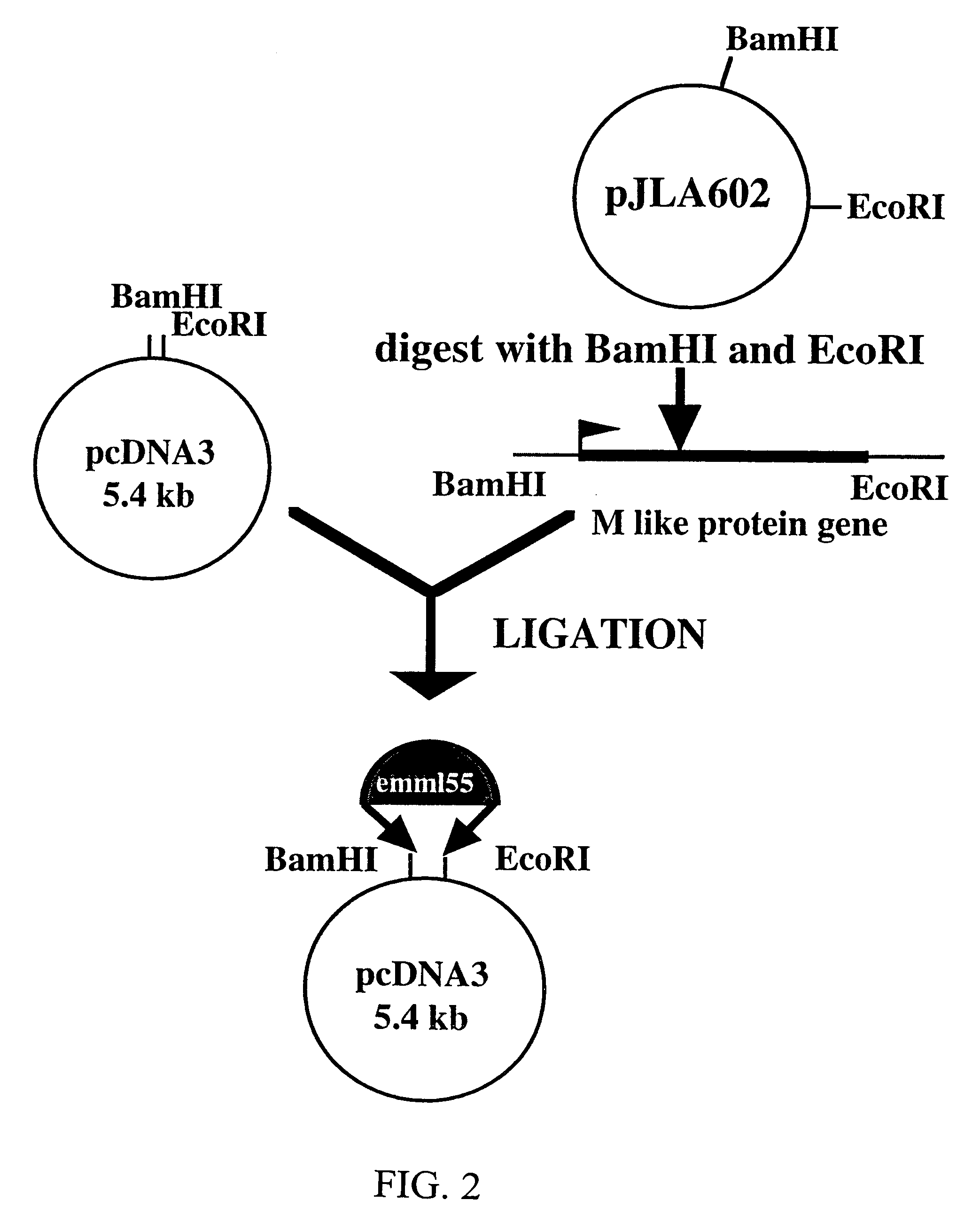
Construction and Analysis of the pCR II / emmL 55

[0080]In order to subclone the emmL 55 gene into pSVK 3, emmL 55 was amplified from the parental vector, pJLA 602 and restriction sites necessary for subcloning were added to the ends of the amplified product. Agarose gel electrophoresis of the emmL 55 PCR product showed that the size of amplified gene was 1.6 kb and the amount of the amplified product was 8 ng per 1 μl of the TE buffer.

[0081]One Shot INFVαF′E coli. competent bacterial cells were then transformed with pCR II / emmL 55. Bacterial clones were screened for the presence of the recombinant construct by white / blue selection. One clone showed the expected size 5.5 kb and was chosen for restriction analysis. The pCR II / emmL 55 construct, when digested with Eco RI and Xho I restriction enzymes, released the emmL 55 gene. Agarose gel electrophoresis of the digested product show the expected band pattern, 1.6 kb for emmL 55 and 3.9 kb for the pCR II vector. The amplified emmL 55 gen...

example 3

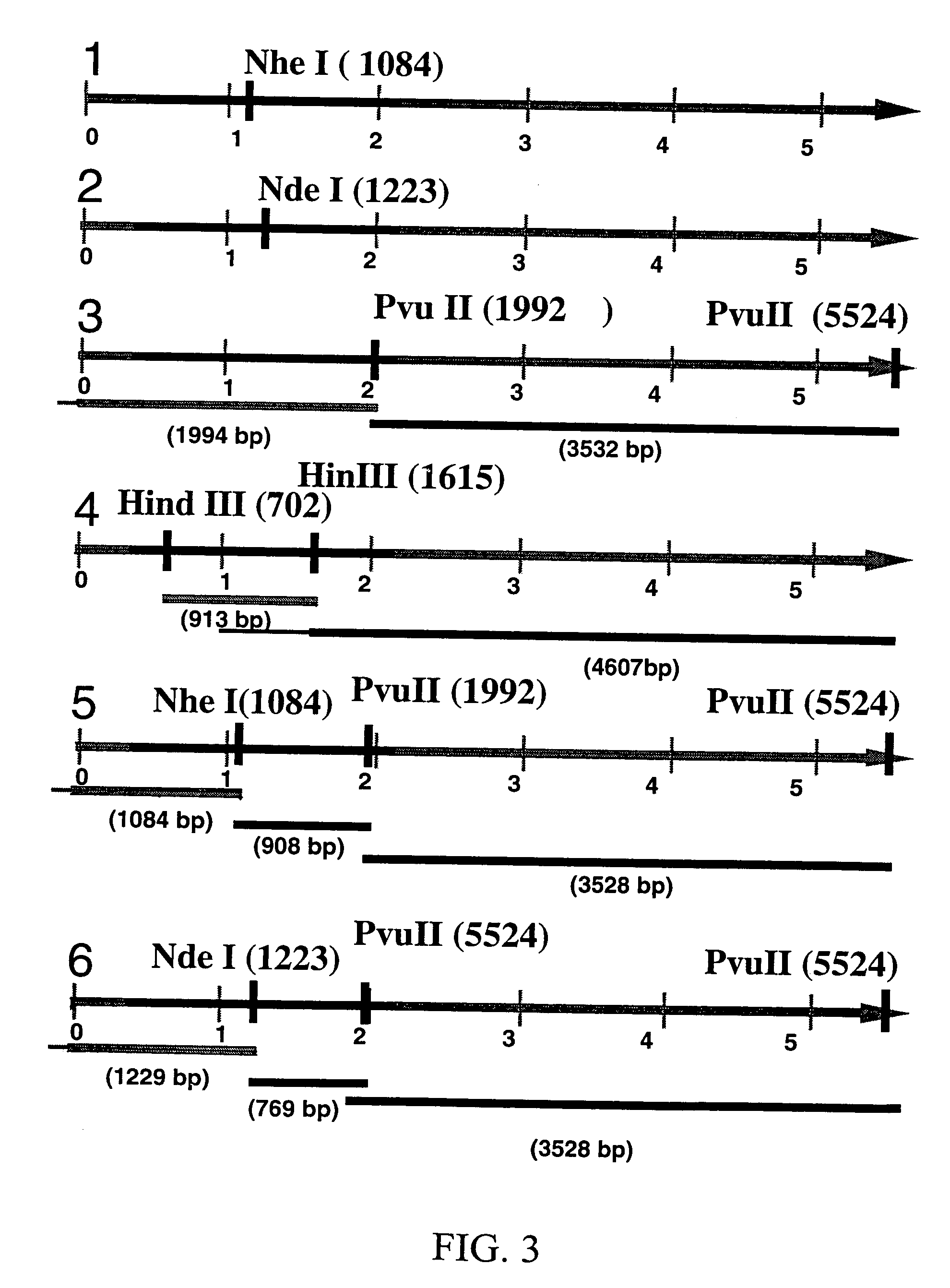
Construction and Analysis of pSVK 3 / emmL 55

[0082]From the 20 analyzed bacterial clones, transformed with the pSVK 3 / emmL 55, four clones with 5.5 kb size were selected and digested with restriction enzymes. Three out of four clones transformed with pSVK 3 / emmL 55 exhibited the expected banding pattern and one was used for the subsequent analysis. pSVK 3 / emmL 55 when digested with Nhe I and with Nde I generated a 5.5 kb band as expected. Digestion with Pvu II yielded a 2 kb and a 3.5 kb band and digestion with Hind III generated two bands, 0.9 kb and 4.6 kb. Double digestion with Pvu II and Nhe I resulted in three bands of approximately 1 kb, 2 kb and 5.5 kb pairs and digestion with Pvu II and Nde I generated three bands, 0.77 kb, 1.2 kb, and 3.5 kb (FIG. 3). Restriction analysis of pSVK 3 / emmL 55 revealed that the recombinant construct had the expected size (5.5 kb) and the expected map.

[0083]In order to confirm that the correct product was amplified, sequence analysis was performed...


PUM

PropertyMeasurementUnit
pHaaaaaaaaaa
final volumeaaaaaaaaaa

Abstract

Novel methods are disclosed for treating oncological disorders in an individual or animal using a superantigen expressed in tumor cells. A gene encoding a superantigen, such as an M-like protein of group A streptococci, can be introduced into a tumor cell in order to make the tumor cell more immunogenic in the host. Also contemplated are methods wherein a cell expresses a superantigen or superantigens, and immunogenic or immunostimulatory proteins, such as foreign MHC, cytokines, porcine-derived hyperacute rejection antigen, Mycobacterium-derived antigens, and the like. The subject invention also pertains to cells transformed with polynucleotides encoding a superantigen and foreign MHC antigen, cytokines, and other immunogenic or immunostimulatory proteins. Transformed cells according to the subject invention are then provided to an individual or animal in need of treatment for an oncological disorder. The immune response to tumor cells transformed according to the present invention inhibits in vivo tumor growth and results in subsequent tumor regression. The subject invention also pertains to cell lines transformed with genes encoding a superantigen and, optionally, a foreign Class II MHC antigen and / or a cytokine.

Description

CROSS-REFERENCE TO A RELATED APPLICATIONS[0001]This application is a continuation of U.S. application Ser. No. 09 / 950,374 filed Sep. 10, 2001, now abandoned, which is a continuation of U.S. application Ser. No. 09 / 394,226, filed Sep. 13, 1999, now abandoned, which is a continuation of International Application No. PCT / US99 / 00787, filed Jan. 14, 1999, which claims priority from U.S. provisional application Ser. No. 60 / 071,497, filed Jan. 14, 1998, now abandoned.BACKGROUND OF THE INVENTION[0002]While a place for somatic gene therapy in the treatment of inherited single gene disorders is no longer disputed, the potential of gene therapy for the treatment of malignancies may not be readily apparent. Because most forms of cancer have been shown to be complex, multifactorial, and multigenic in nature, there are many conceptual and technical obstacles which must be overcome in order to approach this disease at the genetic level. Yet, it is the molecular nature of tumorigenesis, i.e., the a...

Claims


Application Information

Patent Timeline
Patent Type & Authority Patents(United States)
IPC IPC(8): C12N15/83C12N15/00C12N15/85C12N5/00C12N5/02C12N5/06A61K38/16A61K39/00A61K48/00C12N5/08
CPCA61K38/164A61K39/0011A61K39/092A61K2039/5156A61K2039/5152A61K48/00
Inventor LAWMAN, MICHAEL J. P.LAWMAN, PATRICIA
Owner MORPHOGENESIS
Why Eureka
Try Eureka