Method for remyelinating a demyelinized lesion due to injury in the brain or spinal cord

a demyelinized lesion and remyelinating technology, applied in the field of remyelinating a demyelinized lesion due to injury in the brain or spinal cord, can solve the problem of relatively invasive collection of tissues containing neural stem cells from the cerebrum, and achieve the effects of improving the efficiency of therapy, enhancing cell differentiation, and improving the proliferation rate of cells

Inactive Publication Date: 2006-08-29
MITSUI SUMITOMO INSURANCE CARE NETWORK CO LTD RECEIVES 5 +2
View PDF9 Cites 8 Cited by
  • Summary
  • Abstract
  • Description
  • Claims
  • Application Information

AI Technical Summary

Benefits of technology

[0011]Thus, an objective of the present invention is to provide cellular material that is useful in the treatment of neurological diseases, and which can be prepared safely and readily. Another objective of the present invention is to provide a method for treating neurological diseases, preferably a method for autotransplantation therapy, using the cellular material.
[0021]Cells in a cell fraction of the present invention do not have to be proliferated with any trophic factors (then again they can proliferate in the presence of trophic factors). Thus, these cells are simple and practical from the standpoint of the development of autotransplantation technique for the diseases in the neural, and are very beneficial in medical industry. In general, a cell fraction of the present invention is derived from vertebrate, preferably from mammal (for example, mouse, rat, human, etc.).
[0033]Transplantation of any of these cell fractions containing mesodermal stem cells, mesenchymal stem cells, stromal cells, or AC133-positive cells into demyelinated spinal cord can lead to efficient remyelination of the demyelinated region. In particular, the above-mentioned cell fraction containing mesenchymal stem cells can engraft favorably and differentiate into neural cells such as neurons or glia when transplanted into a stroke model or a cerebral infarction model.
[0035]The present invention also provides compositions for treating neurological diseases, which comprise a cell fraction or cells of the present invention. It is possible to transplant the cell fractions or cells of the present invention without modification. However, in order to improve the efficiency of therapy, they may be transplanted as compositions combined with various additives or introduced with genes. The preparation of compositions of the present invention may comprise, for example, (1) addition of a substance that improves the proliferation rate of cells included in a cell fraction of the present invention or enhances the differentiation of the cells into neural cells, or introducing a gene having the same effect; (2) addition of a substance that improves the viability of cells in a cell fraction of the present invention in damaged neural tissues, or introducing a gene having the same effect; (3) addition of a substance that, inhibits adverse effects of damaged neural tissue on the cells in a cell fraction of the present invention, or introducing a gene having the same effect; (4) addition of a substance that prolongs the lifetime of donor cells, or introducing a gene having the same effect; (5) addition of a substance that modulates the cell cycle, or introducing a gene having the same effect; (6) addition of a substance to suppress the immunoreaction or inflammation, or introducing a gene having the same effect; (7) addition of a substance that enhances the energy metabolism, or introducing a gene having the same effect; (8) addition of a substance that improves the migration of donor cells in host tissues, or introducing a gene having the same effect; (9) addition of a substance that improves blood flow (including inductions of angiogenesis), or introducing a gene having the same effect; (10) addition of a substance that cure the infectious diseases, or introducing a gene having the same effect, or (11) addition of a substance that cure the tumors, or introducing a gene having the same effect, but is not limited thereto.
[0038]According to the present invention, cells derived from bone marrow cells of a recipient can be transplanted as donor cells (autotransplantation therapy) This has the following advantages: (1) low risk of rejection for the transplantation; and (2) no difficulty in using immunosuppressant. When autotransplantation therapy is arduous, then cells derived from other person or nonhuman animal may be used. Cells frozen for storage are also usable. The donor cells may be derived from cord blood.
[0040]Transplantation of cells into a patient can be performed, for example, by first filling a syringe with cells to be transplanted. Herein, the cells are suspended in an artificial cerebrospinal fluid or physiological saline. Then, the damaged neural tissue is exposed by surgery, and, with a needle, directly injecting the cells into the lesion. Due to high migrating potential of cells contained in a cell fraction of the present invention, they can migrate in the neural tissues. Hence, cells can be transplanted into a region adjacent to the lesion. Moreover, injection of the cells into the cerebrospinal fluid is also expected to be efficacious. In the case of the injection of the cells into the cerebrospinal fluid, the cells can be injected into a patient by typical lumbar puncture, without surgical operation only under local anesthetization. Thus, the patient can be treated in patient's sickroom (not in an operation room), which makes the method preferable. Alternatively, intravenous injection (including any systemic transplantations such as intravenous, intraarterial, selective intraarterial administration) of the cells is also expected to be effective. Thus, transplantation can be carried out by a procedure based on typical blood transfusion, which is advantageous in that the treatment can be performed in patient's sickroom.

Problems solved by technology

However, while not causing neurologic deficits, collecting tissues containing neural stem cells from cerebrum is relatively invasive.

Method used


Image

  • Method for remyelinating a demyelinized lesion due to injury in the brain or spinal cord
  • Method for remyelinating a demyelinized lesion due to injury in the brain or spinal cord
  • Method for remyelinating a demyelinized lesion due to injury in the brain or spinal cord

Examples


example 1

Preparation of Bone Marrow Cells and Schwann Cells

[0046](1) Bone Marrow Mononuclear Cells

[0047]Mouse bone marrow cells (10 μl) were obtained from the femur of adult LacZ (a structural gene of P-galactosidase) transgenic mice (The Jackson Laboratory, Maine, USA). The collected sample was diluted in L-15 medium (2 ml) containing 3 ml Ficoll, and centrifuged at 2,000 rpm for 15 minutes. Cells were collected from the mononuclear cell fraction, and suspended in 2 ml serum-free medium (NPMM: Neural Progenitor cell Maintenance Medium). Following centrifugation (2,000 rpm, 15 min), the supernatant was removed, and precipitated cells were collected and re-suspended in NPMM.

[0048](2) Schwann Cells

[0049]Primary Schwann cell cultures were established from the sciatic nerve of neonatal mouse (P1–3) according to the method of Honmou et al. (J. Neurosci., 16(10): 3199–3208, 1996). Specifically, cells were released from sciatic nerve by enzymatic and mechanical treatment. 8×10⁵ cells per plate were...

example 2

Experimental Animal Preparation and Transplantation

[0050](1) Preparation of Demyelinated Rat Model

[0051]Experiments were performed on 12 week old Wistar rats. A localized demyelinated lesion was created in the dorsal columns using X-ray irradiation and ethidium bromide injection (EB-X treatment) Specifically, rats were anesthetized with ketamine (75 mg / kg) and xylazine (10 mg / kg) i.p., and a surface dose of 40 Grays of X-ray was irradiated using Softex M-150 WZ radiotherapy machine (100 kV, 1.15 mA, SSD 20 cm, dose rate 200 cGy / min) on the spinal cord caudal to the tenth thoracic spine level (T-10) through a 2×4 cm opening in a lead shield (4 mm thick). Three days after X-ray irradiation, rats were anesthetized as above, and aseptic laminectomy of the eleventh thoracic spine (T-11.) was conducted. A demyelinating lesion was generated by the direct injection of ethidium bromide (EB) into the dorsal columns via a glass micropipette whose end was drawn. 0.5 μl saline containing 0.3 mg / ...

example 3

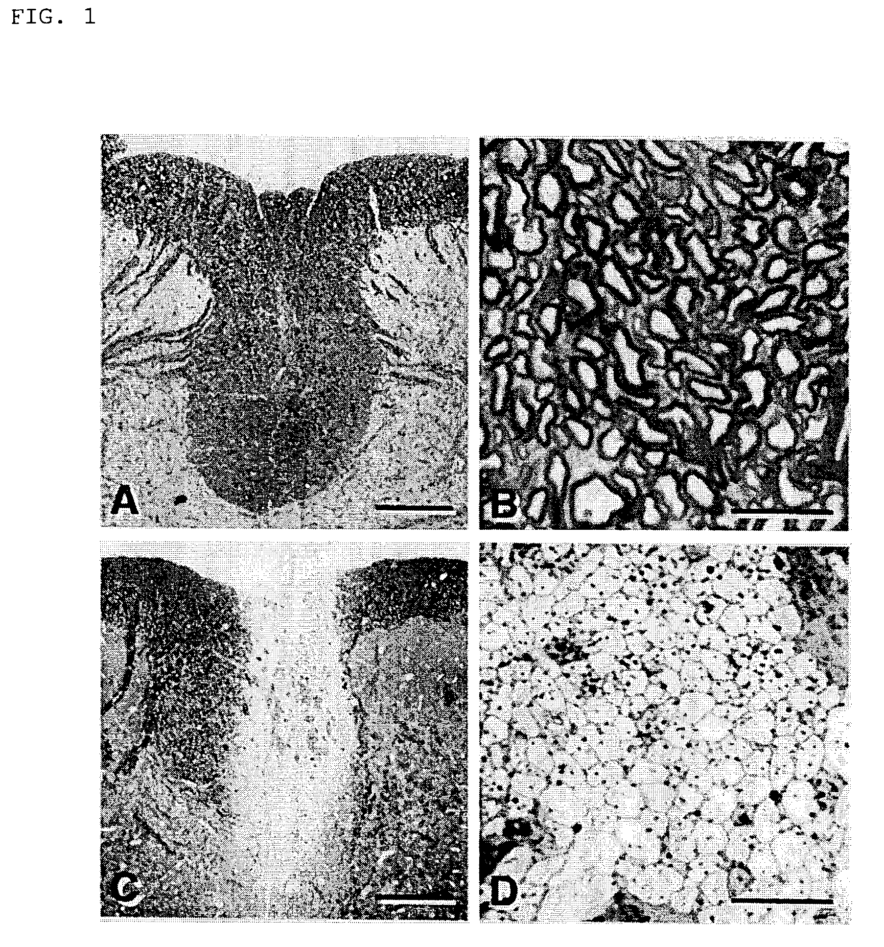
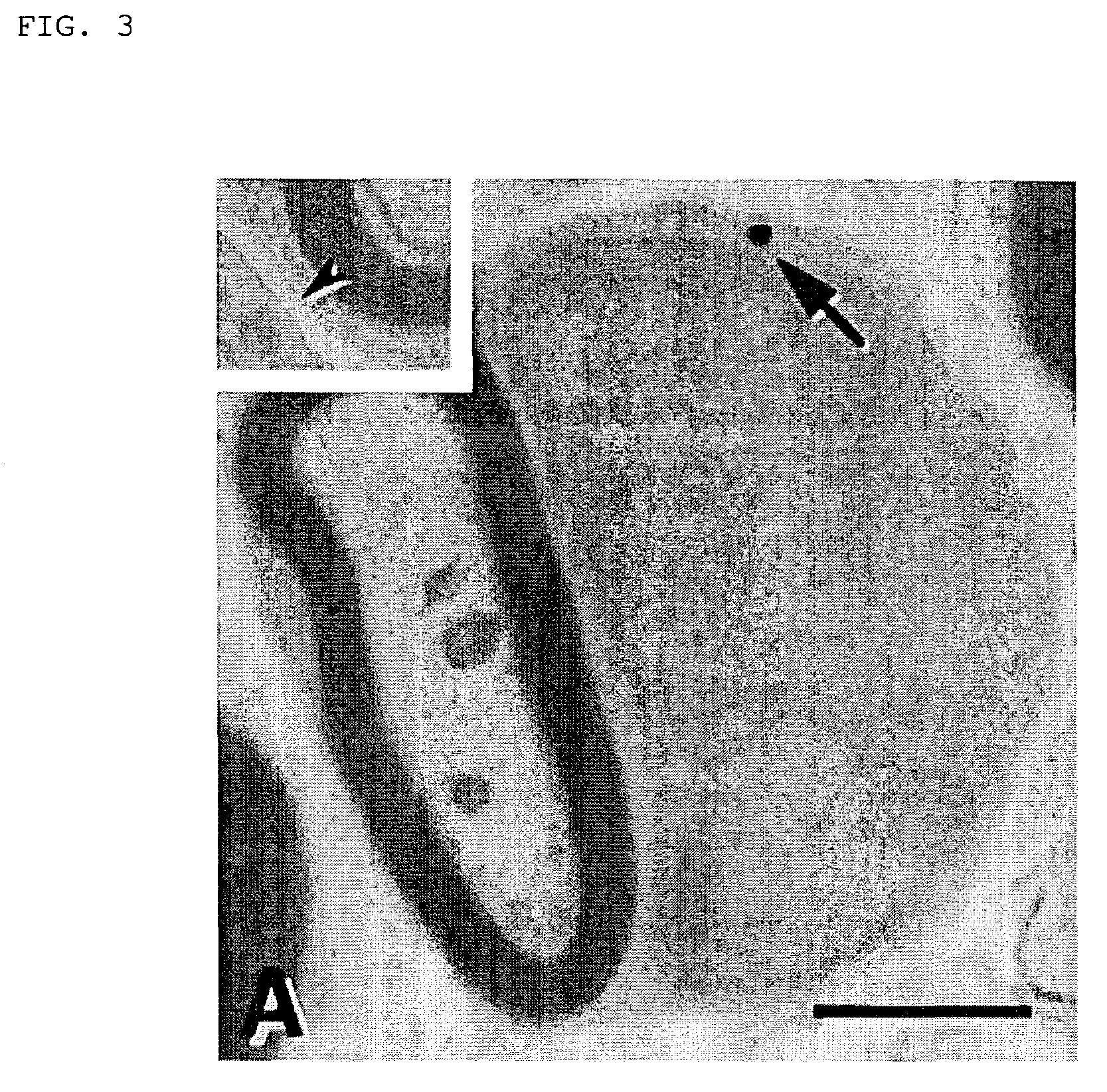
Histological Examination

[0054]Rats were deeply anesthetized by the administration of sodium pentobarbital (60 mg / kg, i.p.), and perfused through the heart cannula, first, with phosphate-buffer solution (PBS) and then with a fixative containing 2% glutaraldehyde and 2% paraformaldehyde in 0.14 M Sorensen's phosphate buffer, pH 7.4. Following in situ fixation for 10 minutes, the spinal cord was carefully excised, cut into 1 mm segments and kept in fresh fixative. The tissue was washed several times in Sorensen's phosphatebuffer, post-fixed in 1% OsO4 for 2 hours at 25° C., dehydrated by elevating the concentration of the ethanol solution, passed through propylene oxide and embedded in EPON. Then, the tissue was cut into sections (1 μm), counterstained with 0.5% methylene blue and 0.5% azure II in 0.5% borax, and examined under light microscope (Zeiss: Axioskop FS). Ultrathin sections were counterstained with uranyl and lead salts, and examined with JEOL JEM1200EX electron microscope (...


PUM

PropertyMeasurementUnit
thickaaaaaaaaaa
thickaaaaaaaaaa
thickaaaaaaaaaa

Abstract

Demyelinated axons were remyelinated in the demyelinated rat model by collecting bone marrow cells from mouse bone marrow and transplanting the mononuclear cell fraction separated from these bone marrow cells.

Description

[0001]This application is a continuation-in-part of International Application Number PCT / JP01 / 05456, filed on Jun. 26, 2001.TECHNICAL FIELD[0002]The present invention relates to cells derived from bone marrow cells, cord blood cells, or embryonic hepatic tissues that can differentiate into neural cells, and cell fractions containing such cells. It is expected that these cells and cell fractions can be used to treat neurological diseases, particularly in autologous transplantation therapy.BACKGROUND ART[0003]Transplantation of oligodendrocytes (i.e., oligodendroglia) (Archer D. R., et al., 1994. Exp. Neurol. 125:268–77; Blakemore W. F., Crang A. J., 1988. Dev. Neurosci. 10:1–11; Gumpel M., et al. 1987. Ann. New York Acad. Sci. 495:71–85) or myelin-forming cells, such as Schwann cells (Blakemore W. F., 1977. Nature 266:68–9; Blakemore W. F., Crang A. J., 1988. Dev. Neurosci. 10:1–11; Honmou O. et al., 1996. J. Neurosci. 16:3199–208), or olfactory ensheating cells (Franklin R. J. et al...

Claims


Application Information

Patent Timeline
Patent Type & Authority Patents(United States)
IPC IPC(8): C12N5/00A01N63/00C12N5/06A61K35/12A61K35/14A61K35/28A61K35/407A61K35/44A61K35/51A61P25/00A61P25/08A61P25/28A61P35/00C12N5/0775C12N5/078C12N5/079
CPCC12N5/0622C12N5/0675A61K35/28C12N5/0663A61K35/12A61K2035/124C12N2506/1353A61P9/10A61P25/00A61P25/02A61P25/08A61P25/18A61P25/28A61P35/00C12N5/0662C12N5/0664C12N5/0665C12N5/0666C12N5/0667C12N5/0668C12N5/06C12N5/0618A61K9/0019A61K35/51C12N5/0676
Inventor HONMOU, OSAMUHASHI, KAZUOUEDE, TEIJI
Owner MITSUI SUMITOMO INSURANCE CARE NETWORK CO LTD RECEIVES 5
Why Eureka
Try Eureka