Methods to culture circovirus
- Summary
- Abstract
- Description
- Claims
- Application Information
AI Technical Summary
Benefits of technology
Problems solved by technology
Method used
Image
Examples
example 1
Preparation of Porcine Retinal Cells Transfected with Human Adenovirus E1 Gene Sequences (VIDO R1 Cells)
[0029]Primary cultures of porcine embryonic retina cells were transfected with 10 μg of plasmid pTG 4671 (Transgene, Strasbourg, France) by the calcium phosphate technique. The pTG 4671 plasmid contains the entire E1A and E1B sequences (nts 505–4034) of HAV-5, along with the puromycin acetyltransferase gene as a selectable marker. In this plasmid, the E1 region is under the control of the constitutive promoter from the mouse phosphoglycerate kinase gene, and the puromycin acetyltransferase gene is controlled by the constitutive SV40 early promoter. Transformed cells were selected by three passages in medium containing 7 μg / ml puromycin, identified based on change in their morphology from single foci (i.e., loss of contact inhibition), and subjected to single cell cloning. The established cell line was first tested for its ability to support the growth of E1 deletion mutants of HAV...
example 2
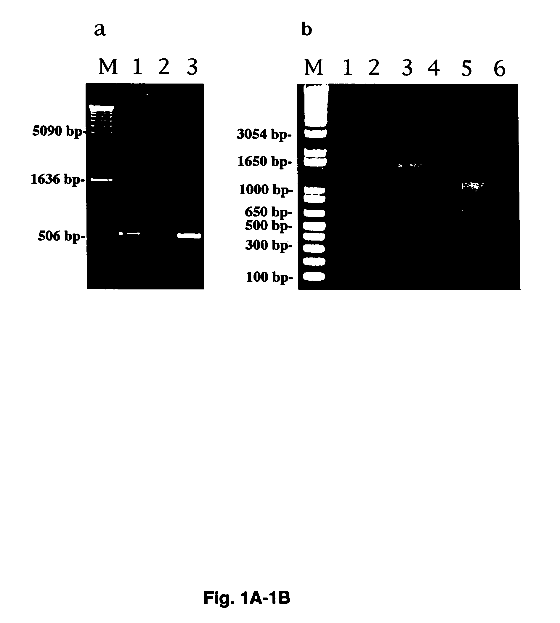
[0031]Example 2 provides a description of the molecular cloning of full-length PCV2 genome.
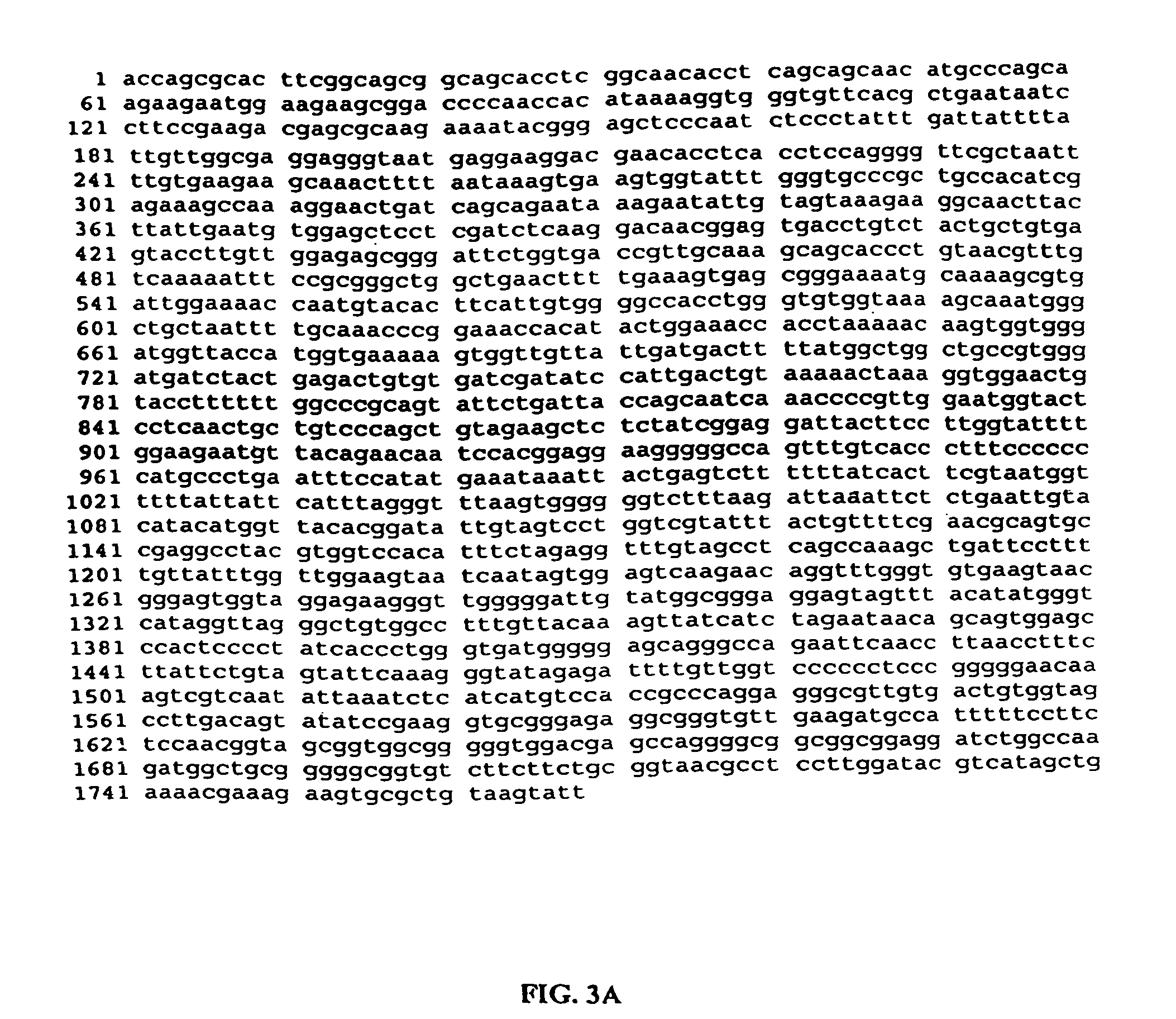
[0032]Initially, PCV2 DNA was amplified by PCR from total DNA extracted from a piglet with PMWS. The cloning of the full-length PCV2 genome DNA into vector pBluescript II KS(+) (Stratagene) by polymerase chain reaction (PCR) was described in Liu et al. (2000). J. Clin. Microbiol. 38:3474–3477). The PCV2 sequence was submitted to GenBank (Accession no. AF086834). The full-length PCV2 genome DNA is released from the resulting plasmid upon SacH digestion.
example 3
[0033]Example 3 describes the transfection of VIDO R1 cells, as described in Example 1, with a plasmid containing the PCV2 genome as constructed in Example 2.
Material and Methods
Cell Culture
[0034]Fetal porcine retina cell line, VIDO R1, as described in Example 1 and Vero cells (ATCC) were maintained at 37° C. with 5% CO2 in Eagles based MEM media supplemented with 10% or 5% heat-inactivated fetal bovine serum (FBS), respectively.
Transfection and Infection
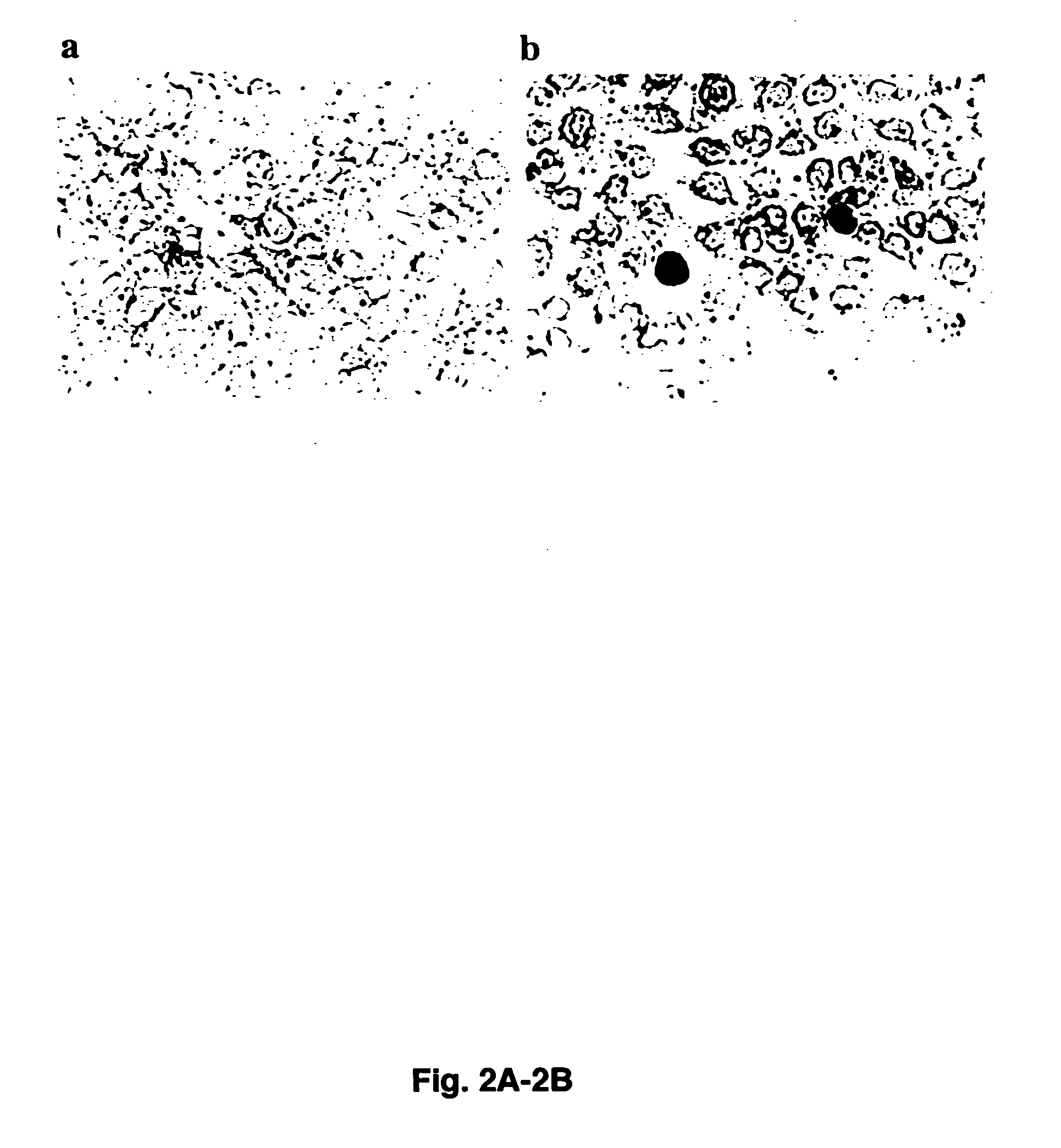
[0035]Monolayers of VIDO R1 cells grown in a six-well dish were transfected with cloned PCV2 DNA using Lipofectin according to the manufacturer's recommendations (GIBCO BRL). Prior to transfection, PCV2 full-length genome was released from the plasmid by digestion with SacII (Liu, Q., et al., 2000, J. Clin. Microbiol. 38:3474–3477). For infection, the transfected VIDO R1 cells were subjected to three cycles of freezing (−70° C.) and thawing (37° C.). The lysate was then clarified by centrifugation and used to infect fresh VIDO R1 ce...
PUM
| Property | Measurement | Unit |
|---|---|---|
| diameters | aaaaa | aaaaa |
| temperature | aaaaa | aaaaa |
| compositions | aaaaa | aaaaa |
Abstract
Description
Claims
Application Information
 - R&D
- Intellectual Property
- Life Sciences
- Materials
- Tech Scout
- Unparalleled Data Quality
- Higher Quality Content
- 60% Fewer Hallucinations
Browse by: Latest US Patents, China's latest patents, Technical Efficacy Thesaurus, Application Domain, Technology Topic, Popular Technical Reports.
© 2025 PatSnap. All rights reserved.Legal|Privacy policy|Modern Slavery Act Transparency Statement|Sitemap|About US| Contact US: help@patsnap.com


