Shear mode diagnostic ultrasound

a technology of ultrasound and ultrasound focusing, applied in the field of ultrasound diagnostic and therapeutic applications using shear waves, can solve the problems of transcranial procedures, strong attenuation, distortion caused by irregularities in the shape, density, sound speed, etc., and achieve the effect of improving the prediction of ultrasound focusing through the skull, simple devices, and high gain

Active Publication Date: 2007-02-13
THE BRIGHAM & WOMEN S HOSPITAL INC
View PDF24 Cites 78 Cited by
  • Summary
  • Abstract
  • Description
  • Claims
  • Application Information

AI Technical Summary

Benefits of technology

[0009]It has been discovered that ultrasound beams can be effectively, coherently transmitted through bone via shear waves. This can be accomplished, for example, by increasing the angle between the beam and the normal to the bone surface beyond the critical angle (approximately 20°) that causes complete reflections of the longitudinal waves. At angles between approximately 25° and 60°, shear waves are generated and propagated through the bone and at the inner surface converted again into longitudinal waves that propagate into the soft tissues on the other side of the bone. This finding has several implications: First, the shear waves have roughly the same propagation velocity in the bone as the longitudinal waves have in soft tissues. Thus, the wave front may not be significantly distorted by the variable thickness of bone as it is for longitudinal waves due to the higher speed of sound in bone (2500–3000 ms / s). It is possible to focus ultrasound beams though bone without performing complex patient-specific aberration corrections. For example, diagnostic ultrasound imaging of the brain can be performed as well as ultrasound brain treatments and diagnosis with relatively simple devices. A second implication is that the entrance angle of the beam can be increased and can reach larger brain volumes with higher gain than using longitudinal waves. Third, predictions of ultrasound focusing through a skull can be significantly improved by combining shear wave propagation modeling with longitudinal propagation models. Further, undesired reflections from bone layers may be reduced or eliminated, making diagnostic information easier to analyze.
[0010]Embodiments of the invention may provide one or more of the following capabilities and can be used for one or more of the following uses. The invention can be used in ultrasound diagnosis and / or therapy and can aid methods that rely on transmitting ultrasound through bone. In particular, the invention can be applied to systems that seek to image blood flow through bone, or can be an enhancement / add-on for existing ultrasound imaging and / or therapeutic devices, or in a stand-alone device for transbone ultrasound therapy and / or applied to techniques that rely on opening of the blood-brain barrier (BBB) using ultrasound or any other diagnostic or therapeutic procedures conducted using ultrasound. Focusing of ultrasound could be greatly simplified, and phase distortion correction could be reduced and possibly eliminated. The tissue volume that can be currently reached with ultrasound can be increased. In addition, embodiments of the invention can detect cavities in bony structures and / or detection / determination of cavity contents with undesired reflections from bone surfaces being reduced or eliminated.
[0028]Various aspects of the invention may provide one or more of the following capabilities. Ultrasound can be propagated through bone, e.g., the skull, while experiencing reduced distortion and increased signal strength, allowing clearer and more accurate brain images, and better focusing for therapy applications compared to previous techniques. Unwanted echoes from overlying bone layers may be reduced, making analysis of diagnostic information received from target tissue easier. Bone cavities and / or their contents may be detected. Transbone shear mode propagation can be used for a number of imaging problems including vessel detection, tumor detection, tissue morphology, and hemorrhaging in the brain. Transbone ultrasound can be provided with reduced distortion and / or higher location accuracy than previous techniques.

Problems solved by technology

The primary setbacks for transcranial procedures are strong attenuation and the distortion caused by irregularities in the skull's shape, density, and sound speed.
Modeling of shear waves has been dismissed as being either of insignificant amplitude or, if significant, that the resulting beam would be incoherent and hard to predict.
The absence of significant information on the skull bone's elastic wave speeds also has inhibited its consideration in modeling.
Similar issues exist with respect to ultrasound propagation through other bony structures.

Method used


Image

  • Shear mode diagnostic ultrasound
  • Shear mode diagnostic ultrasound
  • Shear mode diagnostic ultrasound

Examples


Embodiment Construction

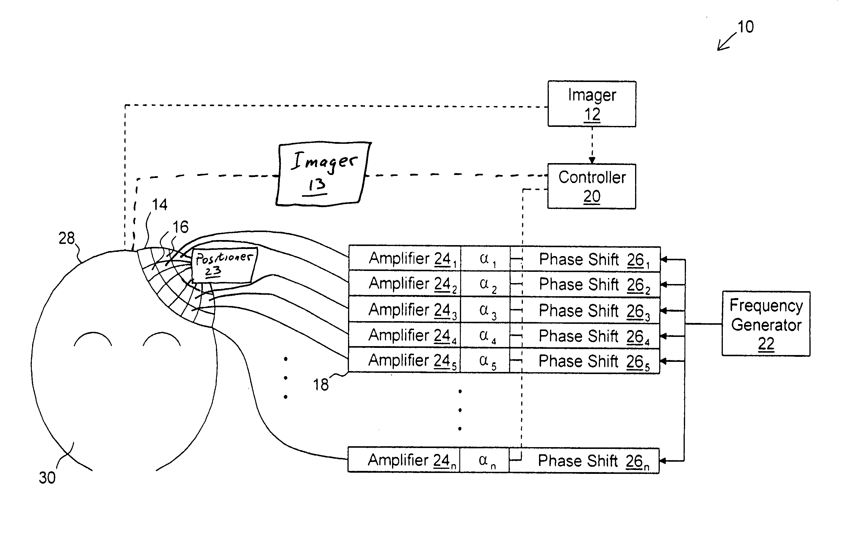
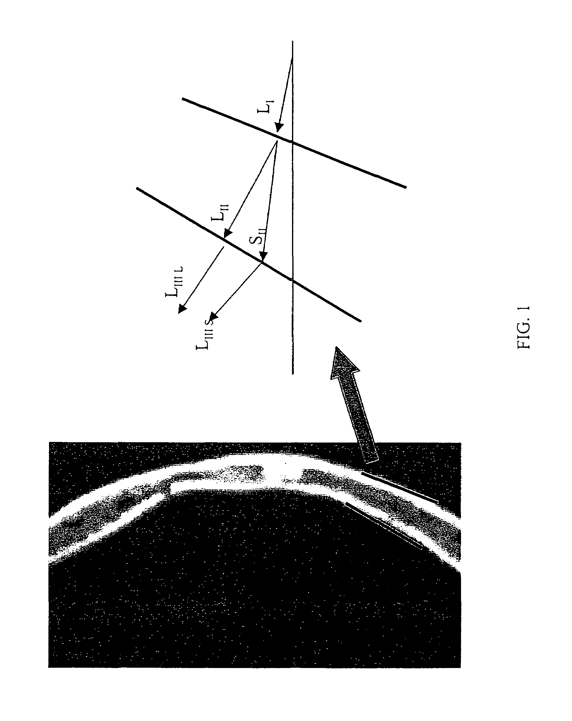
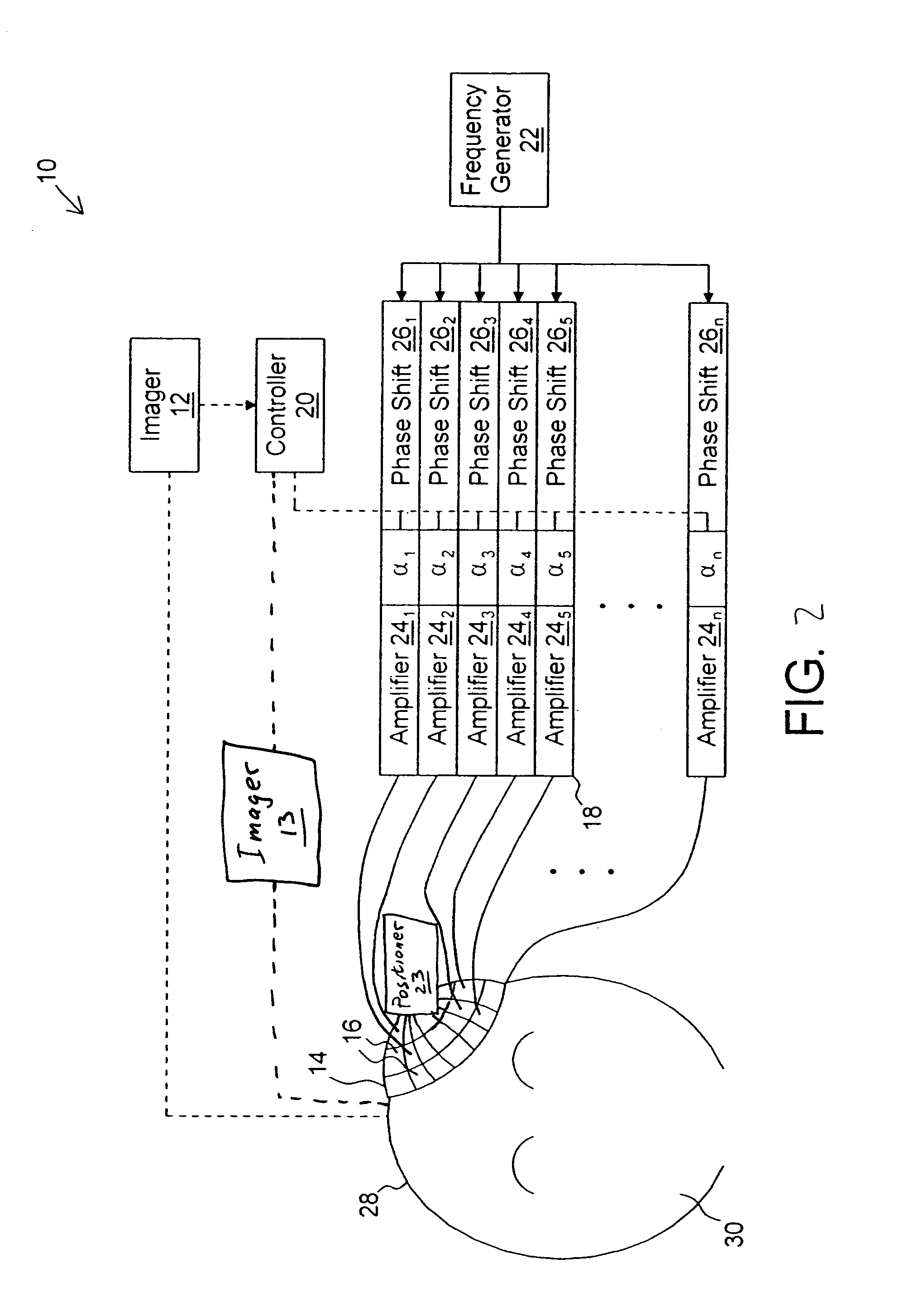
[0049]Embodiments of the invention provide techniques for transskull and other transbone propagation that deliberately induces a shear mode in bone. For transskull propagation, incident waves experience a mode conversion from an incident longitudinal wave into a shear wave in the bone layers and then back to a longitudinal wave in the brain. The skull's shear speed may provide a better acoustic impedance match, less refraction, and less phase alteration than its longitudinal counterpart. Using a shear wave, ultrasound may be focused in the brain. Longitudinal waves will not be induced in the bone if the ultrasound is incident upon the bone at an angle beyond Snell's critical angle. Numerical analysis is provided and demonstrations of the phenomena were studied with plastic phantoms and using an ex vivo human skull. Embodiments of the invention may be used for various applications, including therapeutic and diagnostic applications, which are discussed separately below. Other embodime...


PUM


Abstract

A method of diagnosing a subject by delivering ultrasound signals using shear waves includes applying a portion of an ultrasound mainbeam to a bone surface at an incident angle relative to the surface of the bone to induce shear waves in the bone, energy in the shear waves forming a substantial part of energy of first ultrasound waves at a desired region in the subject through the bone, detecting at least one of reflected and scattered energy of the applied ultrasound mainbeam, and analyzing the detected energy for a diagnostic purpose.

Description

CROSS-REFERENCE TO RELATED ACTIONS[0001]This application claims the benefit of U.S. Provisional Application No. 60 / 463,589 filed Apr. 17, 2003 and entitled “Transskull Shear Mode Ultrasound” which is incorporated here by reference.STATEMENT AS TO FEDERALLY-SPONSORED RESEARCH[0002]This invention was made at least in part with Government support under Grant No. NIH CA76550, awarded by the National Institutes of Health. The Government has certain rights in this invention.FIELD OF THE INVENTION[0003]The invention relates to ultrasound diagnostic and therapeutic applications using shear waves.BACKGROUND OF THE INVENTION[0004]Since its inception in the early 1980's, transcranial ultrasound Doppler imaging has demonstrated an ability to measure blood flow, hemorrhages, and perfusion in the brain. Recent research has also examined the possibility of diagnosing certain degenerative disorders such as Parkinson's and depression. These approaches use transcranial Doppler sonography (TCD), or th...

Claims


Application Information

Patent Timeline
Patent Type & Authority Patents(United States)
IPC IPC(8): A61B8/00A61B8/06A61B17/22A61N7/00
CPCA61B8/06A61B8/54A61B17/22004A61B8/485A61B8/0808A61N7/02A61N2007/0065A61N2007/0095
Inventor HYNYNEN, KULLERVOCLEMENT, GREGORY T.
Owner THE BRIGHAM & WOMEN S HOSPITAL INC
Why Eureka
Try Eureka