Antigen fragments for the diagnosis of Toxoplasma gondii
a technology of toxoplasma gondii and antigen fragments, applied in the field of medicine, can solve the problems of severe foetal disease in mammals, general asymptomatic and self-limiting human infection,
- Summary
- Abstract
- Description
- Claims
- Application Information
AI Technical Summary
Benefits of technology
Problems solved by technology
Method used
Image
Examples
example 1
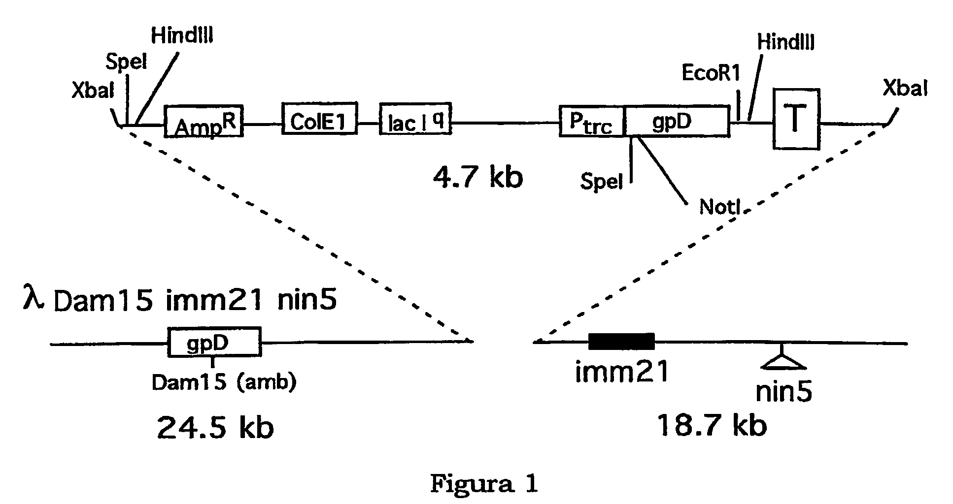
Construction of the Vector λKM4
[0048]This technique is described in international patent application No. PCT / IT01 / 00405, filed on 26 Jul. 2001 and incorporated herein for reference purposes, explicitly mentioning the references cited therein. FIG. 1 represents the map of the vector λKM4. The plasmid pNS3785 (Sternberg and Hoess, 1995, Proc. Natl. Acad. Sci. USA., 92:1609–1613) was amplified by inverse PCR using the synthetic oligonucleotides 5′-TTTATCTAGACCCAGCCCTAGGAAGCTTCTCCTGAGTAGGACAAATCC-3′ (SEQ ID No 1) bearing the sites XbaI and AvrII (underlined) for the subsequent cloning of the lambda phage, and 5′-GGGTCTAGATAAAACGAAAGGCCCAGTCTTTC-3′ (SEQ ID No 2) bearing the site XbaI. In inverse PCR a mixture of Taq DNA polymerase and Pfu DNA polymerase was used to increase the fidelity of the DNA synthesis. Twenty-five amplification cycles were performed (95° C.-30 sec, 55° C.-30 sec, 72° C.-20 min). The autoligation of the PCR product, previously digested with XbaI endonuclease gave ri...
example 2
[0086]Using the vector λKM4 of Example 1, a library of DNA fragments of known Toxoplasma gondii genes was constructed.
[0087]Cells of Toxoplasma gondii (106 parasites, strain ME49) were grown in vitro in monkey kidney cells (“VERO” African green monkey cells) using DMEM culture medium containing 10% foetal bovine serum, 2 mM glutamine and 0.05 mg / ml gentamicin (Gibco BRL, Canada). To have both forms of the parasite (tachyzoites and bradyzoites) present in the cell cultures, an experimental protocol was used based on the change in pH of the culture medium (Soete et al., 1994, Experimental Parasitology, 78, 361–370). The parasites were collected after complete lysis of the host cells and purified by filtration (filter porosity 3 μm) followed by centrifuging. 2 μg of mRNA were isolated from 5×10⁶ parasites using the “QuickPrep Micro mRNA Purification Kit” (Amersham Pharmacia Biotech, Sweden) and following the manufacturer's instructions. cDNA was synthesised from 200 ng of poly(A)+ RNA ...
example 3
[0109]By using the same strategy described in Example 2, a gene collection of DNA encoding for protein products of the Toxoplasma gondii microneme family was used to construct a “microneme-display library”.
[0110]For the construction of the microneme-library the following genes were amplified by means of PCR with specific oligonucleotides:[0111]1—MIC2 (Wan et al, 1997, Mol. Biochem. Parasitol. 84: 203–214) was obtained from single strand cDNA using the oligonucleotides 5′-ATGAGACTCCAACCGAGGCC-3′ (SEQ ID No 69) and 5′-CTGCCTGACTCTTTCTTGGACTG-3′ (SEQ ID No 70);[0112]2—M2AP (Rabenau et al., 2001, Mol. Microbiol. 41: 537–547) was obtained from single strand cDNA using the oligonucleotides 5′-GGAAAGTTGGAAATCCGGCGGC-3′ (SEQ ID No 71) and 5′-CGCCTCATCGTCACTCGGC-3′ (SEQ ID No 72)[0113]3—MIC4 (Brecht et al., 2001, J. Biol. Chem. 276:4119–412) was obtained from single strand cDNA using the oligonucleotides 5′-ATGAGAGCGTCGCTCCCGG-3′ (SEQ ID No 73) and 5′-GTGTCTTTCGCTTCAAGCACCTG-3′ (SEQ ID No 74...
PUM
| Property | Measurement | Unit |
|---|---|---|
| volume | aaaaa | aaaaa |
| volumes | aaaaa | aaaaa |
| volumes | aaaaa | aaaaa |
Abstract
Description
Claims
Application Information
 - R&D
- Intellectual Property
- Life Sciences
- Materials
- Tech Scout
- Unparalleled Data Quality
- Higher Quality Content
- 60% Fewer Hallucinations
Browse by: Latest US Patents, China's latest patents, Technical Efficacy Thesaurus, Application Domain, Technology Topic, Popular Technical Reports.
© 2025 PatSnap. All rights reserved.Legal|Privacy policy|Modern Slavery Act Transparency Statement|Sitemap|About US| Contact US: help@patsnap.com


