Methods and compositions for preparing tissue samples for RNA extraction

a tissue sample and rna extraction technology, applied in the field of histology and molecular biology, can solve the problems of compromising the ability to extract intact rna and/or reverse transcription using rna from fixed samples, and achieving the effect of reducing carryover or contamination

Active Publication Date: 2007-07-31
APPL BIOSYSTEMS INC
View PDF7 Cites 22 Cited by
  • Summary
  • Abstract
  • Description
  • Claims
  • Application Information

AI Technical Summary

Benefits of technology

[0023]In some embodiments of the invention, methods include a) contacting a tissue section with a first set of one or more solutions comprising no more than about 50% water; b) contacting the tissue section with a stain in a solution comprising no more than about 50% water; c) then contacting the tissue section with a second set of one or more solutions comprising no more than about 50% water, wherein the tissue section is not contacted with a solution that is more than about 50% water. The term “contacting” is used according to their plain and ordinary meaning to refer to exposing or submerging of the tissue section to or in a solution. “Dipping” the sample or section into a solution and ...

Problems solved by technology

Linear amplification is generally required for microarray analysis on thin tissue sections, since the amount of RNA needed for conversion to cDNA in order to carry out microarray hybridization is several micrograms, and this amount of RNA cannot generally be obtained from this type of sample.
Treatment of tissue with aldehyde fixatives such as formalin and paraformaldehyde causes chemical crosslinking of nucleic acid, which compromises the ability to extract intact RNA and/or to carry out reverse transcription using RNA from the fixed samples.
Successful extraction of intact RNA from frozen sections is however also challenging, because the e...

Method used


Image

  • Methods and compositions for preparing tissue samples for RNA extraction
  • Methods and compositions for preparing tissue samples for RNA extraction
  • Methods and compositions for preparing tissue samples for RNA extraction

Examples


example 1

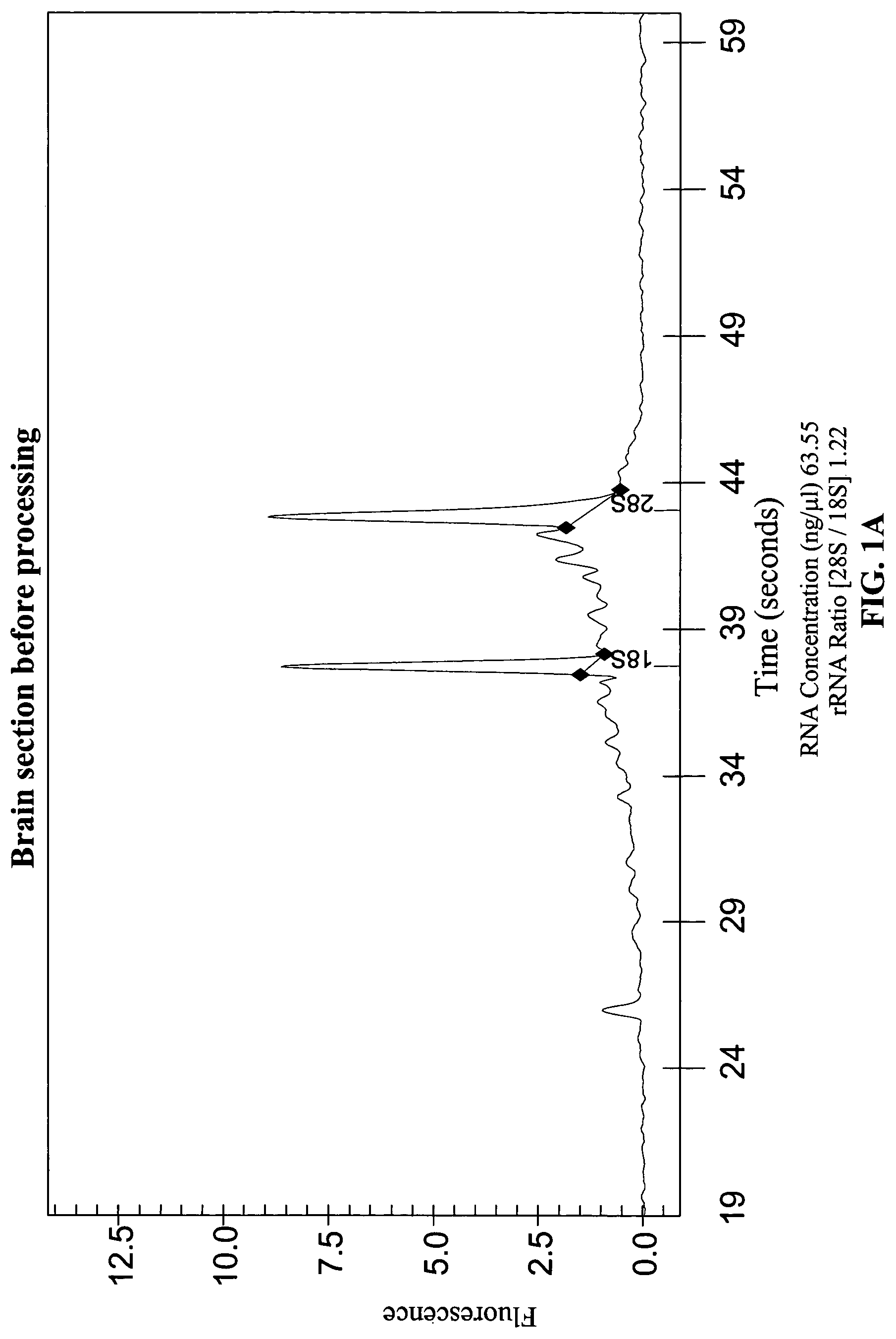
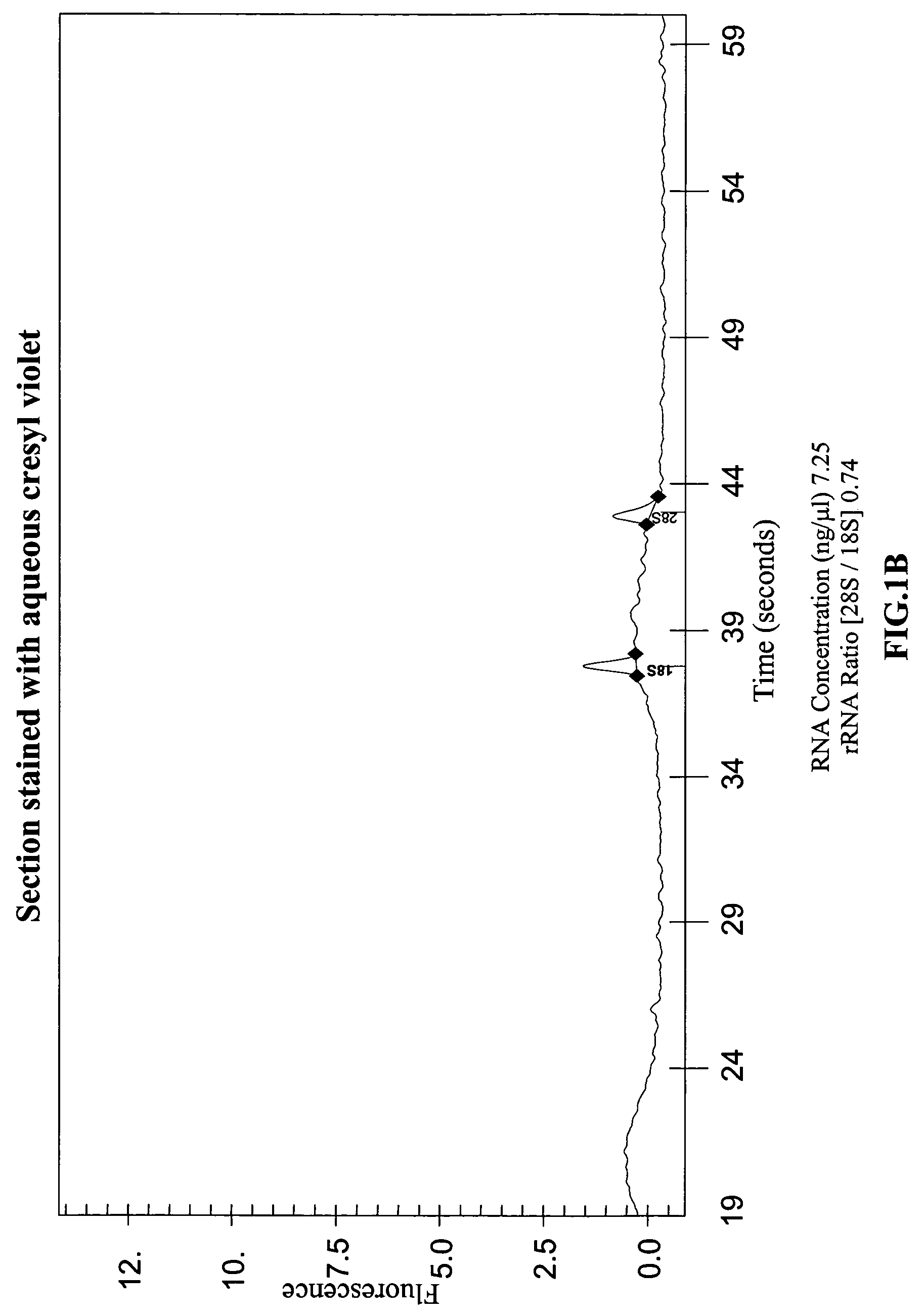
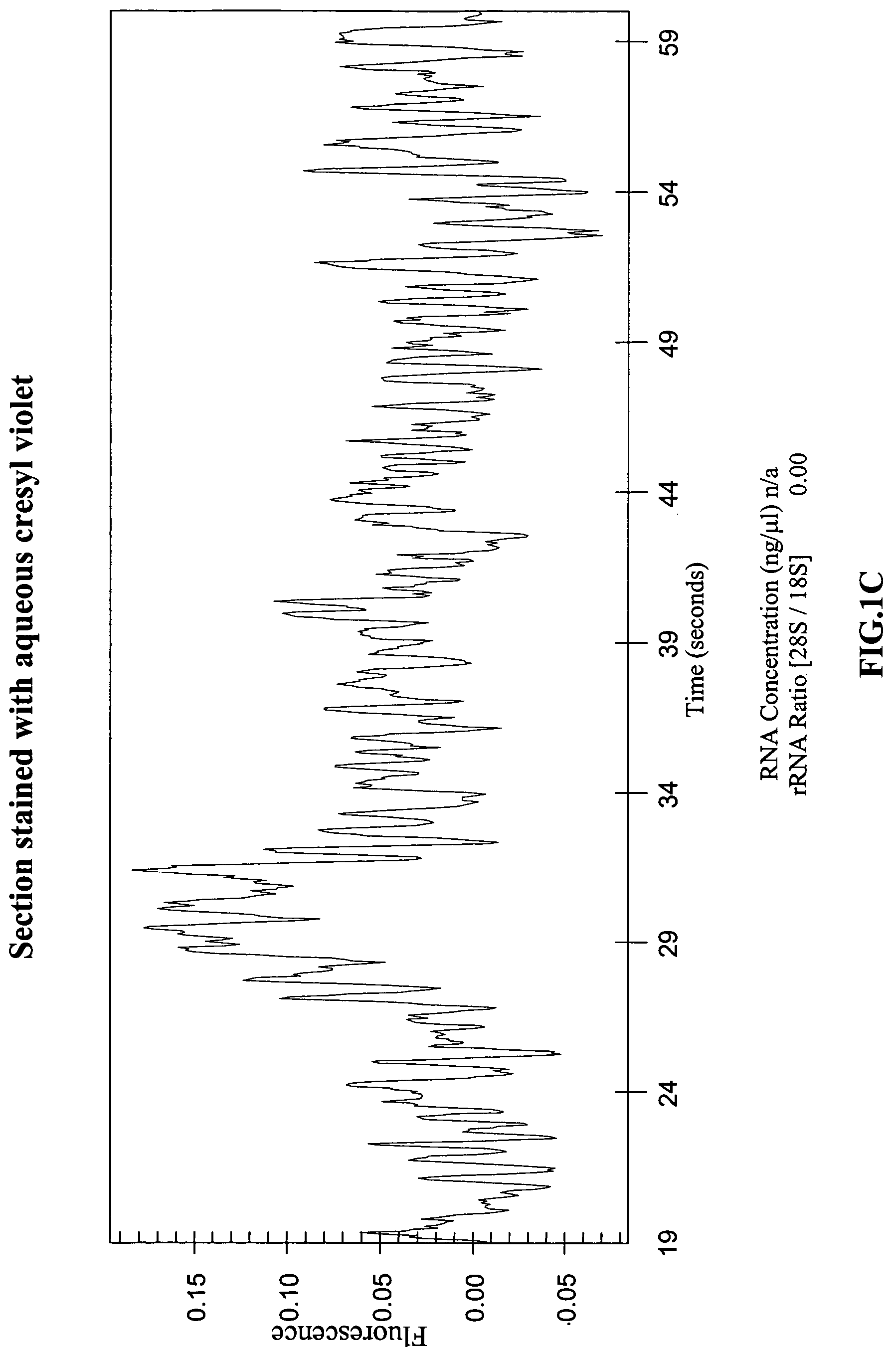
RNA Isolated from Tissue Sections Processed Using Published Procedures is Degraded

[0066]The effect of the various steps used to prepare tissue samples for LCM, and of the LCM process itself, on the yields and quality of RNA recovered was systematically investigated. Protocols described in the literature were used to process various mouse tissues and human tissues were obtained from the Co-operative Human Tissue Network (CHTN) and used to study the effects. The steps used to prepare frozen tissues for LCM and subsequent RNA recovery can be divided into four stages: 1) embedding and sectioning; 2) pre-staining fixation and hydration; 3) staining; and 4) post-staining processing and dehydration. The protocol recommended by Arcturus, Inc. for carrying out these steps is described in a technical bulletin, “Optimized Protocol for Preparing and Staining LCM Samples from Frozen Tissue and Extraction of High-Quality RNA” (found on the world wide web at arctur.com / images / pdf / HistoGene_Applica...

example 2

RNA Degradation is Not Due to RNase in Solutions and Surfaces

[0068]Since RNA degradation was observed in stained tissue, it was investigated whether this was due to RNase contamination of the reagents used. Histological stains such as hematoxylin / eosin, cresyl violet, and toluidine blue are typically made by dissolving powdered stains in water or in phosphate buffer. To rule out RNase contamination of the reagents used to make the stains, synthetic radiolabeled RNA transcripts were incubated for six hours with distilled water and with aqueous solutions of several histological stains, and then analyzed the transcripts on denaturing polyacrylamide gels. The stains tested included commercially available premixed solutions of eosin Y, Harris hematoxylin, and bluing reagent (ThermoShanndon RapidChrome Frozen Section Staining kit) and powdered stains including toluidine blue, cresyl violet, and hematoxylin (EM Science, Sigma and Aldrich). These experiments showed that the RNA degradation ...

example 3

Optimization of Pre-Staining Steps

[0070]Whether the 50% and 75% ethanol pre-staining steps could be eliminated was tested. Omitting these ethanol steps and incubating the sections in 95% ethanol directly before staining resulted in extremely poor tissue morphology, in fact it was impossible to bring the stained sample into sharp focus. The water component of the 50% and 75% ethanol solutions is probably required to dissolve the OCT embedding matrix which otherwise interferes with visualizing the tissue.


PUM


Abstract

The present invention concerns the methods and compositions for preparing a tissue section or biological sample, particularly to preserve RNA in the section or sample, by not exposing or contacting the sample or section to a solution that is composed of mostly water. Tissue sections can be fixed, stained, and dehydrated for subsequent manipulation, including laser capture microdissection (LCM) for further analysis using methods and/or compositions of the invention.

Description

[0001]The government may own rights in the present invention pursuant to grant number R43-CA88699 from National Institutes of Health / National Cancer Institute.BACKGROUND OF THE INVENTION[0002]1. Field of the Invention[0003]The present invention relates generally to the fields of histology and molecular biology. More particularly, it concerns compositions and methods for preparing a tissue section that allows RNA in the section to be recovered more abundantly and in a more intact form than previous methods in which significant degradation occurred.[0004]2. Description of Related Art[0005]Histological staining of thin tissue sections mounted on slides is frequently used to improve the ability to distinguish specific subregions and cellular structures during microscopic examination. Different stains, optimized for visualization of different structures in different types of tissue, for example liver, brain, kidney, tumor biopsies, etc. are used by pathologists and researchers for specif...

Claims


Application Information

Patent Timeline
IPC IPC(8): G01N1/30G01N33/48
CPCG01N1/30
Inventor GOLDRICK, MARIANNAGONZALES, JUANITA C.
Owner APPL BIOSYSTEMS INC
Why Eureka
Try Eureka