Methods for quenching pathogen inactivators in biological materials

a technology of pathogens and biological materials, applied in the direction of extracellular fluid disorder, lighting and heating equipment, furnaces, etc., can solve the problems of no licensed test for the presence of bacteria or protozoans in blood, the transmission of disease by blood products and other biological materials remains a serious health problem, and the prevalence of blood supply, so as to reduce unwanted side reactions and reduce unwanted side reactions

Inactive Publication Date: 2007-11-13
CERUS CORP
View PDF48 Cites 22 Cited by
  • Summary
  • Abstract
  • Description
  • Claims
  • Application Information

AI Technical Summary

Benefits of technology

[0014]The methods disclosed herein are advantageous, since in the two phase system, the quencher is administered to the first phase and substantially is not capable of traversing the exterior membrane of the pathogen, and thus is substantially not present in the second phase, i.e., the interior of the pathogen. The pathogen inactivating compound is administered, for example, to the first phase, and is capable of traversing the membrane of the pathogen, prior to formation of the electrophilic group. Upon formation of the electrophilic group in situ, the pathogen inactivating compound substantially is no longer capable of traversing the membrane. Thus, quenching selectively occurs in the first phase, while in the second phase, i.e., the interior of the pathogen, the pathogen inactivating compound reacts with nucleic acids of the

Problems solved by technology

The transmission of disease by blood products and other biological materials remains a serious health problem.
In addition to the viral hazard, there are currently no licensed tests to screen for the presence of bacteria or protozoans in blood intended for use in transfusions.
The risk also exists that a hitherto unknown pathogen may become prevalent in the blood supply and present a threat of disease transmission, as in fact occurred before the recognition of the risk of HIV transmission via blo

Method used


Image

  • Methods for quenching pathogen inactivators in biological materials
  • Methods for quenching pathogen inactivators in biological materials
  • Methods for quenching pathogen inactivators in biological materials

Examples


example 1

Reaction of Phosphate Ion with Pathogen Inactivating Compounds

[0112]The reactivity of phosphate ion with pathogen inactivating compounds was demonstrated. Reactivity was found to increase for higher pH values, at higher temperatures and for higher phosphate ion concentrations.

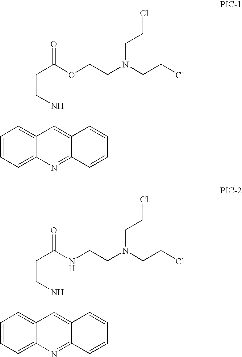
[0113]Upon incubation of 100 μM PIC-1 with 25 mM phosphate buffer of increasing pH values at room temperature (RT), a large increase of the rate of decomposition was observed. At pH 2.2 a t1 / 2 of 450 minutes was observed compared to a t1 / 2 of 25 minutes at pH≈6. Reaction of PIC-1 with phosphate ions produces at least two observable phosphate intermediates as determined by HPLC. The diphosphate ester of PIC-1 further hydrolyzes at the frangible ester. LC / MS analysis of the reaction mixture between the PIC-1 and phosphate ions was conducted. The species observed under the reaction conditions (pH=7, phosphate buffer) were the diphosphate ester, the monophosphate ester of the diol, and the phosphate ester of the ch...

example 2

Reaction of Thiol and Other Nucleophiles with Pathogen Inactivating Compounds

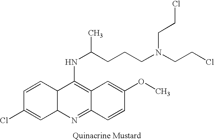
[0116]The reactivity of the thiol containing compound glutathione with pathogen inactivating compounds was studied. Reactivity was found to increase at higher pH values, at higher temperatures and for higher concentrations of thiol. 1 mM quinacrine mustard was incubated in 25 mM HEPES (N-[2-hydroxyethylpiperazine-N′-[2-ethanesulfonic acid], Sigma, St. Louis, Mo.), at pH 7 with 4 mM glutathione (GSH). Additionally, 100 μM PIC-1 was incubated in 25 mM HEPES at pH 7 with 4 mM GSH at room temperature. The reactions of quinacrine mustard and PIC-1 with glutathione occurred with a t1 / 2 of 50 minutes and 32 minutes, respectively. The PIC-1 / GSH bis adduct was identified by mass spectroscopy. LC / MS analysis of the reaction mixture with glutathione at early time points identified the glutathione monoadduct / aziridinium intermediate.

[0117]The reaction of the thiols with the pathogen inactivating compounds was also foll...

example 3

Study of Red Blood Cell Membrane Penetration of Glutathione

[0119]The ability of glutathione to penetrate red blood cell membranes was studied, as a model membrane system for the envelopes of viruses, since both would be devoid of any active transport systems for this peptide. The partitioning of the GSH inside and out of red cells (Sacramento Blood Center packed red blood cells, HCT 55-65%) was evaluated by determination of the amounts of thiol groups in the inside and the outside of the cells after addition of exogenous glutathione. Table 2 shows the total amount of glutathione (intracellular and supernatant) and the intracellular amount of glutathione of red blood cells as a function of time with and without addition of 3 mM GSH. The concentration of GSH was calculated based on the absorbance at 412 nm using a Beckman DU20 (Beckman, Irvine, Calif.) single wavelength spectrometer. The results show that the amount of sulfhydryl groups in both compartments stays constant.

[0120]

TABLE ...


PUM


Abstract

Methods are provided for quenching undesired side reactions of pathogen inactivating compounds in biological materials comprising red blood cells. In a particular embodiment, methods are provided for quenching undesired side reactions of a pathogen inactivating compound that includes a functional group which is, or which is capable of forming, an electrophilic group. In this embodiment, the material is treated with the pathogen inactivating compound and a quencher, wherein the quencher comprises a nucleophilic functional group that is capable of covalently reacting with the electrophilic group. The electrophilic group on the pathogen inactivating compound is preferably a non-radical cationic group. In one embodiment, the pathogen inactivating compound includes a nucleic acid binding ligand and a mustard group, wherein the mustard group is capable of reacting in situ to form the electrophilic group. Preferred quenchers are thiols, such as glutathione. The methods permit inhibition of the modification of red blood cells in red blood cell containing materials during pathogen inactivation.

Description

CROSS-REFERENCE TO RELATED APPLICATIONS[0001]This application is a continuation of U.S. patent application Ser. No. 09 / 912,031, filed Jul. 23, 2001, which issued as U.S. Pat. No. 6,709,810, which is a continuation of U.S. patent application Ser. No. 09 / 110,776, filed Jul. 6, 1998, which issued as U.S. Pat. No. 6,270,952, which claims the benefit of U.S. Provisional Application No. 60 / 070,597, filed Jan. 6, 1998, the disclosures of which is incorporated herein by reference in their entirety.TECHNICAL FIELD[0002]This application relates to methods of treating biological materials, such as blood products, with a nucleophilic compound to quench reactive electrophilic compounds in the material.BACKGROUND ART[0003]The transmission of disease by blood products and other biological materials remains a serious health problem. While significant advances in blood donor screening and blood testing have occurred, viruses such as hepatitis B (HBV), hepatitis C (HCV), and human immunodeficiency vi...

Claims


Application Information

Patent Timeline
IPC IPC(8): A01N1/02A01N63/00A61K35/14A61L2/00A61M1/36C12N5/08
CPCA61L2/0088A61P7/08
Inventor COOK, DAVIDSTASSINOPOULOS, ADONIS
Owner CERUS CORP
Why Eureka
Try Eureka