X-ray image processing

a technology of x-ray images and x-ray images, applied in image enhancement, image analysis, instruments, etc., can solve the problems of poor signal-to-noise ratio, poor accuracy, mammographic images pose a tough challenge, etc., and achieve the effect of improving the accuracy of the hint representation and the calculation of the hint representation

Inactive Publication Date: 2008-01-01
SIEMENS MEDICAL SOLUTIONS USA INC
View PDF16 Cites 56 Cited by
  • Summary
  • Abstract
  • Description
  • Claims
  • Application Information

AI Technical Summary

Benefits of technology

[0037]The present invention is concerned with improvements in the calculation of the hint representation, which

Problems solved by technology

This huge cost and the poor accuracy in diagnosis (8-25% of cancers are missed and 70-80% of open surgical biopsies turn out to be benign), have led to increased interest in applying computer-aided techniques.
As an application of image processing, mammographic images pose a tough challenge because they have poor signal-to-noise ratio.
This is largely because the images exhibit complex textures, and because there is a compromise between radiation dose and image quality.
Worse, abnormalities appear as quite subtle, irregular, often non-local differences in intensity and the images are inevitably cluttered due to superimposition in the image of features separated in the breast.
Further, the background varies greatly between different breasts, and there is relatively weak control of the imaging acquisition.
Unfortunately, while there have been proposals for the application of image processing to mammography, the vast majority have been of limited scope and incorporate only general non-mammography-specific image processing considerations.
This involves great dangers.
Calcifications present particular interest and problems.
It is, however, a challenge for image analysis programs.
Further, dust and dirt entering the imaging system can create artifacts that mimic the appearance of microcalcifications and thus can be a cause of false positives bot

Method used


Image

  • X-ray image processing
  • X-ray image processing
  • X-ray image processing

Examples


Embodiment Construction

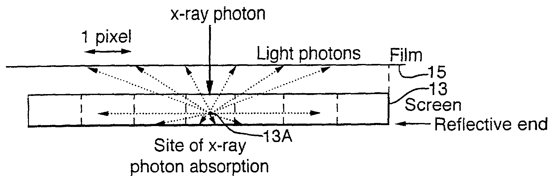
[0085]An embodiment of the invention will now be described by going through the steps necessary to calculate the hint representation. These can be summarized as follows:[0086](1) Convert pixel value P(x, y) to film density D(x, y) using the digitiser calibration data;[0087](2) Remove digitiser blur;[0088](3) Convert film density D(x, y) to energy imparted to intensifying screen Eimppse(x, y) using film-screen calibration data;[0089](4) Compensate Eimppse(x, y) for intensifying screen glare;[0090](5) Compensate Eimppse(x, y) for the anode-heel effect and diverging x-ray beam;[0091](6) Estimate the thickness of the compressed breast;[0092](7) Estimate the scattered radiation Eimps(x, y);[0093](8) Estimate the extra-focal radiation Eimpe(x, y) components;[0094](9) Compute the measured primary energy Eimpp:

Eimpp(x, y)=Eimppse(x, y)−Eimps(x, y)−Eimpe(x, y)[0095](10) Compare this measured primary energy with the theoretical primary energy calculated by equation (2) for different values of...


PUM


Abstract

A method of enhancing and normalizing x-ray images, particularly mammograms, by correcting the image for digitizer blur, glare from the intensifying screen and the anode-heel effect. The method also allows the calculation of the compressed thickness of the imaged breast and calculation of the contribution to the mammograms of the extra focal radiation. The correction of the image for glare from the intensifying screen allows the detection of noise, such as film shot noise, in the image, and in particular the differentiation between such noise and microcalcifications.

Description

CROSS-REFERENCE TO RELATED APPLICATION[0001]This application is the national stage of international application no. PCT / GB00 / 00617, filed Feb. 21, 2000, which designates the United States and which claims priority from GB 9904692.2, filed Mar. 1, 1999.BACKGROUND[0002]The present invention relates to improvements in the processing of digitised x-ray images (particularly of the breast—termed mammograms or mammographic images), and more particularly to the enhancement of such images to assist clinicians in making accurate diagnoses based on them.[0003]X-ray imaging is used as a basis for many medical techniques and, in particular, mammography continues to be the examination of choice for early detection of breast cancer in post-menopausal women and is the basis for national screening programmes.[0004]Early detection of breast cancer greatly improves mortality rates, perhaps by as much as 25%. For this reason, mammographic examinations are nowadays performed on about 25 million women an...

Claims


Application Information

Patent Timeline
IPC IPC(8): G06K9/00G06T5/00
CPCG06T5/002G06T5/003G06T2207/10116G06T2207/30068
Inventor BRADY, JOHN MICHAELHIGHNAM, RALPH
Owner SIEMENS MEDICAL SOLUTIONS USA INC
Why Eureka
Try Eureka