Viral fibers

a technology of fibers and fibers, applied in the field of fibers, can solve the problems of reducing the specific surface area of fibers, reducing the surface reactivity, and reducing the amount of fibers

Inactive Publication Date: 2008-02-19
BOARD OF RGT THE UNIV OF TEXAS SYST +1
View PDF20 Cites 23 Cited by
  • Summary
  • Abstract
  • Description
  • Claims
  • Application Information

AI Technical Summary

Benefits of technology

[0008]The invention provides a plurality of methods of fabricating virus-based micro- and nanometer scale fibers that, generally, can be in broad terms a mimic of the spinning process of silk spiders. In some embodiments of the invention, wet spinning and electrospinning were used to fabricate virus- based micro- and nanofibers, respectively. In some embodiments, the resulting fibers showed nematic ordered morphologies due to flow forces. In additional embodiments, the virus was blended with a synthetic carrier polymer, poly(vinyl pyrrolidone) (PVP), to improve its processing ability. The resulting virus blended PVP fibers were continuous and could be formed into non-woven fabrics mats that retain their ability to infect bacterial hosts.
[0015]The present invention further provides a method of converting virus particles to fiber form in which the virus particles retain their virus structure in the solid state comprising the step of spinning the viral particles into fiber form while controlling concentration, viscosity, and optional use of electric field and blending agent to control the fiber form and retain the virus structure after spinning. The concentration can be controlled so that a liquid crystalline phase can be formed for spinning. The concentration can be sufficiently high during spinning to avoid electrospraying.
[0016]The present invention also provides experimental conditions which provide the fibers with useful properties such as, for example, the ability to be specifically bound to the conjugate material after fiber spinning or retain the ability to specifically bind to the conjugate material after fiber spinning, as well as retain the ability to infect a host after fiber spinning. For example, concentrations can be adjusted, viscosity can be adjusted, solvents can be altered or replaced, electric fields can be applied, and use of blending materials can be varied.
[0022]The present invention provides numerous advantages over the prior art. For example, the present invention provides a method of forming fibers on a micrometer or nanometer scale engineered for specific applications, including a wide variety of different applications ranging from military to biomedical. Genetic engineering can be used to engineer the application at an unprecedented level of structural control ranging down to the nanoscale. In another advantage, the viruses used in making the fibers of the present invention may be engineered to express polypeptides having one or more amino acid oligomers on the exterior surface of the viruses. These amino acid oligomers may serve a variety of functions. For example, the amino acid oligomers may be cell adhesion factors, antibodies, or have specificity for inorganic molecules. Fibers provided by the present invention have a variety of application, including but not limited to, scaffolds for tissue engineering, sensors for detecting environmental toxins, reaction sites for chemical reactions, and filters. For nanofibers, particularly when the fiber diameter is about 100 nm or less, the materials can have exceptionally high levels of specific surface area which enables a high proportion of atoms on the fiber surface. This can provide quantum efficiency, nanoscale effect of unusually high surface energy, surface reactivity, high thermal and electrical conductivity, and high strength.
[0023]In sum, virus based micro- and nanofibers can be fabricated using wet-spinning and electrospinning processes. M13 viruses in the wet-spun fibers were aligned parallel to the fiber long axis. Electrospun fibers, composed of fragment of M13 viruses and its subunits, were also fabricated by suspending M13 viruses in HFP. Although the viral structure was partially disrupted, these results indicated that the novel biomaterials could be used to expand the dimension of the engineered viruses into endless fibers which can be used to nucleate useful semiconductor nanofibers or to selectively bind desired materials. Additionally, uniform nanofibers fabricated by blending M13 virus with PVP might provide useful biological functions and highly sensitive catalytic functions in future biomedical applications and biosensors.

Problems solved by technology

However, above an upper concentration limit, the solution at times becomes too viscous to be pulled by electrostatic forces.

Method used


Image

  • Viral fibers
  • Viral fibers
  • Viral fibers

Examples


working examples

[0105]The invention is further described with use of the following non-limiting working examples.

[0106]A. Virus

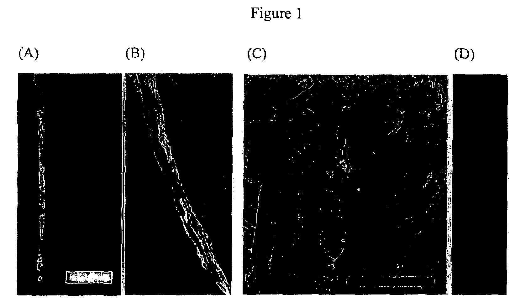
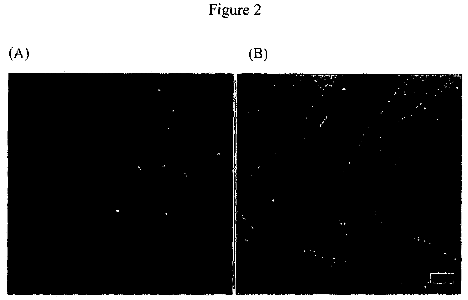
[0107]Anti-streptavidin M13 bacteriophage possessing an engineered peptide sequence, N′-TRP ASP PRO TYR SER HIS LEU LEU GLN HIS PRO GLN-C′ (SEQ ID NO.:1), in its pIII coat protein (virus) was used as a basic building block to fabricate the micro- and nanoscale fibers. The virus was selected from the PhD-12 phage display library (New England Biolabs, Inc. Beverly, Mass.) for affinity to streptavidin (5). The virus was amplified and purified according to phage library manufacturer instructions and suspended in tris buffered saline (TBS; 50 mM Tis, 150 mM NaCl, pH 7.5).

[0108]B. Conjugate Material.

[0109]The bacteriophage was used with or without conjugation with R-phycoerythrin (eBioscience, CA) previously reported [5]. Conjugation was at the pIII subunit.

[0110]C. Wet Spinning

[0111]The M13 virus suspension (˜100 mg / ml) extruded through ˜20 um capillary tube into 37.3% aqueous g...


PUM

PropertyMeasurementUnit
wt. %aaaaaaaaaa
lengthaaaaaaaaaa
cross sectional diameteraaaaaaaaaa

Abstract

Long rod shaped M13 viruses were used to fabricate one dimensional (1D) micro- and nanosized diameter fibers by mimic the spinning process of the silk spider. Liquid crystalline virus suspensions were extruded through the micrometer diameter capillary tubes in cross-linking solution (glutaraldehyde). Resulting fibers were tens of micrometers in diameter depending on the inner diameter of the capillary tip. AFM image verified that molecular long axis of the virus fibers were parallel to the fiber long axis. Although aqueous M13 virus suspension could not be spun by electrospinning, M13 viruses suspended in 1,1,1,3,3,3-hexafluoro-2-propanol were spun into fibers. After blending with highly water soluble polymer, polyvinyl 2-pyrolidone (PVP), M13 viruses was spun into continuous uniform virus blended PVP (virus-PVP) fibers. Resulting virus-PVP electrospun fibers showed intact infecting ability to bacterial hosts after suspending in the buffer solution.

Description

CROSS-REFERENCE TO RELATED PATENT APPLICATIONS[0001]This application claims benefit to provisional application 60 / 510,862 filed Oct. 15, 2003 to Belcher et al., which is hereby incorporated by reference in its entirety.[0002]This invention was developed under the following grants from the federal government: Grant No. DAAD19-03-1-0088, from the Army Research Office. The government has certain rights in the invention.INTRODUCTION[0003]The introduction section provides reference to a number of technical publications which are found at the end of the specification and can be used as a guide to one skilled in the art to practice the present invention. No admission is made that any of these references are indeed prior art.[0004]Efforts to mimic the unique structures and specific functions of natural systems have provided various useful tools and materials in nanoscience [1-14]. Biosystems produce highly programmed, self-assembled, self-templated structures [6-11]. For example, a small pe...

Claims


Application Information

Patent Timeline
Patent Type & Authority Patents(United States)
IPC IPC(8): C12N7/01D01F8/18D02G3/04A61K9/70A61K35/76A61K39/12C12N7/00C12N7/02C12Q1/70D01D5/00D01F1/10D04H1/728G01N
CPCA61K35/76B82Y5/00C12N7/00C12Q1/70D01D5/0053D01F1/10D01F9/00A61K47/48176A61K47/48238D01D5/0007C12N2795/14111C12N2795/14122Y10T428/2927A61K47/58A61K47/62Y10T442/2525
Inventor BELCHER, ANGELA M.LEE, SEUNG-WUK
Owner BOARD OF RGT THE UNIV OF TEXAS SYST
Why Eureka
Try Eureka