

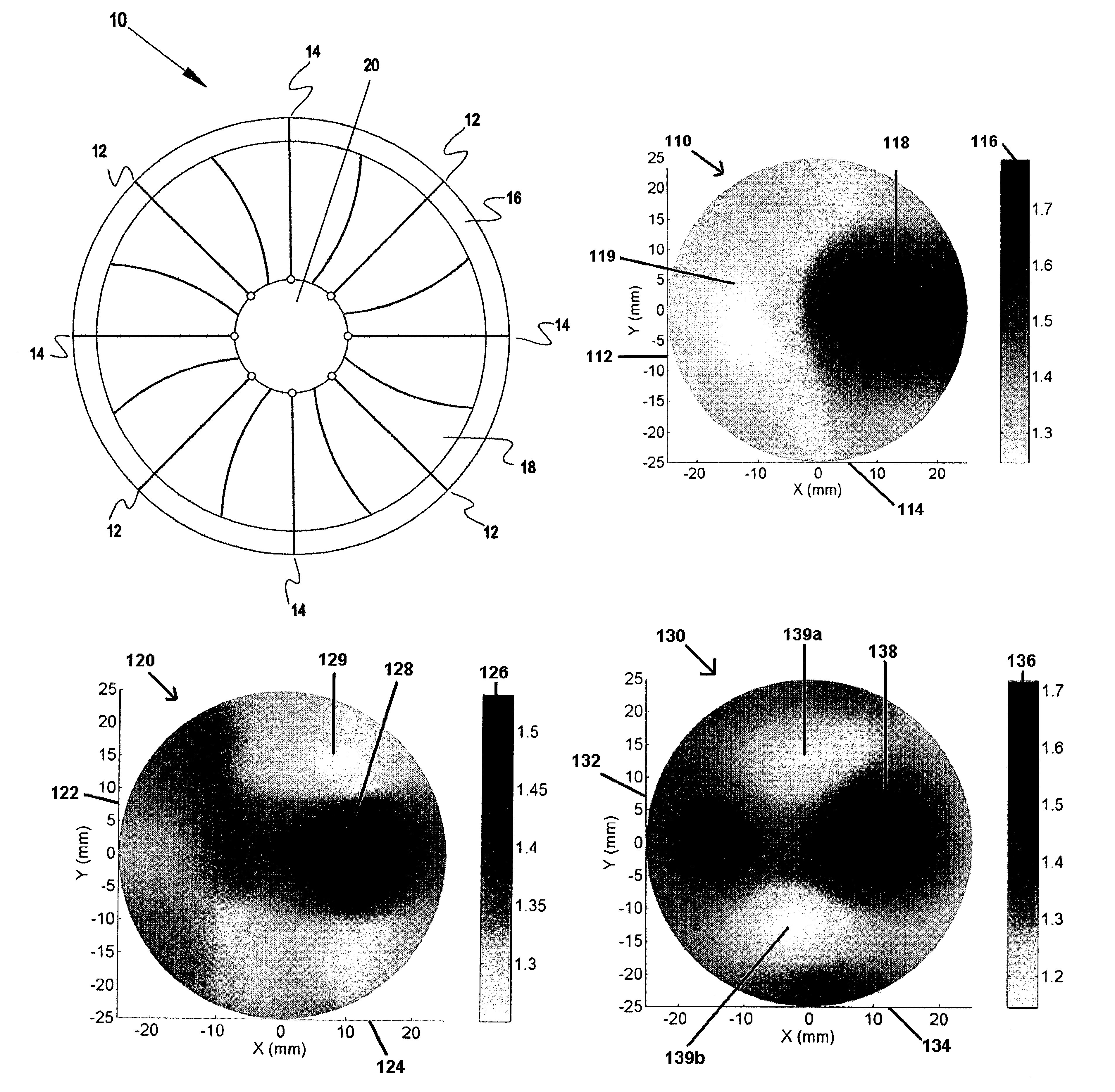
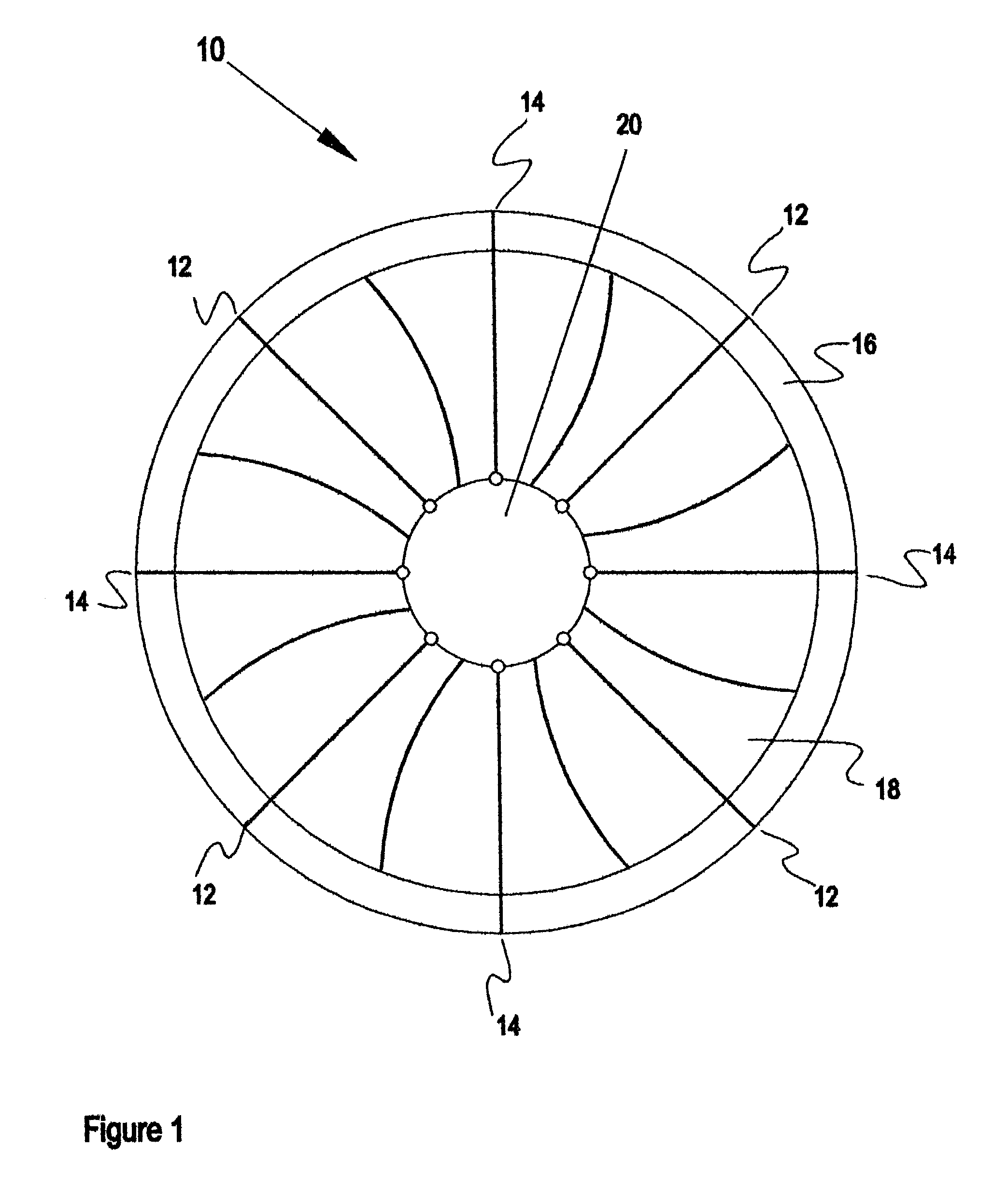
Reconstructed refractive index spatial maps and method with algorithm
- Summary
- Abstract
- Description
- Claims
- Application Information
AI Technical Summary
Benefits of technology
Problems solved by technology
Method used
Image

Examples
example 1
[0031]Phantom measurements in vitro using tissue-like materials with different contrasts of reconstructed refractive index between targets (simulated tumors or abnormal tissue) and background material illustrate the application of the reconstruction algorithm and total system in identifying abnormal tissue. A background lipid emulsion solution (Intralipid) was used to mimic tissue scattering (reduced scattering coefficient (μs′)=1.0 mm−1). India ink (absorption coefficient=0.007 mm−1) was used to simulate tissue absorption. Agar powder (2%) was used to solidify the Intralipid / India ink solution yielding a background material with a refractive index close to that of water (n=1.33 and λ=785 nm, where n is the refractive index and λ the wave length of the light used). Targets in the background material represented one of two cases: essentially void holes (air, n=1.0) or mixtures of sugar (glucose, 60%) and Intralipid / India ink solution (n=1.47).
[0032]Five phantom situations were evalua...
example 2
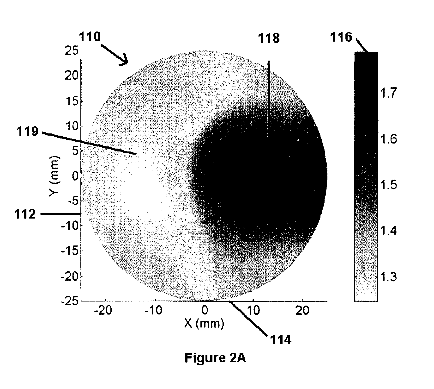
[0039]The ultimate utility of the invention is realized in the production of reconstructed refractive index spatial maps, the images of which allow the identification of tumorous tissue and distinguishes between benign and malignant tumors. FIGS. 3A and 3B illustrate clinical applications of the system and methods of this invention following the same theories of Example 1.
[0040]FIG. 3A illustrates the image of a reconstructed refractive index spatial map utilizing near infrared light in the examination of breast tissue of a 39 year-old, female patient. The image 200 reveals a relatively light, well defined area 210 below and to the left of the center of the field, compared with the background material 220. This suggests a tumorous mass at the indicated position at least in part within the plane of the spatial map of reconstructed refractive index. Low refractive index is characteristic of certain types of malignant tumors. The image of the spatial map and conclusion that a malignant...
PUM
 Abstract
Description
Claims
Application Information