
Medical device and method of embolizing bronchus or bronchiole

a technology of bronchus and bronchiole, which is applied in the field of medical devices and methods of embolishing bronchus or bronchioles, to achieve the effects of facilitating puncturing manipulation, improving workability, and maintaining swelling

Active Publication Date: 2008-04-15
OLYMPUS CORP
View PDF4 Cites 98 Cited by
  • Summary
  • Abstract
  • Description
  • Claims
  • Application Information

AI Technical Summary

Benefits of technology

[0024]In this case, the injection material preferably contains fibrin adhesive and collagen. In addition, it is preferable to adopt an injection material having the property of expanding by absorbing water inside a living body. When particles are contained as solid components in the injection material, the particles have the effect of maintaining the swellings for a long time. In this case, preferred examples of the particles contain a component of high biocompatibility, such as silicone, metal particles such as titanium particles, apatite, and β-TCP. Addition of a curing agent to the injection material after the injection also has the effect of ensuring embolization and maintaining the swellings for a long time.
[0025]The above-described puncturing manipulation is facilitated by the use of a cap element to be attached to the leading end of the endoscope. The cap element has a through-hole from which the puncture portion of the catheter projects and a sheath for guiding the puncture portion of the catheter from the leading opening of the channel of the endoscope into the through-hole. In this case, it is convenient that the through-hole be provided on a side of the cap. Furthermore, in the case where a balloon is provided on the side of the cap opposite to the side in which the through-hole is provided, expansion of the balloon makes the inside wall of the bronchus and the cap adhere closely to each other, thereby improving workability.

Problems solved by technology

The lung tissue of a part affected by lung emphysema suffers overexpansion, and cannot achieve sufficient deflation accompanied by expiration.
Although there may be some cases where the symptoms of the patient are relieved by such internal medicine treatment, the advance of the patient's morbid state cannot be stopped.
Since this removal imposes a large burden on the patient, it takes a long time for the patient to recover after the surgical operation.
In the case where lung-emphysema-affected parts and normal lung tissues exist in disorder, it is difficult to separate only morbid tissues, so that there is a case in which even normal lung tissues must be removed.
In addition, there is also a case where the shape of a serosa, which contains a pulmonary external surface varies, hinders the inflation of the remaining portion of the lung.
As a result, there is a case where the function of the remaining normal lung tissue of the patient cannot be fully retained.
In addition, since the line of a removed part differs from a natural state and is exposed to nonuniform pressure, there is a case where the tissue of the removed part is broken and pneumothorax occurs.
However, the removal of lung tissue imposes a large burden on the body of the patient and, therefore, is difficult to practice repeatedly.
However, lung transplantation imposes a very large burden on the body of the patient, and entails problems peculiar to transplantation treatment such as immunological problems and the necessity of securing donors.
Neither lung volume reduction surgery nor lung transplantation can be easily practiced, because of the problem of requiring a large surgical operation and hence a huge cost for treatment.
Lung volume reduction surgery and lung transplantation, which are practiced as surgical treatment of lung emphysema, have the following problems:(1) Since lung volume reduction surgery and lung transplantation need a large surgical operation, a large scar is left on the body of a patient.(2) Neither lung volume reduction surgery nor lung transplantation can be repeatedly practiced on the same patient.(3) Lung volume reduction surgery cannot be practiced, if an affected part does not exist at a position where it can be easily removed.(4) There is a case where lung volume reduction surgery cannot fully ensure the function of the remaining portion of the lung.(5) Both lung volume reduction surgery and lung transplantation require huge costs.(6) Pneumothorax may occur after a surgical operation.(7) There are large obstacles to be surmounted, such as the necessity of securing donors and a rejection of a transplanted piece.
However, PCT WO98 / 48706 has the problem that since the embolization element for giving treatment to lung emphysema is extremely small in size, it is difficult to manufacture the embolization element having the complicated locking element.

Method used


Image

  • Medical device and method of embolizing bronchus or bronchiole
  • Medical device and method of embolizing bronchus or bronchiole
  • Medical device and method of embolizing bronchus or bronchiole

Examples


first embodiment

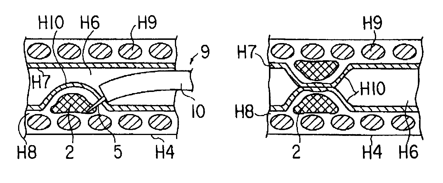
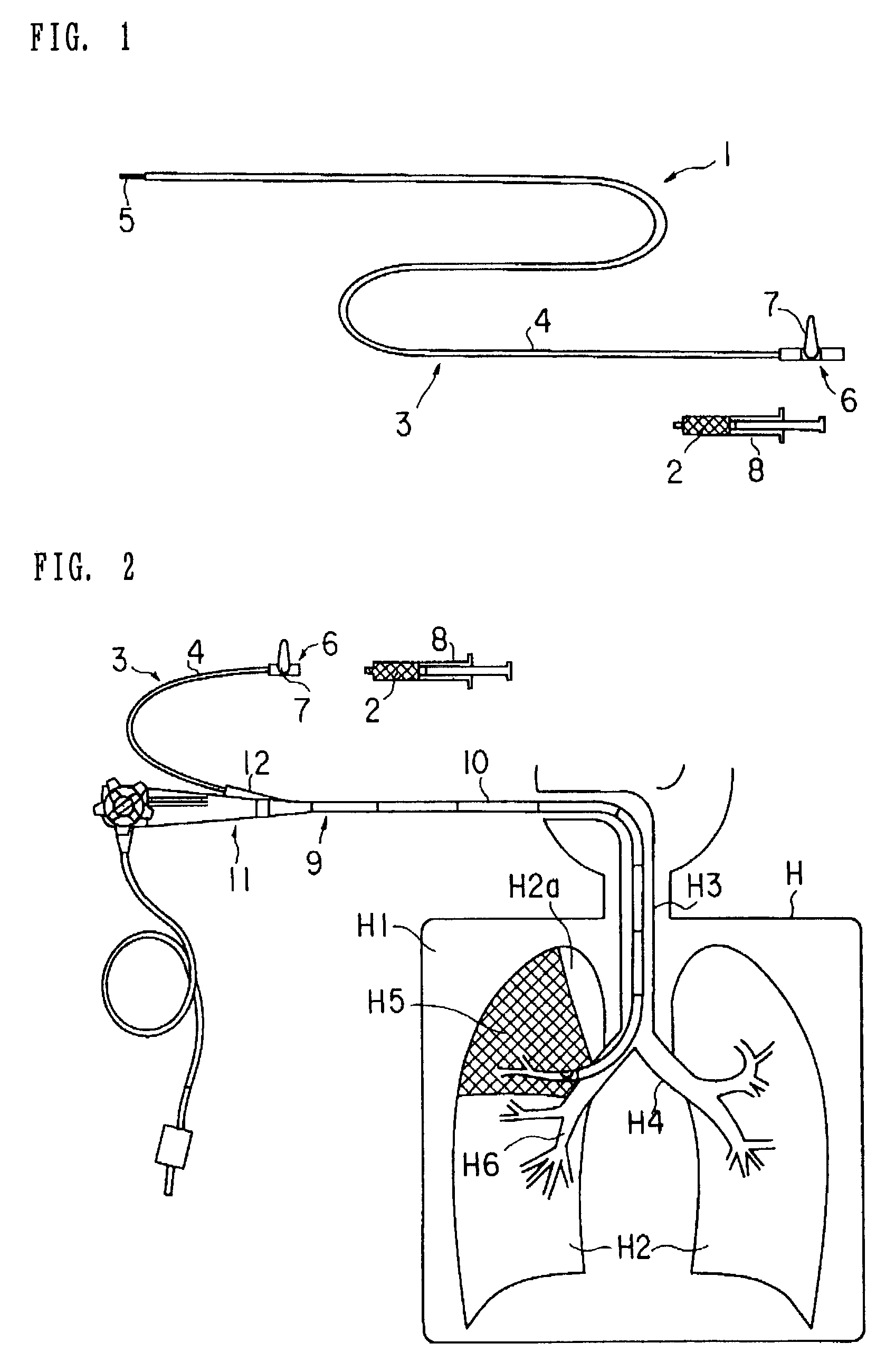
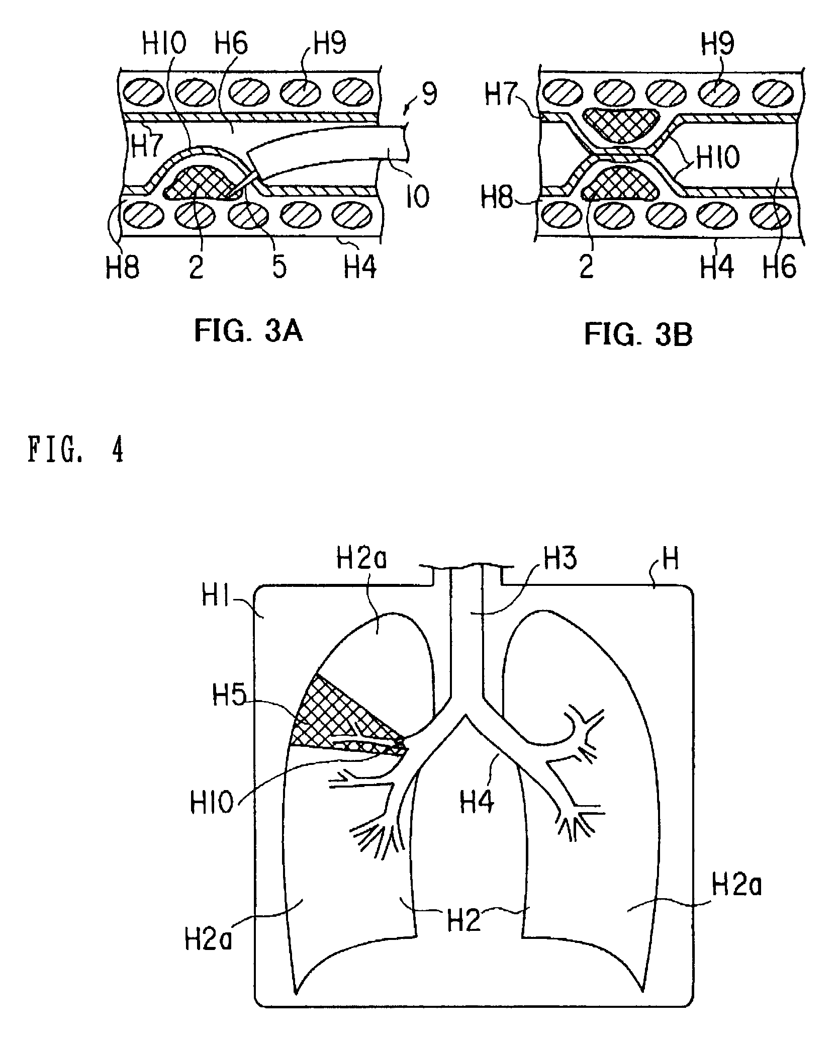
[0059]As shown in FIG. 2, during treatment of lung emphysema in a pleural cavity H1 of a patient H, a bronchoscope 9 is used in combination with the bronchus embolization device 1 of the This bronchoscope 9 is provided with an elongated inserting portion 10 to be inserted into the body of the patient H. A manipulation portion 11 is provided in a proximal portion of the inserting portion 10 on the operator side.

[0060]Furthermore, a channel 10c (refer to FIG. 7) through which to insert a treatment element is provided in the interior of the inserting portion 10 of the bronchoscope 9. A leading opening portion of this channel 10c is provided in a leading portion of the inserting portion 10. The manipulation portion 11 on the operator side is provided with a channel port portion 12, which communicates with a proximal portion of the channel 10c. The injection device 3 is to be inserted into the channel 10c of the bronchoscope 9 from the channel port portion 12.

[0061]The operation of the ...

second embodiment

[0069]Namely, the injection material 2 of the second embodiment has a construction in which a particulate substance 22 which is a solid component is suspended in a liquid base material 21 as shown in FIG. 5A. The constituent material of the injection material 2 is made of a material having good biocompatibility, which does not exhibit toxicity nor stimulus to a living tissue when the material is injected into the submucosa H8 of the bronchus H4. The particulate substance 22 contains a solid component of high biocompatibility, such as silicone, metal particles (such as titanium particles), apatite or β-TCP (tricalcium phosphate).

[0070]In the second embodiment, the constituent material of the injection material 2 used in the bronchus embolization device 1 for embolizing the bronchus H4 and giving treatment for lung emphysema has a construction in which the particulate substance 22, which is a solid component, is suspended in the liquid base material 21. Consequently, the second embodi...

sixth embodiment

[0110]The delivery device 83 is provided with a holding element 84 for holding the placing object 82 and an elongated sheath 85 disposed at the outside of the holding element 84. The holding element 84 is provided with a holding portion 87, such as grasper jaws, for holding the placing object 82 at a leading portion of an elongated shaft 86. In addition, a handle 88 for manipulating the holding portion 87 is provided at a proximal portion of the shaft 86. In this delivery device 83, the holding portion 87 at the leading end of the holding element 84 can move into and out of the sheath 85, and can be made to hold and release the placing object 82 by manipulating the handle 88 on the operator side. Incidentally, in the sixth embodiment, the delivery device 83 is constructed to serve also as a retrieving device, which retrieves the placing object 82.

[0111]The operation of the sixth embodiment having the above-described construction will be described below. In the sixth embodiment, as s...


PUM


Abstract

To provide a medical device and a method both of which can suitably embolize a bronchus in a target part by embolizing the bronchus with (at least a part of) a living tissue during surgical treatment of lung emphysema. For example, an injection material is injected into a submucosa by the use of a syringe, and in the injected part, a swelling swollen into the internal cavity of the bronchus is formed to tightly seal the living tissue of the bronchus or a bronchiole, thereby embolizing a target bronchus.

Description

CROSS-REFERENCE TO RELATED APPLICATION[0001]This application is based upon and claims the benefit of priority from the prior Japanese Patent Application No. 2001-220004, filed Jul. 19, 2001, the entire contents of the application are incorporated herein by reference.BACKGROUND OF THE INVENTION[0002]1. Field of the Invention[0003]The invention relates to a method and a medical device both of which are used for embolizing a bronchus or a bronchiole, for example, during treatment of lung emphysema.[0004]2. Description of the Related Art[0005]In general, lung emphysema is a morbid change, which is mainly formed by inhalation of harmful substances due to smoking or the like and mainly features wide destruction in peripheral airways and alveoli. The formation of such morbid change is chronic and progressive, and the respiratory function of a patient whose lung emphysema is in an advanced stage is disordered to a remarkable extent. Respiratory disorder due to lung emphysema entails, as its...

Claims


Application Information

Patent Timeline
IPC IPC(8): A61M31/00A61M29/00A61B17/12A61B17/00A61B17/24A61M25/00
CPCA61B17/12013A61B17/12022A61B17/12104A61B17/12136A61B17/12181A61B17/12186A61B2017/1205A61B2017/22065
Inventor KAJI, HINAKOKAJI, KUNIHIDE
Owner OLYMPUS CORP
Learn More