

Products and methods for single parameter and multiparameter phenotyping of cells
a single parameter and cell technology, applied in the field of single parameter and multiparameter phenotyping and cell immunophenotyping, can solve the problems of inability to correct subtyping of tumors (and proper assignment to treatment protocols), high degree of suspicion, and low degree of suspicion
- Summary
- Abstract
- Description
- Claims
- Application Information
AI Technical Summary
Benefits of technology
Problems solved by technology
Method used
Image

Examples
example 1
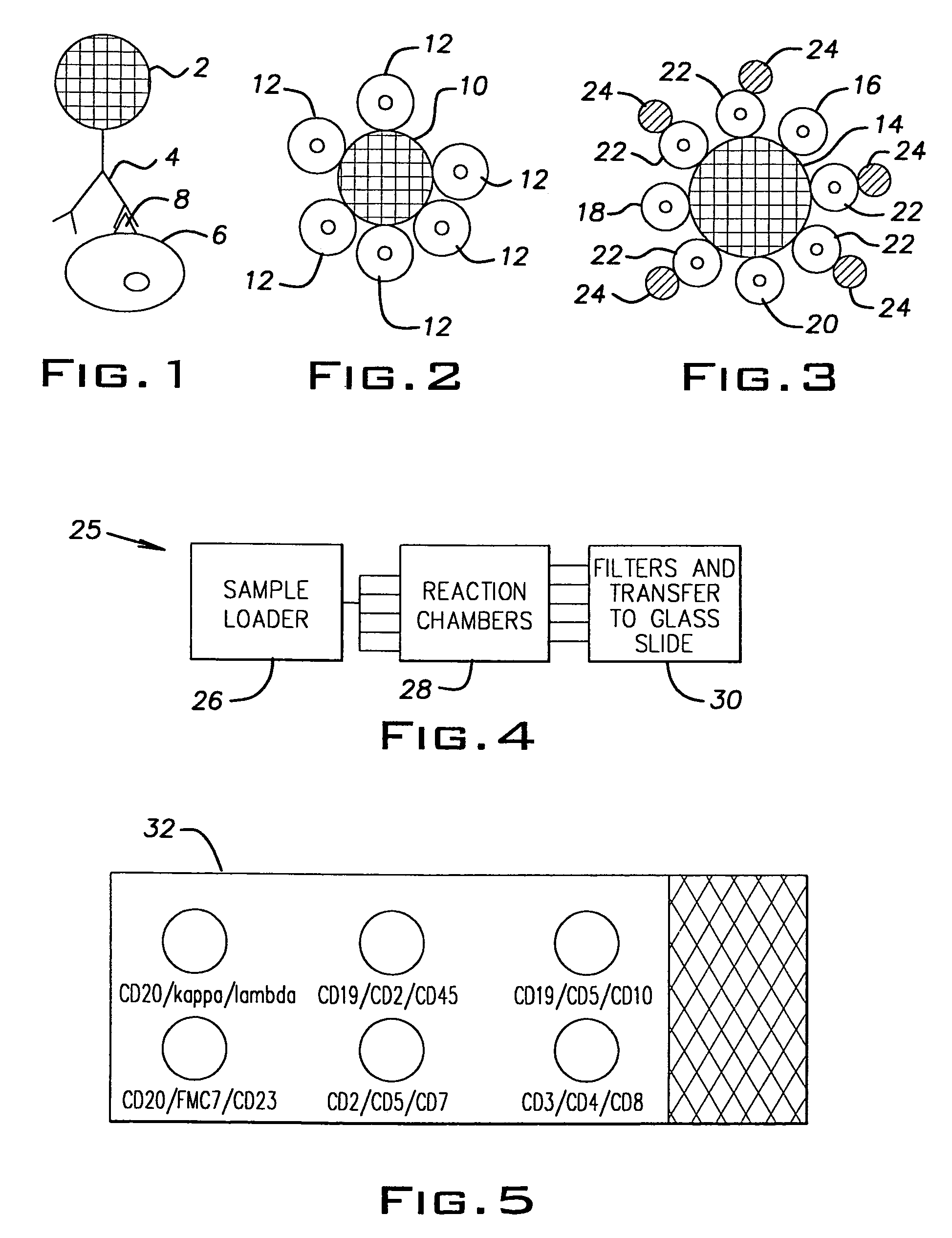
[0063]A 30 year old man presented with pancytopenia and splenomegaly. Examination of the peripheral smear confirmed the pancytopenia. In addition, scattered cells were present that showed bland cytological characteristics, with a monocytoid appearance. The nuclei of these cells were round to oval, with a single intermediate nucleolus. There was abundant blue-gray cytoplasm that showed numerous cytoplasmic projections. A bone marrow examination revealed a hypocellular aspirate with similar cells present. Small clusters of abnormal cells were present on the core biopsy. A buffy coat sample of the peripheral smear was suspended in anti-CD20 coated 10-micron colorless beads to distinguish the abnormal cells from monocytes. The suspension was passed through an appropriate filter and the cells were then transferred to a glass slide and stained. A schematic of the resulting slide preparation is demonstrated in FIG. 2. Positive binding of the abnormal cell population to the 10-micron beads ...
example 2
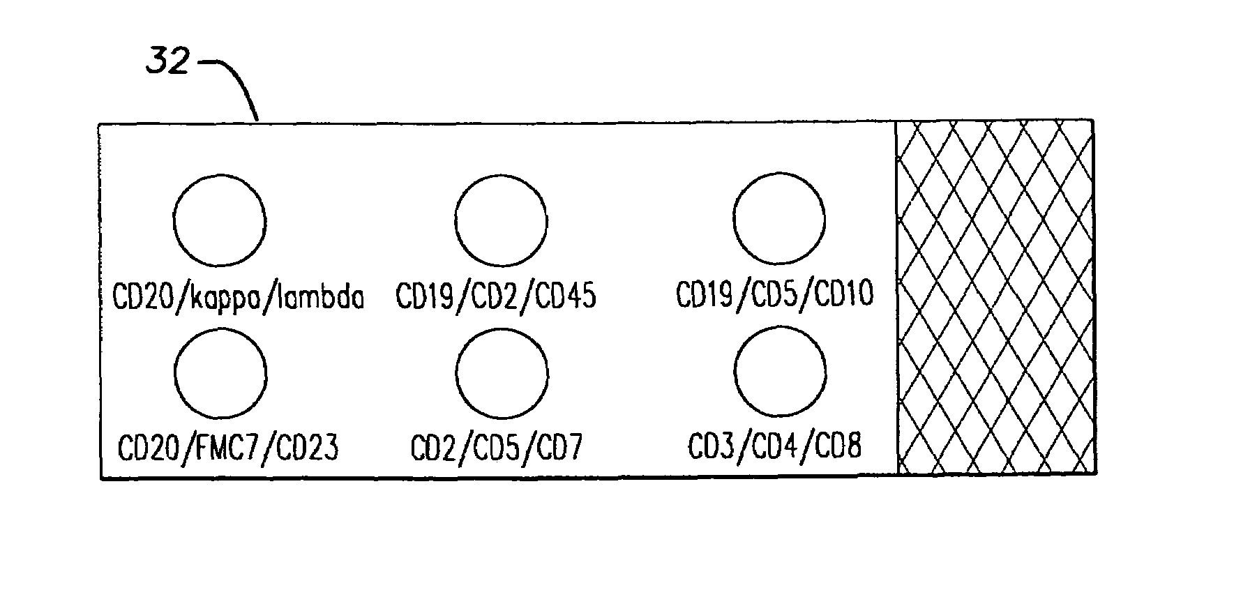
[0064]A 68 year old man with a known history of chronic lymphocytic leukemia (CLL) presented for routine follow up examination. Clinical examination revealed that the patient had a peripheral white cell count of 435,500 cells / ml (normal range 4,300-11,000 cells / ml) which included 87% lymphocytes. Morphologic examination of the peripheral blood smear revealed predominantly an abnormal population of small lymphocytes with a small but significant population of large transformed cells. A suspension of cells in a liquid medium was provided. This sample was analyzed using anti-CD20 coated 10-micron beads, anti-kappa coated colorless 5-micron beads and anti-lambda coated colorless 5-micron beads in two separate tubes. In the procedure, the same sample was placed into each of 2 tubes. To each tube was added anti-CD20 coated 10-micron beads. These strongly bound the B cells. The question then was whether the B cells were kappa, lambda or a combination of both. Therefore, the 5 micron anti-ka...
example 3
[0065]A 19 year old man presented with headache and stiff neck to the emergency. His evaluation included obtaining a sample of cerebral spinal fluid for which emergency pathologist evaluation of the fluid was requested to rule out the presence of “blasts”. Evaluation showed a relatively uniform population of small lymphocytes, and a diagnosis of viral meningitis was suggested. The patient's physician requested flow cytometry to completely rule out the possibility of malignancy. Since excess fluid was available, a small sample was treated with anti-CD20 coated 10-micron beads and anti-kappa and anti-lambda coated 5-micron beads in two separate tubes using essentially the same procedure as described in Example 2 above. The majority of cells did not bind to either the anti-CD20, anti-kappa, or anti-lambda beads, suggesting that the lymphoid population was composed predominantly of T cells. Flow cytometric analysis received two days later confirmed approximately 60% T cells and 40% B ce...
PUM
| Property | Measurement | Unit |
|---|---|---|
| diameter | aaaaa | aaaaa |
| diameter | aaaaa | aaaaa |
| diameter | aaaaa | aaaaa |
Abstract
Description
Claims
Application Information