Autocrine growth factor receptor antibodies and methods
a technology of growth factor receptor and autocrine growth factor, which is applied in the direction of immunoglobulins, peptides, drugs against animals/humans, etc., can solve the problem of limiting the therapeutic usefulness of antibodies developed in animals, and achieve the effect of reducing tumor cell proliferation and inhibiting the biological activity of pcdg
- Summary
- Abstract
- Description
- Claims
- Application Information
AI Technical Summary
Benefits of technology
Problems solved by technology
Method used
Image

Examples
example 1
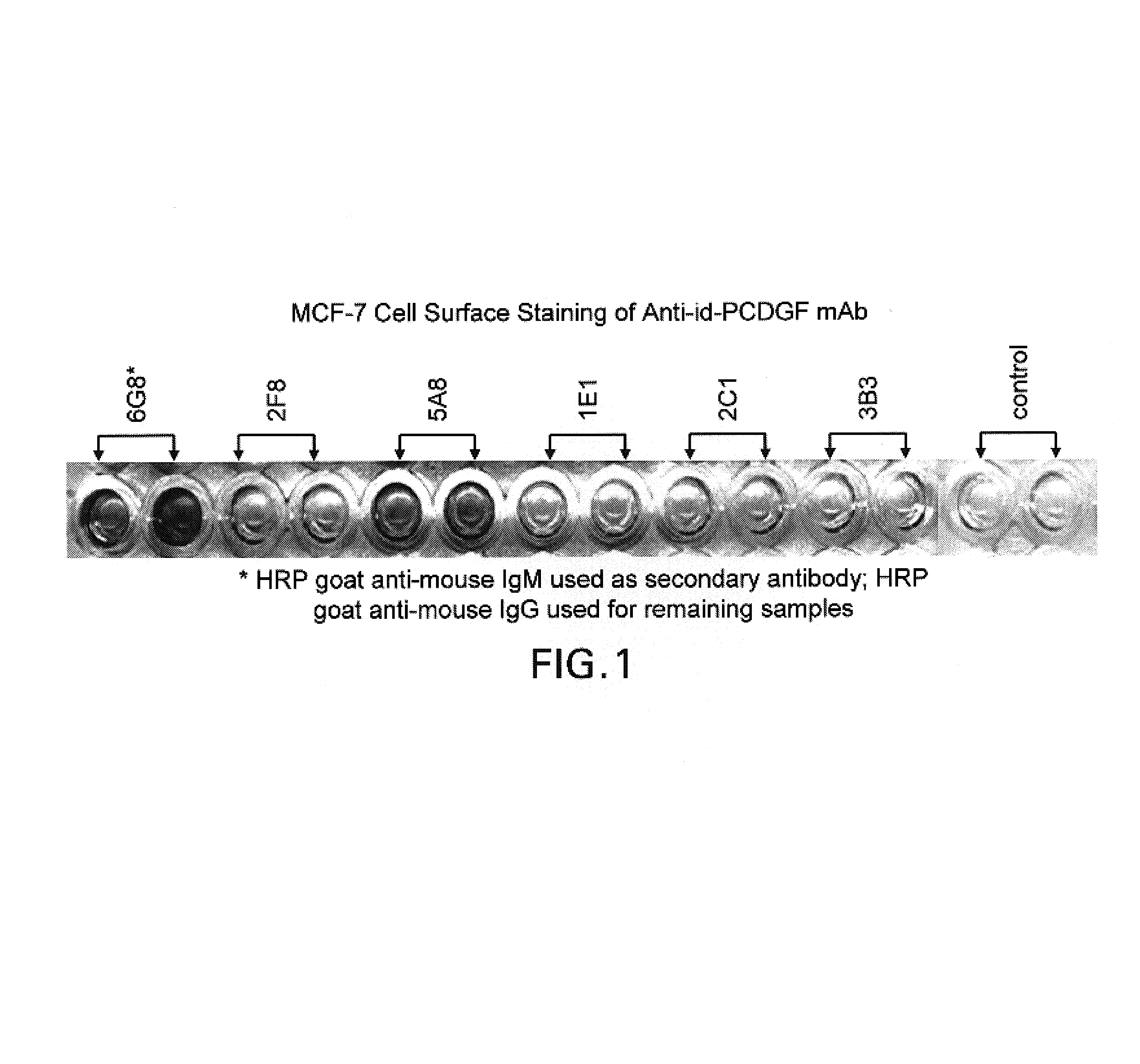
Cell Surface Staining of PCDGF Receptor Antibody on Human Breast Cancer MCF-7 Cell Line
[0046]1×10⁵ / well MCF-7 cells were seeded in 96 well plates and incubated at 37° C., in a 5% CO2 incubator overnight. Purified anti-id-PCDGF mAbs 6G8, 2F8, 1E1, 2C1 and 3B2 were diluted in DMEM, 5% FBS medium at 50 μg / ml. 200 μl / well of each monoclonal antibody (mAb) was added in duplicate wells and incubated at room temperature for 1 hour. Cells were then washed three times in PBS and incubated with a goat anti-mouse IgG or IgM (for 6G8 only) conjugated to horseradish peroxidase (“HRP”) at 1:2000 dilution for 1 hour. After an additional three washes in PBS, a TMB microwell component peroxidase substrate was added to each well. The plates were read in a plate reader set at a wavelength of 620 nanometers. Anti-PCDGF receptor antibodies stained the surface of the MCF-7 cells (FIG. 1).
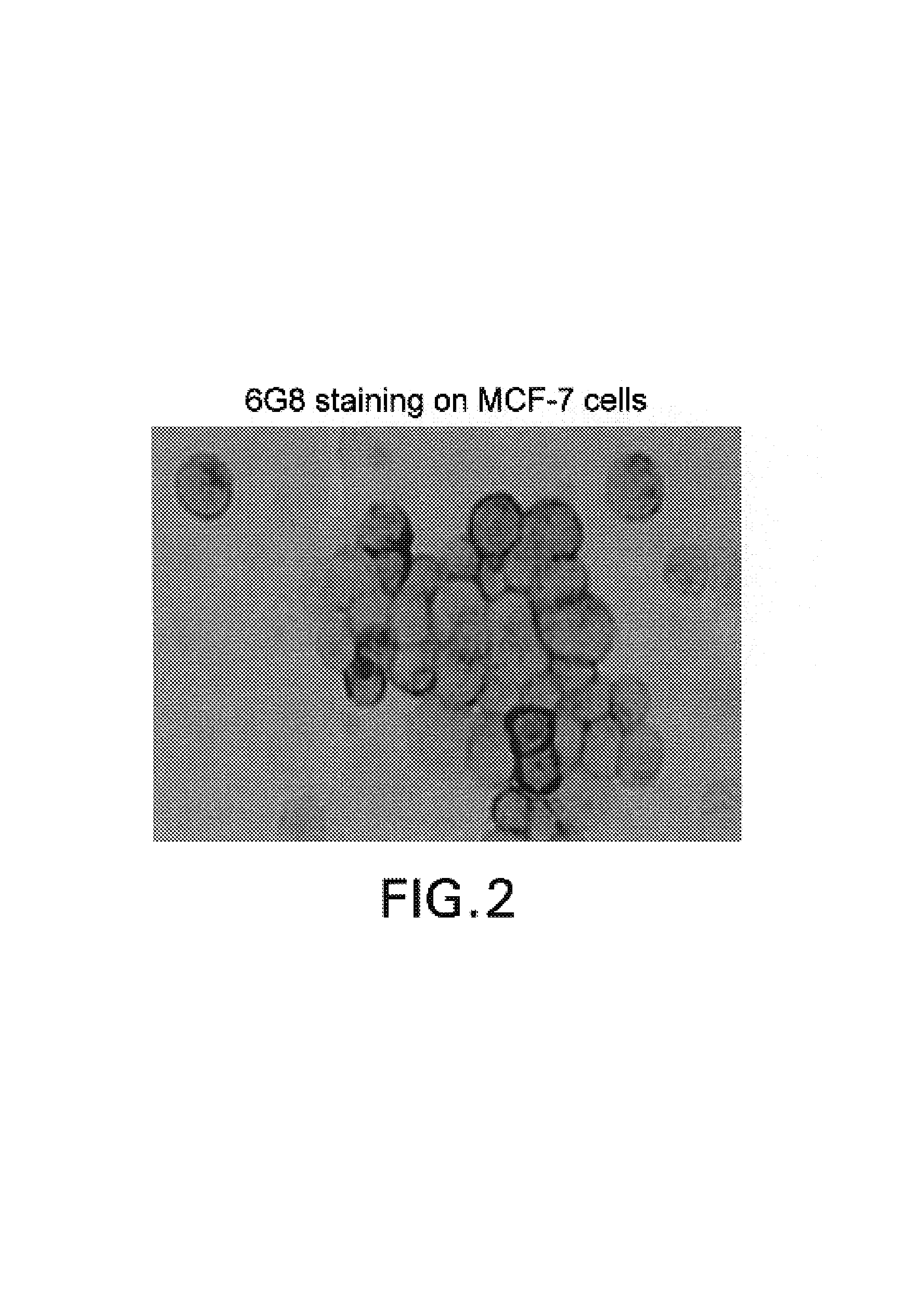
Immunostaining of MCF-7 Cells With Anti-PCDGF Receptor Antibody
[0047]Fixed MCF-7 cells were blocked with 3% BSA / PBS an...
example 2
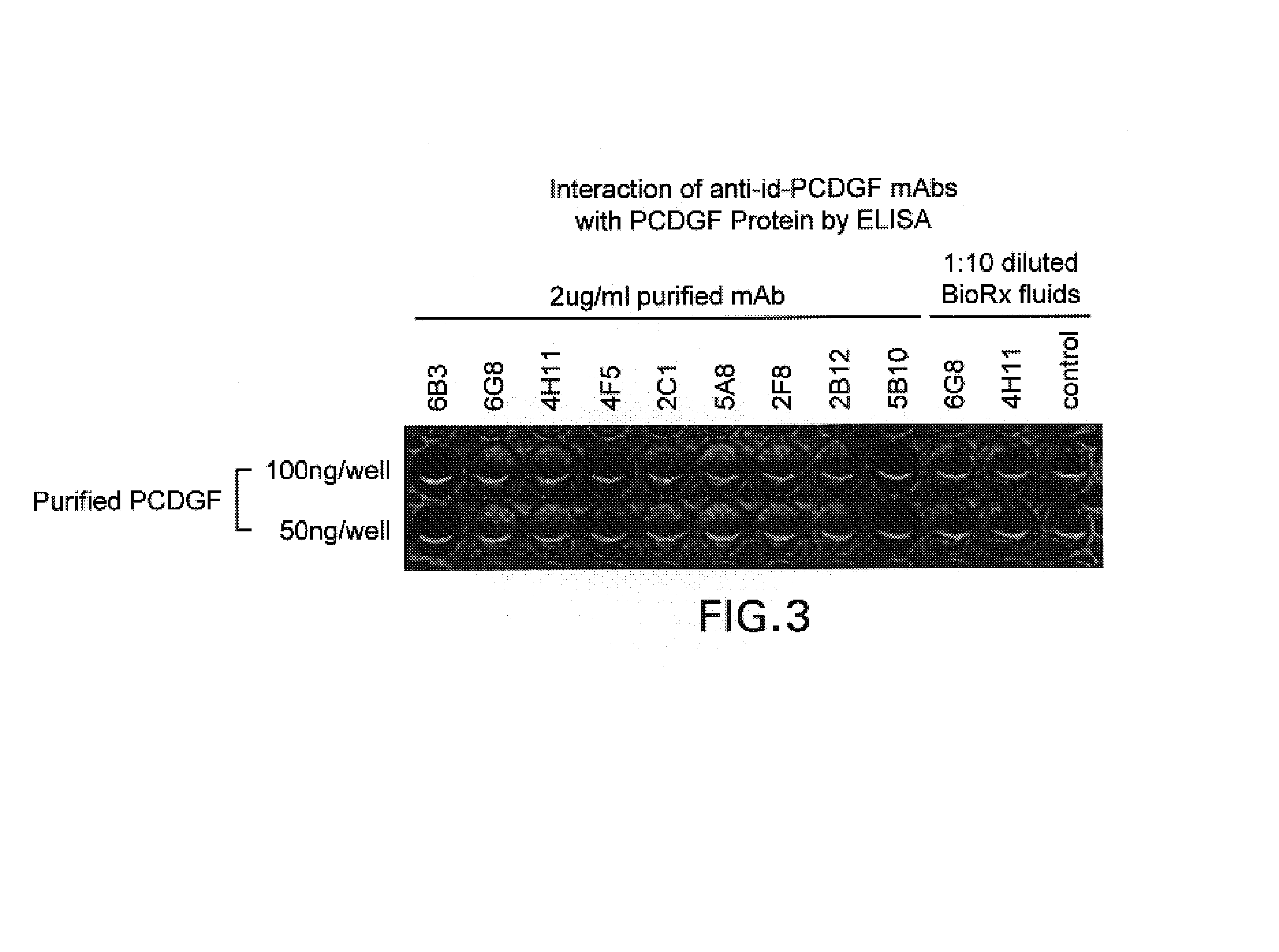
PCDGF Receptor Antibodies which do not Bind to PCDGF
[0049]Purified PCDGF protein was diluted in PBS and coated onto 96 well ELISA plates at a concentration of 100 ng / well and 50 ng / well. The treated plates were incubated overnight at 4° C. After washing 3 times with PBS, the plate was blocked with 5% non-fat milk PBS for 1 hour at room temperature. 2 μg / ml purified or 1:10 diluted BioRx fluids of PCDGF receptor mAb was added to each well and incubated at room temperature for 1 hour. The plate was washed 3 times with PBS and incubated with HRP conjugated goat anti-mouse IgG secondary antibody for another hour. After an additional three washes in PBS, a TMB microwell 1 component peroxidase substrate was added to each well. The plates were read in a plate reader set at a wavelength of 620 nanometers. As shown in FIG. 3, the anti-PCDGF receptor antibodies did not bind PCDGF.
example 3
PCDGF Receptor Antibody 6G8 Inhibits PCDGF Induced Cyclin D1 Expression in Human Breast Cancer MCF-7 Cells
[0050]2×10⁵ / ml MCF-7 cells were plated in 6 well plates in DME / F12 medium plus 5% FBS overnight. The cell culture medium was replaced with phenol-red free DMEM / F12 supplemented with 5% charcoal-stripped FBS and synchronized by treatment with 1 uM Tamoxifen for 48 hours. The cell culture medium was then replaced with serum free, phenol-red free DME / F12 and treated with either 10−9M estradiol (E2), 200 ng / ml PCDGF or 6G8 (50 μg / ml) alone or with E2 or PCDGF for 5 hours. After treatment, cells were lysed with RIPA buffer plus protease inhibitors. 60 μg of whole cell lysates were separated by 10% SDS-PAGE gel, and proteins were electrotransferred onto nitro-cellulose membranes. Western blot detection of cyclin D1 expression was performed using anti-cyclin D1 / 2 clone 5D4 monoclonal antibody (FIG. 4). As shown in FIG. 4, either PCDGF or E2 induced the expression of cyclin D1 expressio...
PUM
| Property | Measurement | Unit |
|---|---|---|
| concentrations | aaaaa | aaaaa |
| concentrations | aaaaa | aaaaa |
| wavelength | aaaaa | aaaaa |
Abstract
Description
Claims
Application Information
 - R&D
- Intellectual Property
- Life Sciences
- Materials
- Tech Scout
- Unparalleled Data Quality
- Higher Quality Content
- 60% Fewer Hallucinations
Browse by: Latest US Patents, China's latest patents, Technical Efficacy Thesaurus, Application Domain, Technology Topic, Popular Technical Reports.
© 2025 PatSnap. All rights reserved.Legal|Privacy policy|Modern Slavery Act Transparency Statement|Sitemap|About US| Contact US: help@patsnap.com