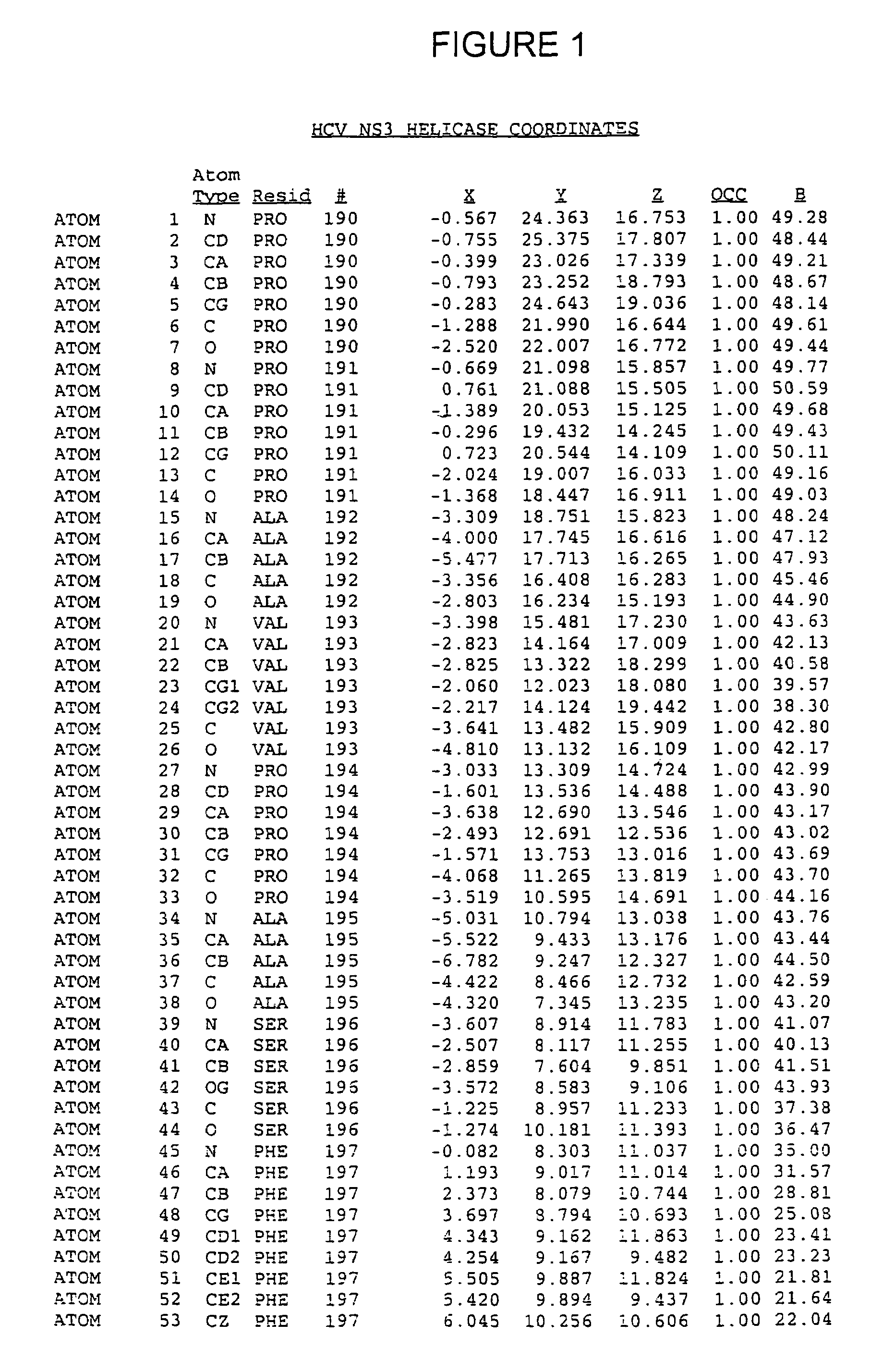
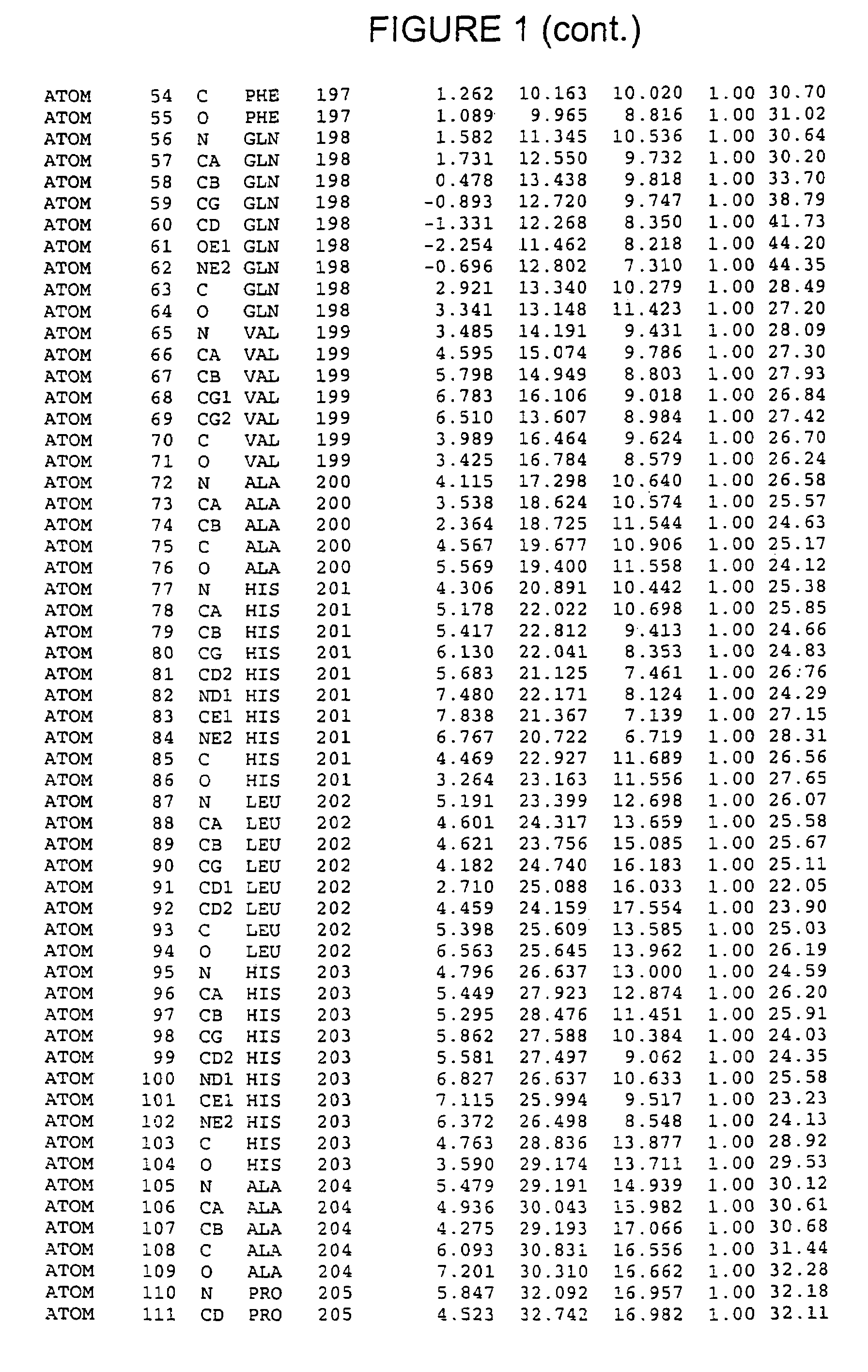
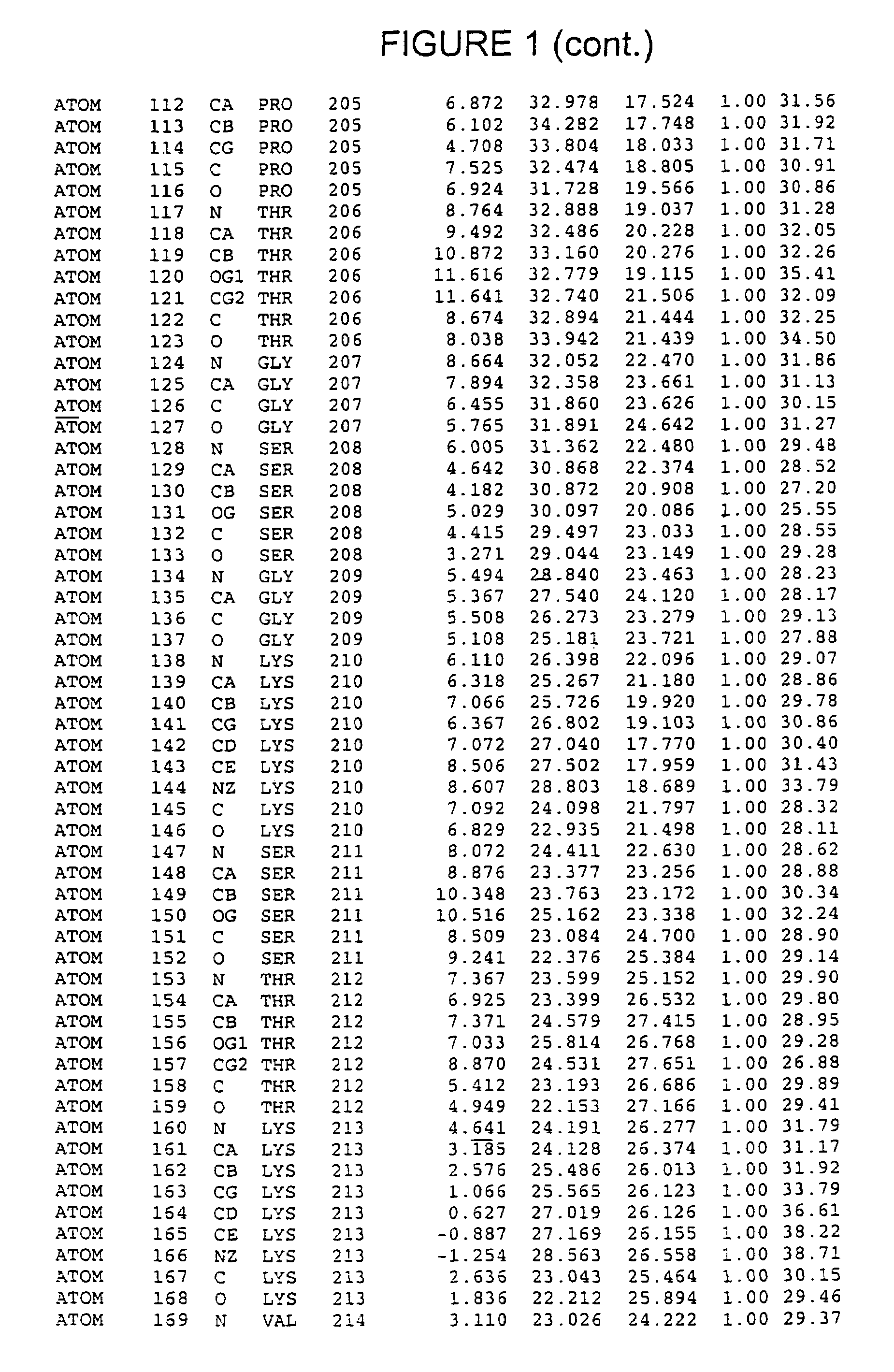
Crystals of hepatitis C virus helicase or fragments thereof comprising a helicase binding pocket
- Summary
- Abstract
- Description
- Claims
- Application Information
AI Technical Summary
Benefits of technology
Problems solved by technology
Method used
Image
Examples
example 1
Cloning and Expression of NS3 Helicase
[0177]The HCV NS3 RNA helicase domain (encoded by nucleotides 502-1896 of SEQ ID NO:6) was subcloned from a cDNA of the HCV H strain [A. Grakoui et al., J. Virol., 67, pp. 1385-95 (1993); C. Lin et al., J. Virol., 68, pp. 8147-57 (1994), the disclosures of which are herein incorporated be reference] into a pET expression vector (Novagen, Madison, Wis.). The resulting plasmid, pET-BS(+) / HCV / NS3-C465-His (SEQ ID NO:4), also contained a methionine start codon, a linker encoded Gly-Ser-Gly-Ser sequence attached the C-terminal threonine of the NS3 helicase domain and a six-histidine tag fused to the C-terminus of the Gly-Ser-Gly-Ser sequence to facilitate protein purification. This plasmid was used as a template for single-stranded DNA-based site-directed mutagenesis essentially as described by (T. A. Kunkel, Proc. Natl. Acad. Sci. USA, 82, pp. 488-492 (1985) and C. Lin et al., Virology, 192, pp. 596-604 (1993), the disclosures of which are herein in...
example 2
Crystallization and Data Collection
[0188]Crystals of the NS3 helicase:dU8 complex were grown by hanging-drop vapor diffusion over wells containing 0.1 M Tris pH 8.0, 0.2 M Li2SO4, 18% Polyethylene glycol 6000, and 8 mM β-mercaptoethanol. Drops were macroseeded within 12 hours after being set up. Crystals grew over the course of 2-3 weeks to dimensions of 0.4×0.4×0.1 mm3. The crystals belong to space group P21212 with unit cell dimensions a=73.1 Å b=117.5 Å, c=63.4 Å, and contain one helicase:dU8 complex per asymmetric unit.
[0189]Heavy atom soaks were carried out by transferring crystals to a solution containing 0.1 M Tris pH 8.0, 0.2 M Li2SO4, 17% Polyethylene glycol 6000, 8 mM β-mercaptoethanol, in addition to the heavy atom in question. Heavy atom soaks with K2WO4 were performed in the absence of Li2SO4.
[0190]Crystals were transferred to a solution containing 0.08 M Tris pH 8.0, 0.2 M Li2SO4, 16% Polyethylene glycol 6000, 8 mM β-mercaptoethanol, and 15% glycerol and immediately fr...
example 3
Phasing, Model Building and Refinement
[0192]Heavy atom positions were located from difference Patterson and anomalous difference Patterson maps and confirmed with difference Fourier syntheses. Heavy atom parameters were refined and phases computed to 2.3 Å using the program PHASES [W. Furey et al., Meth. Enzymol., 277, pp. 590-620, (1997) need full cite]. MIR phases were improved by cycles of solvent flattening [B. C. Wang, Methods Enzymol., 115, pp. 90-112 (1985)] combined with histogram matching [K. Y. J. Zhang et al., Acta Crystallogr., A46, pp. 377-381 (1990)] using the CCP4 crystallographic package [CCP4; C. C. Project, Acta Crystallogr., D50, pp. 760-763 (1994)].
[0193]Model building was carried out using QUANTA96 (Molecular Simulations), and all refinement done in XPLOR [A. T. Brunger, “X-PLOR: A System for X-Ray Crystallography and NMR,” Yale University Press, New Haven, Conn. (1993)], using the free R-value [A. T. Brunger, Nature, 355, pp. 472-475 (1992)] to monitor the cour...
PUM
| Property | Measurement | Unit |
|---|---|---|
| Fraction | aaaaa | aaaaa |
| Fraction | aaaaa | aaaaa |
| Fraction | aaaaa | aaaaa |
Abstract
Description
Claims
Application Information
 - R&D
- Intellectual Property
- Life Sciences
- Materials
- Tech Scout
- Unparalleled Data Quality
- Higher Quality Content
- 60% Fewer Hallucinations
Browse by: Latest US Patents, China's latest patents, Technical Efficacy Thesaurus, Application Domain, Technology Topic, Popular Technical Reports.
© 2025 PatSnap. All rights reserved.Legal|Privacy policy|Modern Slavery Act Transparency Statement|Sitemap|About US| Contact US: help@patsnap.com


