Corneal implant and uses thereof

a technology of corneal implants and corneal blindness, applied in the field of corneal implants, can solve the problems of corneal blindness, corneal opacification, and subsequent impairment, and achieve the effect of poor success rate of the procedur

Inactive Publication Date: 2009-01-13
OTTAVA HEALTH RES INST (CA) +1
View PDF24 Cites 49 Cited by
  • Summary
  • Abstract
  • Description
  • Claims
  • Application Information

AI Technical Summary

Problems solved by technology

Injury, disease or cellular failure can cause opacification of the cornea with subsequent impairment and corneal blindness.
This procedure has a poor success rate in disorders such as autoimmune conditions or chemical injuries.
In industrialized countries, the ageing of the population exacerbates this demand, which is compounded by the shelf-life of donated eyes being only a few days.
Furthermore, in developing countries where the number of cases of cornea blindness is increasingly problematic, healthy donor tissue is rare.
Alternatively, corneal transplants are needed in several third world countries where donor corneas are not culturally acceptable.
Such attempts have often failed because of an absence of healing and permanent attachment between the periphery of the synthetic device and the residual rim of the host cornea.
These devices have been more or less successful (e.g., remain in place for 6 months).
However, the skirt of most of them have low tensile strength leading to suturability problems, inflammatory reaction, and subsequent extrusion of the keratoprosthesis.
However, extrusion is still a major issue, as it is undesirable because it tends to cause clinical complications and product failure.
Although, corneal architecture is normally restored at long term with a layering of the epithelium and endothelium, with pigmentation and vascularization present in the deep layers of the cornea, amniotic membranes are susceptible to infectious contamination and transmission.

Method used


Image

  • Corneal implant and uses thereof
  • Corneal implant and uses thereof
  • Corneal implant and uses thereof

Examples


example 1

Collagen-pNIPAAm Membranes

[0057]A sterile RTT collagen solution of 3.0-3.5 mg / ml (w / v) in acetic acid (0.02N in water) is made, and kept at 4° C. A 4% (w / v) solution of pNIPAAm homopolymer is made in water (ddH2O) and sterilized by autoclaving or by filtering (0.24tm). The 4% solution of pNIPAAm is diluted to a 1% solution with sterile ddH2O. The 0.30-0.35% solution of collagen and the 1% solution of pNIPAAm are mixed (1:1 vol / vol) in a sterile test tube at 4° C. by pumping with a pipette until well dispersed. Cold mixing will avoid any premature gelification or fibrillogenesis of the collagen during this procedure. Collagen-pNIPAAm is then poured into a plastic dish (non-treated culture dish) or a mold (e.g., lens or cornea mold) and left air-drying under sterile conditions in a laminar flow hood for at least 2-3 days at room temperature. After drying to constant weight (˜7% water residue) at room temperature, under laminar flow at relatively constant humidity, the formed membrane ...

example 2

Fibrin-pNIPAAm Membranes

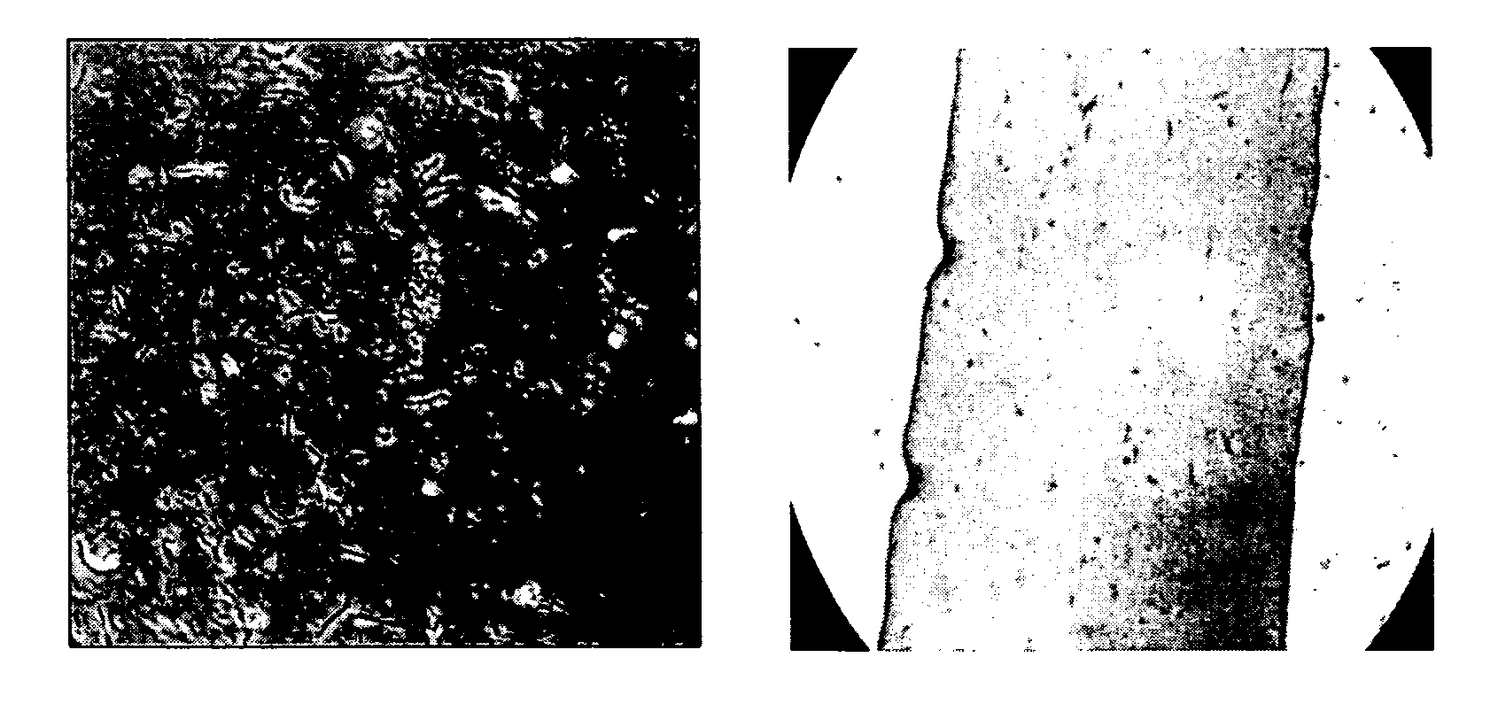
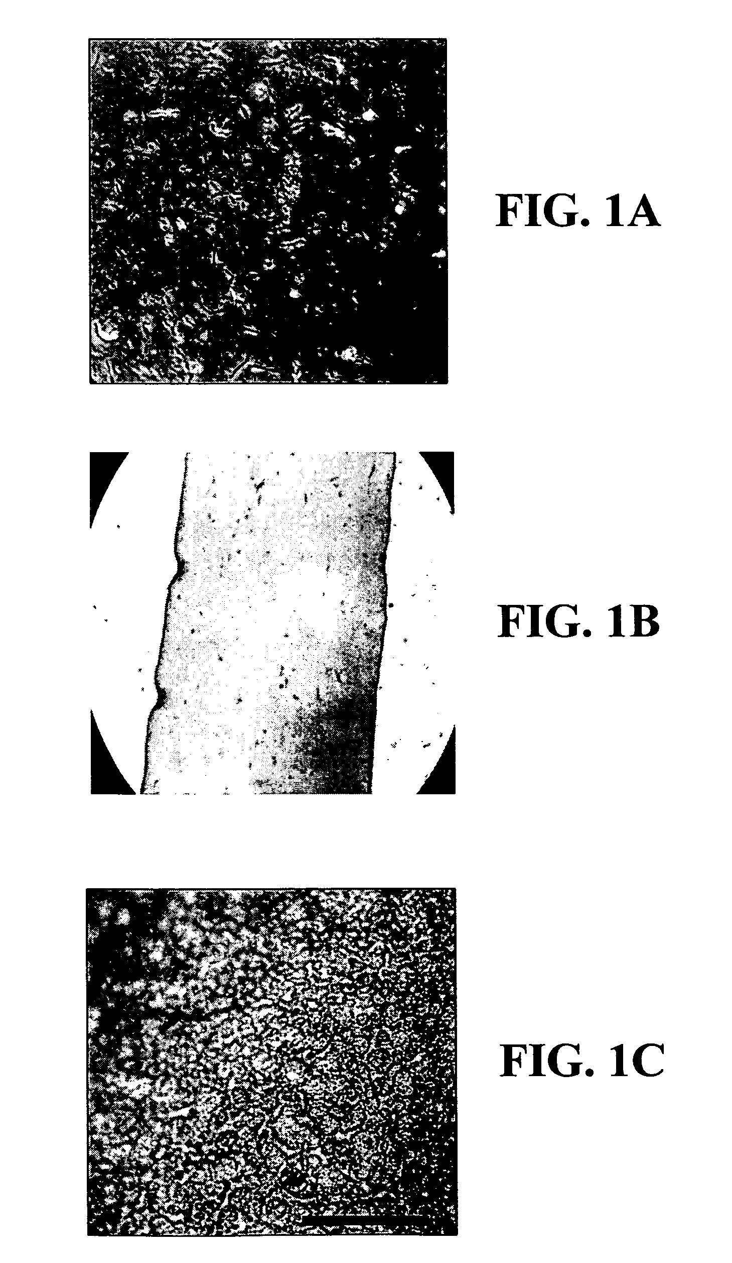
[0059]A sterile fibrinogen (fraction I; type I-S from bovine plasma) solution (3 mg / ml in HBSS) is mixed with a 1% pNIPAAm solution as reported in Example 1. During mixing, thrombin (50 U / ml purchased from Parke Davis) is added at a final concentration of 0.03:1 v / v (thrombin:fibrin), and incubated for 10-15 min at 37° C. to allow gelification (hydrogel) in a culture well. This procedure strengthens the fibrin gel, probably by aiding the development of a filamentous network as shown on FIG. 1C.

example 3

Crosslinked Membranes

[0060]A sterile RTT collagen viscous solution and a 1% (w / v) solution of pNIPAAm are made as described in Example 1. The 0.30-0.35% solution of collagen and the 1% solution of pNIPAAm are mixed (1:1 vol / vol) with a 10% stock solution of 1-(3-dimethylaminopropyl)-3-ethyl carbodiimide hydrochloride (EDC) in water to give a final EDC concentration of 0.5; 0.1; or 0.05% (w / v) in a sterile test tube at 40° C. by pumping with a pipette until well dispersed. The collagen-pNIPAAm-EDC mixture is then poured into a plastic dish (non-treated culture dish) or a mold (e.g., lens or cornea molds) and left air-drying under sterile conditions in a laminar flow hood for at least 2-3 days at room temperature. After drying to constant weight (˜7% water residue), an interpenetrating network of pNIPAAm in cross-linked collagen is formed. Each membrane is removed from the mold after soaking in a sterile buffered solution (e.g., HBSS) at room temperature. Among the three different con...


PUM

PropertyMeasurementUnit
thicknessaaaaaaaaaa
thicknessaaaaaaaaaa
tensile energy to breakaaaaaaaaaa

Abstract

A membrane for corneal implant or keratoprosthesis comprising a biological polymer and a polyacrylamide is described. The mixture of both polymers produces a hydrogel that becomes a transparent film or membrane upon drying. The resulting device and tissue engineered implants are useful for biomedical applications of the cornea, such as tissue repair and transplantation.

Description

CROSS-REFERENCE TO RELATED APPLICATIONS[0001]This application claims the benefit of U.S. provisional Patent Application Ser. No. 60 / 391,942 filed Jun. 28, 2002, which is incorporated by reference herein in its entirety.FIELD OF THE INVENTION[0002]The invention relates to a corneal implant its use for a number of biomedical applications of the cornea, including keratoprosthesis, repair and transplantation. The invention further relates to a method of preparing such a corneal implant.BACKGROUND OF THE INVENTION[0003]The cornea protects the intraocular contents and serves as major optical element of the eye. 75% of the diopteric power of the eye depends on the interface of the cornea and air. Injury, disease or cellular failure can cause opacification of the cornea with subsequent impairment and corneal blindness. Affecting more than 10 million patients worldwide, corneal opacification is often managed by transplantation of human donor tissues. This procedure has a poor success rate in...

Claims


Application Information

Patent Timeline
Patent Type & Authority Patents(United States)
IPC IPC(8): A61F2/00A61F2/14
CPCA61L27/26C08L33/26A61F2/14A61F2/142
Inventor DOILLON, CHARLES J.GRIFFITH, MAYLI, FENGFUSHIMMURA, SHIGETO
Owner OTTAVA HEALTH RES INST (CA)
Why Eureka
Try Eureka