Pharmaceutically enhanced low-energy radiosurgery
a low-energy radiosurgery and drug-enhancing technology, applied in nuclear engineering, liquid/fluent solid measurement, dispersed delivery, etc., can solve the problems of destroying the blood supply to the tumor and its growing periphery, and achieve the effect of greatly enhancing the therapeutic ability of x-rays, especially for tumor treatmen
- Summary
- Abstract
- Description
- Claims
- Application Information
AI Technical Summary
Benefits of technology
Problems solved by technology
Method used
Image
Examples
example 1
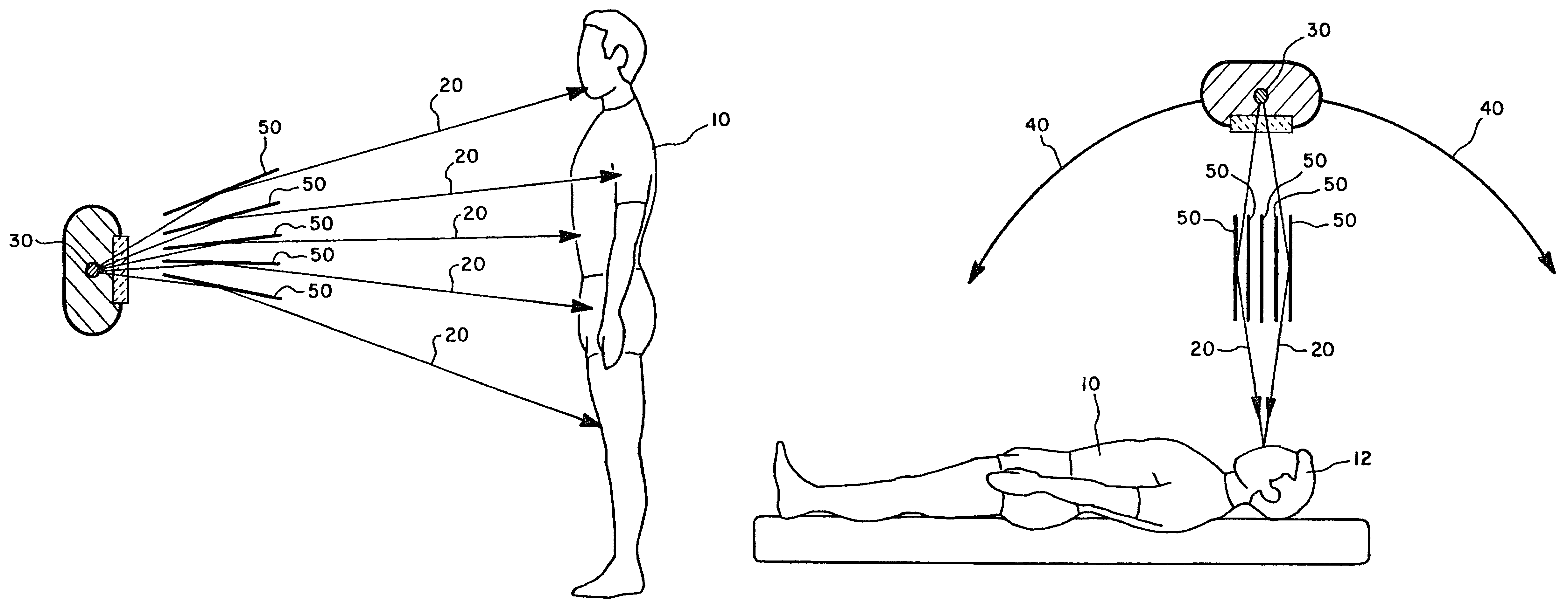
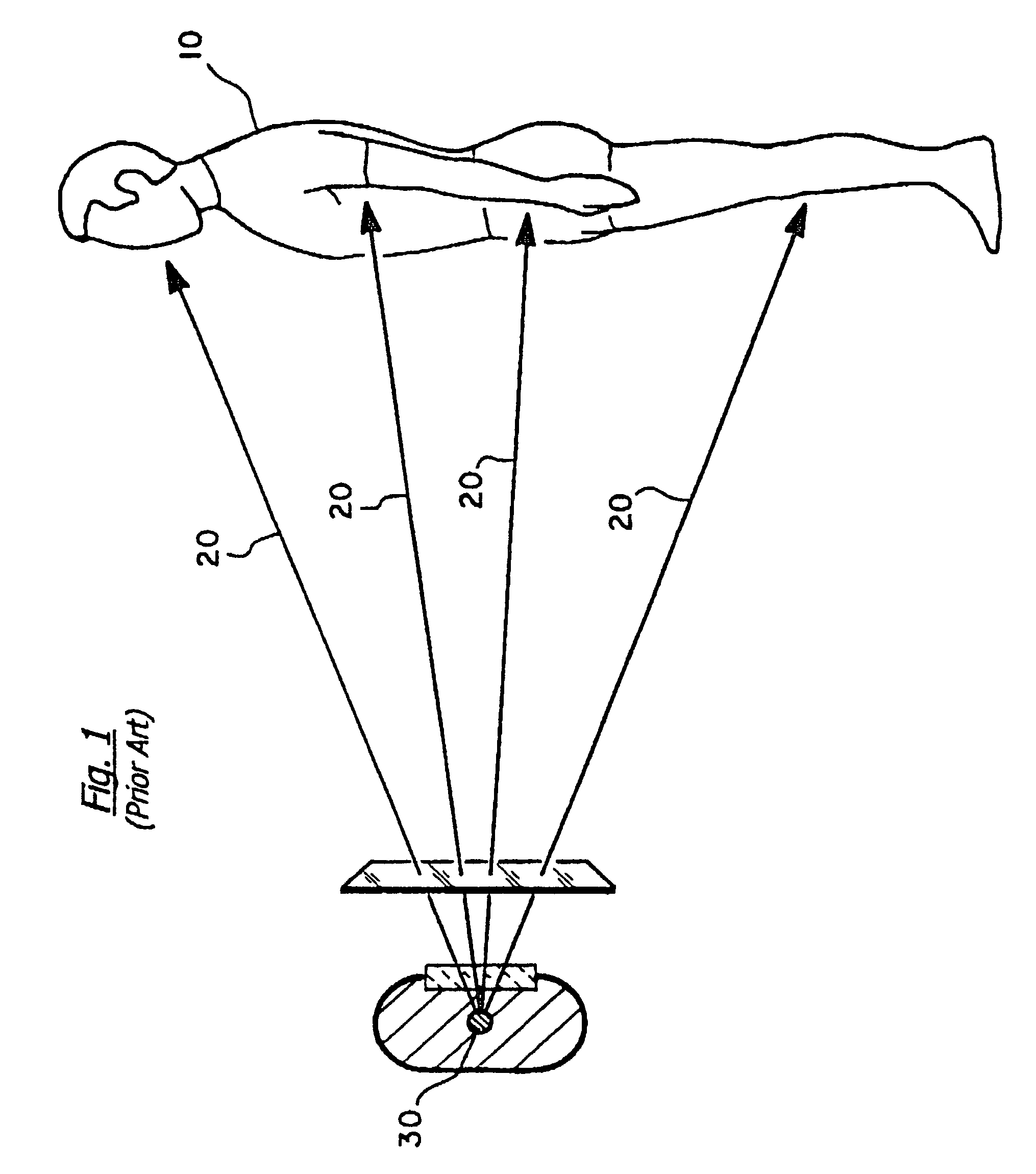
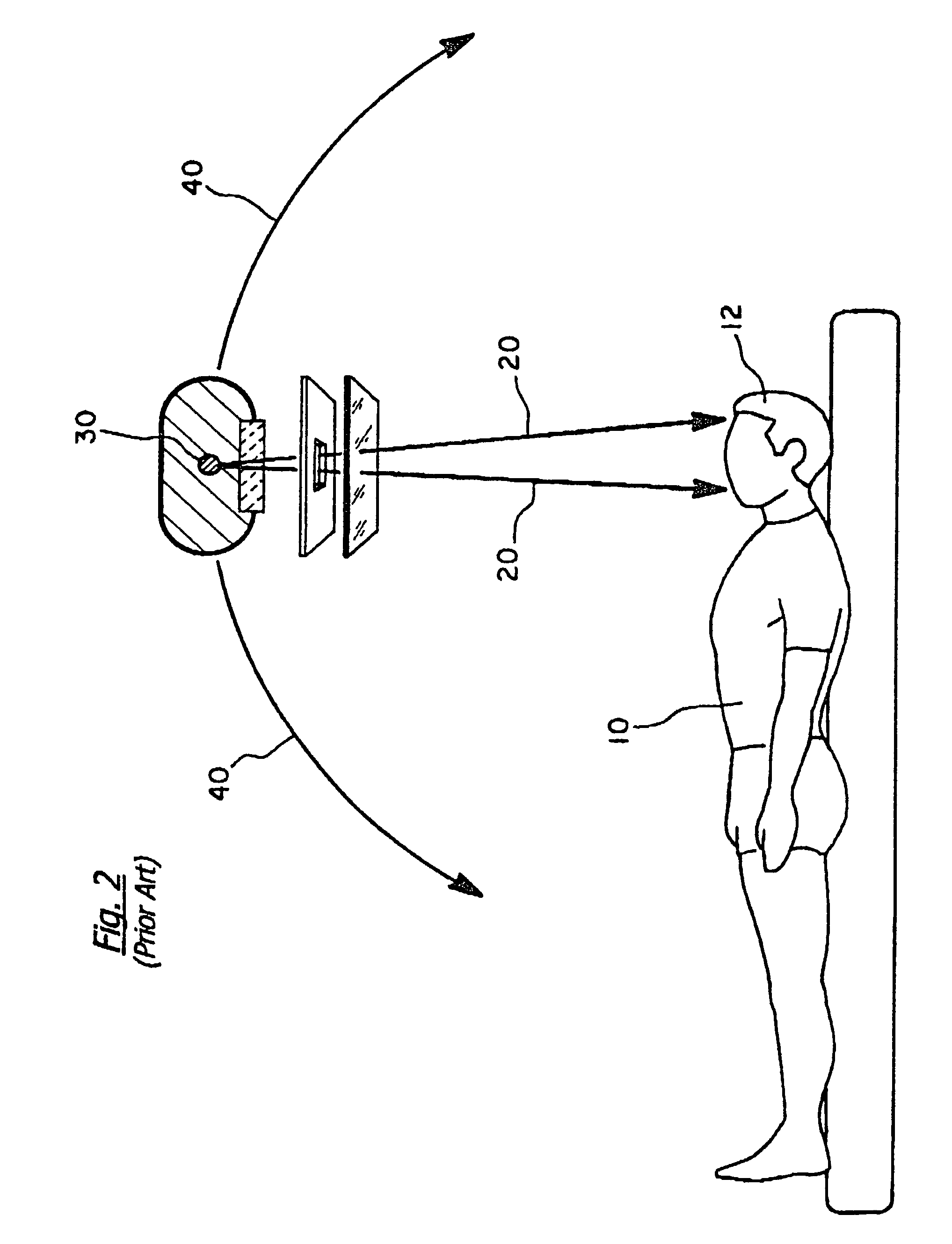
[0088]We use for this example a modern angiography x-ray source 30, which operates at 50 mA and 100 kVp continuously for about 20 minutes. A standard efficiency factor for such a source 30 predicts a flux of 6.5 W / steradian. At a distance of 500 mm, this represents 2.6×10⁻⁵ W / mm2 impacting the patient. Since the beam loses approximately 2% of its flux per millimeter of tissue traveled, the flux of x-rays scattered from the beam is 5.2×10⁻⁷ W / mm3. However, because the cross section is dominated by Compton scattering which, on the average, retains only 20% of the incident flux for ionization, while sending 80% away in scattered radiation, the total density of ionizing energy is about 10−7 W / mm3, or 10 cGy / s at the skin. This falls to 3.5 cGy at a typical tumor depth of 50 mm.
[0089]Iohexol (sold as Omnipaque™ by Nycomed of Princeton N.J.) is a tri-iodinated molecule that remains undissociated in water, and is 35% iodine by weight. When used as a contrast agent for CT imaging, the stand...
example 2
[0093]The second example was modeled in a computer, as illustrated in FIGS. 8 through 14. We created an approximation to a human head, as shown in FIG. 8, a sphere 100 of radius 78 mm, containing 2 mm3 pixels. Each pixel was assigned a composition and density. The outer layer 105 represented skin, followed by an inner layer 110 representing the bone of the skull. The bulk of the volume 115 represents regular body tissue, which represents the brain and its fluids. A 30-mm-diameter tumor 120 was located 50 mm deep (28 mm off center), and was given the same composition as tissue, but could include an additional 2.4% iodine by weight.
[0094]Individual rays (not illustrated) were traced through this model 100 in a Monte Carlo fashion, to quantify the effects of beam shape and energy, and composition of the tumor 120. The first beam featured a 57 keV x-ray beam diverging from a 1 mm spot, 1 meter away. FIG. 9 illustrates the dose distribution resulting from such a beam, which remained fixe...
example 3
[0096]This example sets forth a preferred embodiment including radiosensitization with iodinated contrast agent and orthovoltage radiosurgery of malignant tumors. We treated three patients (on a compassionate-use basis) with iodinated contrast agent and photoelectric radiotherapy. The three patients had failed multiple conventional therapies and all had end stage disease (see Table II for the paramaters of the patients treated by the method of the present invention).
[0097]
TABLE IIParameters of treated patientsAgeGenderPathologySitemtdfxkVpmAsecssddiamcvolHcertoxicrespPatient 154FemaleMelanomaforearm,3001125156704315117005.301back,500112515120043151 0#?401Patient 265MaleNHL*thigh 1225112515720485012019602thigh 22001125103004350521006.2502Patient 331MaleLmyo**abd,22511251036043524.52000601rightabd, left29311251583043585 6002.501*Non-hodgkin's lymphoma**Leiomyosarcomamtd - prescribed minimum tumor dose, the dose to the normal surrounding tissuefx - number of fractionskVp - peak kilovo...
PUM
| Property | Measurement | Unit |
|---|---|---|
| Fraction | aaaaa | aaaaa |
| Fraction | aaaaa | aaaaa |
| Fraction | aaaaa | aaaaa |
Abstract
Description
Claims
Application Information
 - R&D
- Intellectual Property
- Life Sciences
- Materials
- Tech Scout
- Unparalleled Data Quality
- Higher Quality Content
- 60% Fewer Hallucinations
Browse by: Latest US Patents, China's latest patents, Technical Efficacy Thesaurus, Application Domain, Technology Topic, Popular Technical Reports.
© 2025 PatSnap. All rights reserved.Legal|Privacy policy|Modern Slavery Act Transparency Statement|Sitemap|About US| Contact US: help@patsnap.com


