Simple membrane assay method and kit

a technology of simple membrane and assay kit, which is applied in the field of assay method, can solve the problems of serious consequences, false positive information on the patient, and aggravating the condition of the disease to more severe, and achieves the effect of highly reliable and simple test method

Active Publication Date: 2009-08-25
DENKA CO LTD
View PDF26 Cites 3 Cited by
  • Summary
  • Abstract
  • Description
  • Claims
  • Application Information

AI Technical Summary

Benefits of technology

[0007]An object of the present invention is to provide a simple assay method of testing a specimen using a membrane assay method, wherein the method can inhibit the occurrence of false positivity and can detect or quantitate an analyte with high accuracy, and a kit used in such a method.
[0009]That is to say, an object of the present invention can be achieved by a simple membrane assay method for detecting or quantitating an analyte in a specimen sample using an assay device equipped with a membrane bound with a capture-substance to capture the analyte, comprising the steps of filtering the specimen sample using a filter, dropping the filtrate onto the membrane and detecting the presence of the analyte in said specimen sample.
[0010]In addition, an object of the present invention can be achieved by a simple membrane assay kit for detecting the presence of an analyte in a specimen sample, comprising:(1) a filter tube for specimen sample equipped with a filter; and(2) an assay device equipped with a membrane bound with a capture-substance to capture the analyte.
[0019]According to the method of the present invention, in a simple membrane assay method using an assay device equipped with a membrane bound with a capture-substance to capture an analyte, the occurrence of false positivity could be greatly decreased. The rationale for the effect would be considered as follows but not limited thereto. In a simple membrane assay method using an assay device equipped with a membrane bound with a capture-substance to capture an analyte, the specimen is collected by wiping the site where the analyte is predicted to exist, for example, the pharynx, nasal cavity or the like of a patient to suspend into a buffer, or a portion of secretion or excretion containing the analyte such as nasal discharge or urine is collected and diluted with a buffer to prepare a sample for the membrane assay. In this case, besides the materials to be measured, cells peeled off from the sampling site, or components of the secretion or excretion may be contaminated into said sample. Because various biological components including viscous substance such as proteoglycan and glycolipid are contained in such the contaminant, if said sample is added onto a membrane directly, some components may be adhered onto or into the membrane. Particularly membranes whose pore size or the particle retention size is from 0.1 to 10 μm are often used. When a component with a size equivalent to such the pore size or the particle retention size is added, it is considered that the component may block the pores in the membrane and then inhibit the movement of the ingredients in the solution. It is considered that non-specific reactions may occur due to such a phenomenon and then so-called false positivity may be occurred, wherein it is judged to be positive in spite of the absence of the analyte in the sample. Such a false positivity could be inhibited effectively according to the method of the present invention, and then a highly reliable simple test method could be established.

Problems solved by technology

However, when a specimen practically collected from a patient is analyzed by such simple membrane assay method using membranes or filters, so-called “false positive” or “false positivity”, which is a positive judgment made in spite of the absence of an analyte in the specimen, may occur.
The occurrence of false positive reaction during measuring the infection with pathogens leads to mistaken information on the patient.
Therefore, not only the specification of the cause may be delayed, but also an inappropriate treatment may be taken to lead to serious consequence such as aggravation of the condition of a disease to more severe.
Accordingly, suppression of false positive reaction is an extremely important issue in view of the major purpose of use of a simple test method.

Method used


Image

  • Simple membrane assay method and kit
  • Simple membrane assay method and kit
  • Simple membrane assay method and kit

Examples


example

Example 1

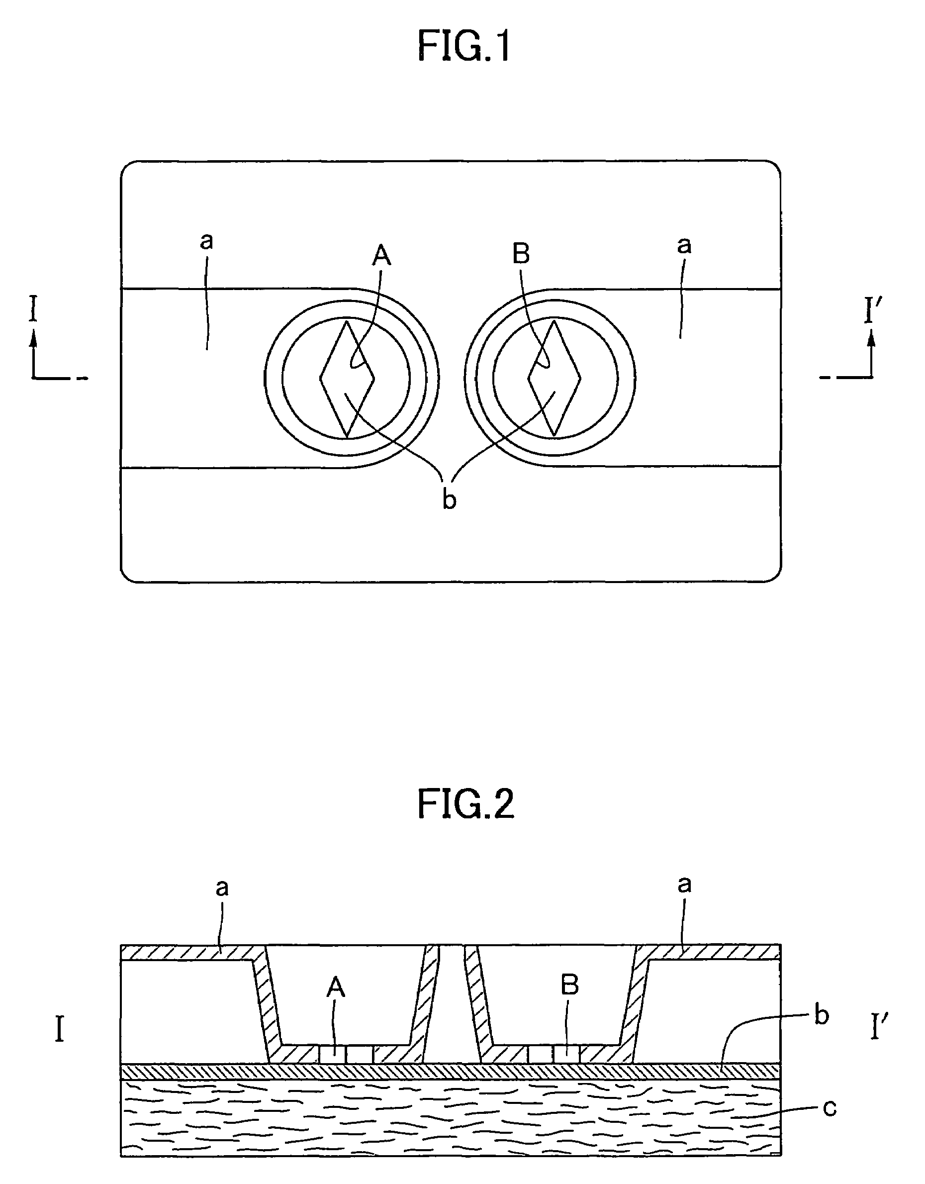
Detection of Influenza Virus by Flow-Through Type Immunochromatographic Assay Method

1. Preparation of Monoclonal Antibody

(1) Preparation of Anti-Influenza a Type Virus NP Monoclonal Antibody (Mouse)

[0061]A spleen was ablated from a BALB / c mouse which had been immunized with purified influenza A type virus antigen and kept for a certain period, and it was fused with mouse myeloma cell (P3×63) by the method of Kohler et al. (Kohler et al., Nature, Vol, 256, pp 495-497 (1975)).

[0062]The obtained fused cell (hybridoma) was kept in an incubator at 37° C. and purification (monocloning) of the cell was performed while confirming the antibody activity of the supernatant by ELISA using influenza A type virus NP antigen solid phase plate.

[0063]Two of the obtained cells were each administered intraperitoneally to a pristane-treated BALB / c mouse, after about 2 weeks, each ascites containing an antibody was collected. IgG was purified from each of the obtained ascites by ammonium sulfat...

example 2

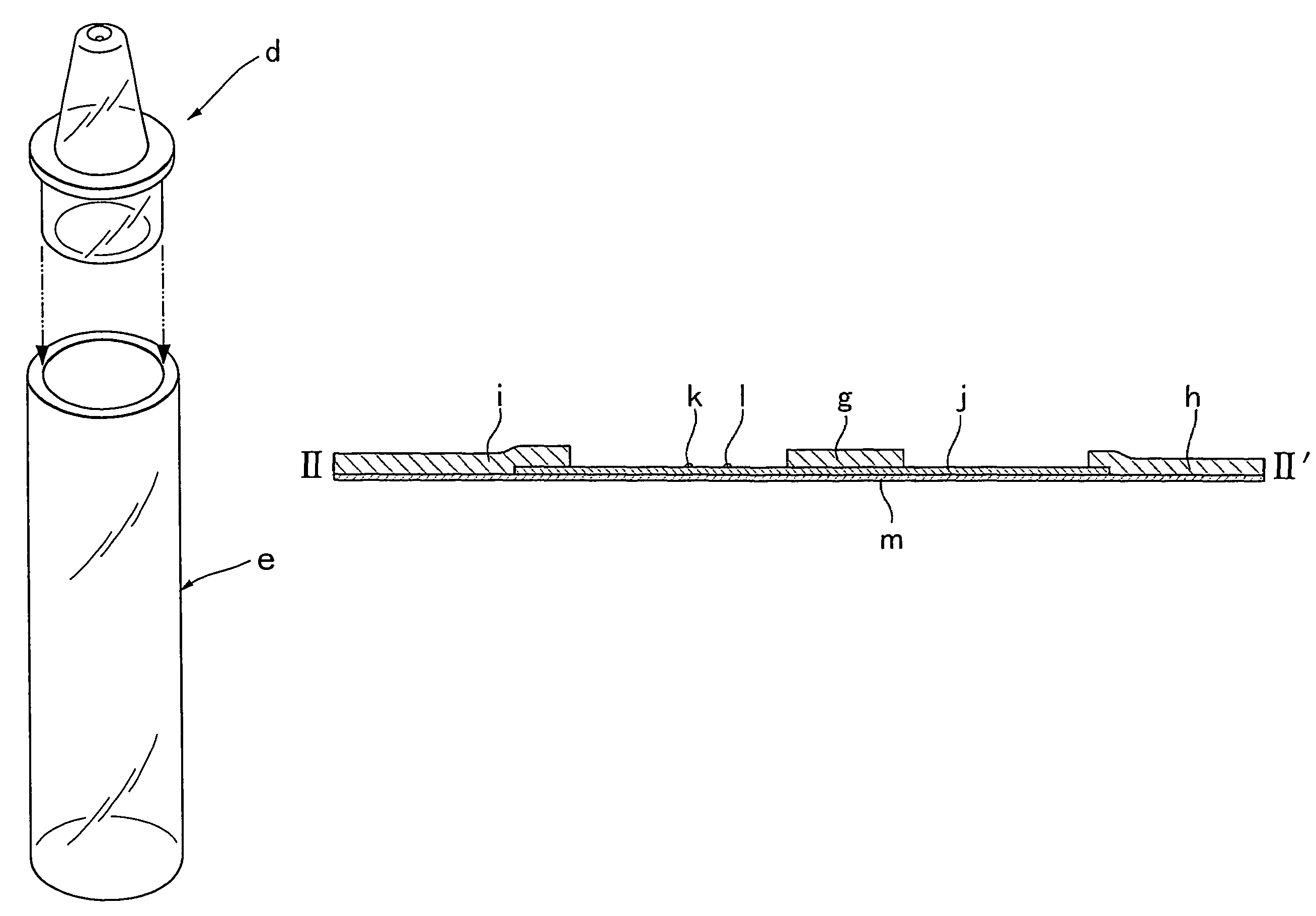
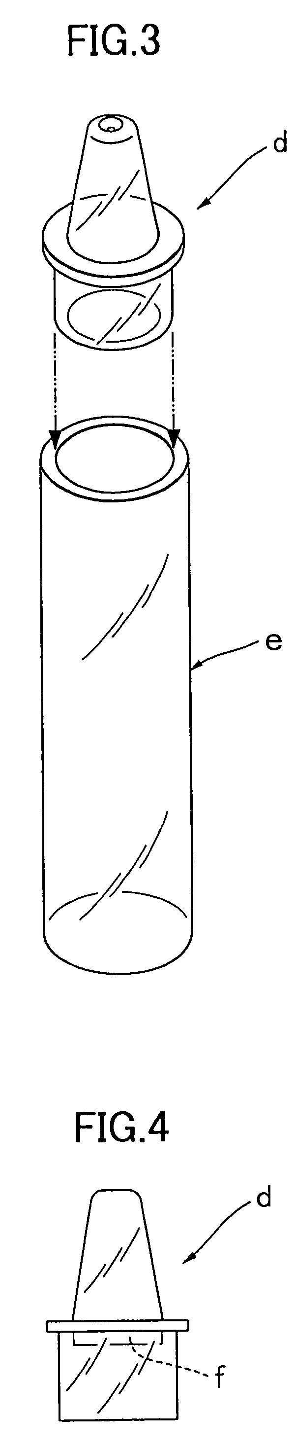
Detection of Influenza Virus by Lateral-Flow Type Immunochromatographic Assay Method

1. Preparation of Monoclonal Antibody

(1) Preparation of Anti-Influenza A virus NP Monoclonal Antibody (Mouse)

[0084]This antibody was prepared in the same manner as the method described in [Example 1] 1.(1).

(2) Preparation of Anti-Influenza B Type Virus NP Monoclonal Antibody (Mouse)

[0085]This antibody was prepared in the same manner as the method described in [Example 1] 1.(2).

2. Preparation of Labeled Anti-Influenza Antibody

(1) Preparation of Labeled Anti-Influenza A Type Antibody

[0086]20 mg of one kind of the purified anti-influenza A type virus NP monoclonal antibodies was dialyzed with 0.1 M acetate buffer (pH 3.8) followed by addition of 10 mg of pepsin and Fab′ digestive treatment was performed for one hour at 37° C. The treated solution was fractionated through ultrogel AcA44 column to yield purified fraction of anti-influenza A type F(ab′)2. The thus obtained fraction was concentrated up to a...


PUM

PropertyMeasurementUnit
pore sizeaaaaaaaaaa
particle retention sizeaaaaaaaaaa
wt %aaaaaaaaaa

Abstract

The present invention is to provide a simple membrane assay method for detecting or quantitating an analyte in a specimen sample using an assay device equipped with a membrane bound with a capture-substance to capture the analyte, comprising the steps of filtering a specimen sample using a filter, dropping the filtrate onto said membrane and detecting the presence of the analyte in said specimen sample, as well as a simple membrane assay kit for detecting the presence of an analyte in a specimen sample, comprising (1) a filter tube, and (2) an assay device equipped with a membrane bound with a capture-substance to capture the analyte. The method or the kit can decrease the occurrence of false positivity and can provide a highly accurate detection of the analyte such as pathogen and antibody in a specimen collected in a medical scene or by an individual.

Description

[0001]This application is a continuation of PCT / JP2003 / 016615 filed Dec. 24, 2003, the entire content of which is hereby incorporated by reference.TECHNICAL FIELD[0002]The present invention relates to an assay method for detecting or quantitating a certain substance in a specimen using an assay device equipped with a membrane, specifically to a simple membrane assay method which can be used clinically and to a kit used for this method.BACKGROUND ART[0003]Recently, there have been developed simple assay reagents or kits wherein various measuring items including the infection with pathogens such as viruses and bacteria, the presence of pregnancy and blood-sugar level can be detected or quantitated in a short time of from a few minutes to several tens minutes using antigen-antibody reactions, enzyme reactions and the like. Proteins composing pathogens, human chorionic gonadotropin (hCG), blood-sugar and the like are the subjects of the detection or quantitation. Most of the simple assa...

Claims


Application Information

Patent Timeline
Patent Type & Authority Patents(United States)
IPC IPC(8): G01N33/53B01L3/00G01N1/28G01N1/34G01N33/569
CPCB01L3/5023G01N1/4077G01N33/569B01L2300/0681B01L2300/069B01L2400/0406Y10S436/81G01N2001/4016Y10S435/81Y10S436/807Y10S436/808Y10S435/805
Inventor SHIMIZU, HIDEHARUWATANABE, TAKESHITAKIZAWA, KAZUYUKIMATSUDA, JUNJISATO, TOSHINORI
Owner DENKA CO LTD
Why Eureka
Try Eureka