Diagnostic markers of stroke and cerebral injury and methods of use thereof

a technology of diagnostic markers and stroke, applied in the field of identification and use of diagnostic markers for stroke and cerebral injury, can solve the problems of high risk factor for the future development of a more severe episode, patient may experience transient ischemic attack, physical obstruction of arterial blood supply to the brain, etc., and achieve the effect of being ready to analyz

Inactive Publication Date: 2009-10-27
BIOSITE INC
View PDF74 Cites 36 Cited by
  • Summary
  • Abstract
  • Description
  • Claims
  • Application Information

AI Technical Summary

Benefits of technology

[0016]In a first aspect, the invention discloses methods for determining a diagnosis or prognosis related to cerebral ischemia, or for differentiating between types of strokes and / or other cerebral injuries such as TIA. These methods comprise analyzing a test sample obtained from a subject for the presence or amount of one or more markers as described hereinafter. These methods can comprise identifying one or more markers, the presence or amount of which is associated with the diagnosis, prognosis, or differentiation of cerebral ischemia. Once such marker(s) are identified, the level of such marker(s) in a sample obtained from a subject of interest can be measured. In certain embodiments, these markers can be compared to a level that is associated with the diagnosis, prognosis, or differentiation of cerebral ischemia. By correlating the subject's marker level(s) to the diagnostic marker level(s), the presence or absence of cerebral ischemia, the probability of future adverse outcomes, etc., in a patient may be rapidly and accurately determined.
[0041]Similarly, a prognosis is often determined by examining one or more “prognostic indicators.” These are markers, the presence or amount of which in a patient (or a sample obtained from the patient) signal a probability that a given course or outcome will occur. For example, when one or more prognostic indicators reach a sufficiently high level in samples obtained from such patients, the level may signal that the patient is at an increased probability for experiencing a future stroke in comparison to a similar patient exhibiting a lower marker level. A level or a change in level of a prognostic indicator, which in turn is associated with an increased probability of morbidity or death, is referred to as being “associated with an increased predisposition to an adverse outcome” in a patient. Preferred prognostic markers can predict the onset of delayed neurologic deficits in a patient after stroke, or the chance of future stroke.

Problems solved by technology

Stroke can be categorized into two broad types, “ischemic stroke” and “hemorrhagic stroke.” Additionally, a patient may experience transient ischemic attacks, which are in turn a high risk factor for the future development of a more severe episode.
Thrombi or emboli can result from atherosclerosis or other disorders, for example, arteritis, and lead to physical obstruction of arterial blood supply to the brain.
Stroke-associated symptom(s) of intracerebral hemorrhage are abrupt, with the onset of headache and steadily increasing neurological deficits.
Prior to rupture, aneurysms may be asymptomatic, or occasionally associated with tension or migraine headaches.
However, headache typically becomes acute and severe upon rupture, and may be accompanied by varying degrees of neurological deficit, vomiting, dizziness, and altered pulse and respiratory rates.
Most TIAs are due to emboli from atherosclerotic plaques, often originating in the arteries of the neck, and can result from brief interruptions of blood flow.
Concomitant with underlying risk factors, patients experiencing TIAs are at a markedly increased risk for stroke.
The similarity of these acute clinical syndromes made it difficult to differentiate them; they were distinguished on the basis of an arbitrary criterion for the duration of symptoms.
However, determining the immediate cause of verebral ischemia and differentiating ischemic from hemorrhagic stroke is difficult.
MRI may be more effective than CT scan in early detection of ischemic stroke, but it is less accurate at differentiating ischemic from hemorrhagic stroke, and is not widely available.
It is, however, an invasive procedure that is also limited by cost and availability.
Moreover, delays in the confirmation of stroke diagnosis and the identification of stroke type limit the number of patients that may benefit from early intervention therapy.
Finally, there are currently no diagnostic methods that can identify a TIA, or predict delayed neurological deficits which are often detected at a time after onset concurrent with the presentation of symptoms.

Method used


Image

  • Diagnostic markers of stroke and cerebral injury and methods of use thereof
  • Diagnostic markers of stroke and cerebral injury and methods of use thereof
  • Diagnostic markers of stroke and cerebral injury and methods of use thereof

Examples


example 1

Blood Sampling

[0258]Blood specimens were collected by trained study personnel using EDTA as the anticoagulant and centrifuged for greater than or equal to 10 minutes. The plasma component was transferred into a sterile cryovial and frozen at −20° C. or colder. Clinical histories were available for each of the patients to aid in the statistical analysis of the assay data.

example 2

Biochemical Analyses

[0259]Markers were measured using standard immunoassay techniques. These techniques involved the use of antibodies to specifically bind the protein targets. A monoclonal antibody directed against a selected marker was biotinylated using N-hydroxysuccinimide biotin (NHS-biotin) at a ratio of about 5 NHS-biotin moieties per antibody. The antibody-biotin conjugate was then added to wells of a standard avidin 384 well microtiter plate, and antibody conjugate not bound to the plate was removed. This formed the “anti-marker” in the microtiter plate. Another monoclonal antibody directed against the same marker was conjugated to alkaline phosphatase using succinimidyl 4-[N-maleimidomethyl]-cyclohexane-1-carboxylate (SMCC) and N-succinimidyl 3-[2-pyridyldithio]propionate (SPDP) (Pierce, Rockford, Ill.).

[0260]Immunoassays were performed on a TECAN Genesis RSP 200 / 8 Workstation. Biotinylated antibodies were pipetted into microtiter plate wells previously coated with avidin ...

example 3

Statistical Analyses

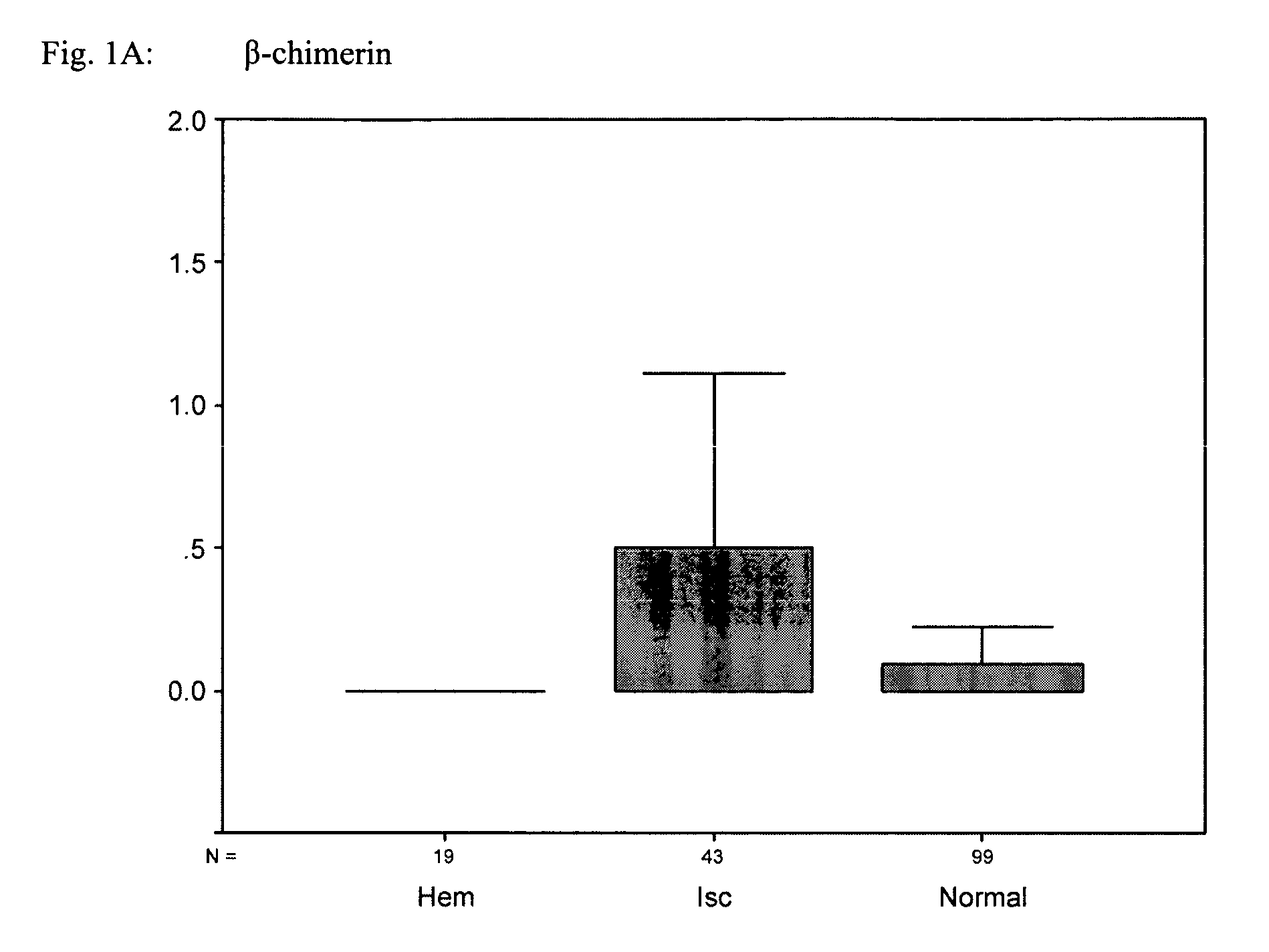
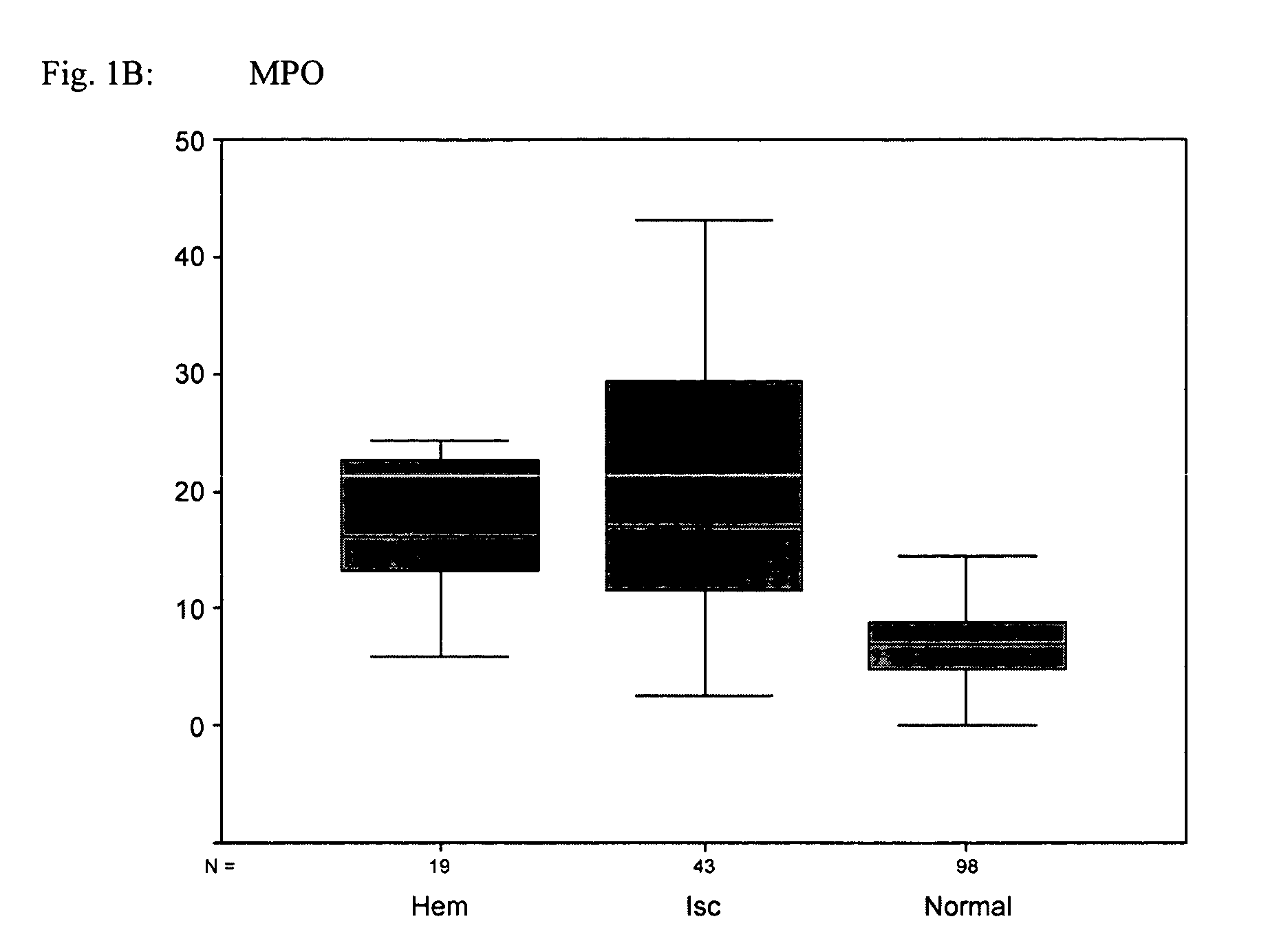
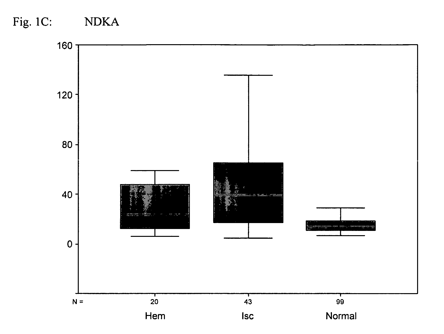
[0261]Three cohorts were classified as ischemic stroke, hemorrhagic stroke, and normal, based on clinical diagnosis. Plasma samples were measured using two-site sandwich immunoassays designed to detect β-chimerin, myeloperoxidase, NDKA, Nogo receptor, RAGE, RNA-BP, and UFDP1H, and a sandwich assay using a single antibody for nitrotyrosine. Units are reported as ng / mL (MPO, RNA-BP, RAGE, Nogo receptor, UFDP1H, β-chimerin, and NDKA,), and nM (nitrotyrosine).

[0262]Exemplary receiver operator (ROC) curve analysis is summarized in the following table:

[0263]

ROC curveSens (at 93%MarkerN(nondiseased)N(disease)Areaspecificity)MPO98610.8862.3%RNA-BP991170.8060.7%RAGE991170.5718.8%NOGO99520.6021.2%ReceptorUFDP1H99580.3912.1%β-chimerin99610.5416.4%NDKA99630.8058.7%Nitrotyrosine19100.6430.0%“nondiseased” group = Age-Matched Normals“diseased” group = Ischemic and Hemorrhagic stroke)

[0264]FIGS. 1A-G provide “box and whisker” plots calculated from the assay data using SPSS 10.1 ...


PUM

PropertyMeasurementUnit
diameteraaaaaaaaaa
concentrationaaaaaaaaaa
molecular massaaaaaaaaaa

Abstract

The present invention relates to methods for the diagnosis and evaluation of stroke and transient ischemic attacks. A variety of markers are disclosed for assembling a panel for such diagnosis and evaluation. In various aspects, the invention provides methods for early detection and differentiation of stroke types and transient ischemic attacks, for determining the prognosis of a patient presenting with stroke symptoms, and identifying a patient at risk for cerebral vasospasm. Invention methods provide rapid, sensitive and specific assays to greatly increase the number of patients that can receive beneficial stroke treatment and therapy, and reduce the costs associated with incorrect stroke diagnosis.

Description

RELATED APPLICATIONS[0001]This application is a continuation-in-part application of U.S. application Ser. No. 10 / 714,078, filed Nov. 14, 2003; which is a continuation-in-part application of U.S. application Ser. No. 10 / 673,077, filed Sep. 26, 2003; which is a continuation-in-part of U.S. application Ser. No. 10 / 371,149, filed Feb. 20, 2003; which is a continuation-in-part application of U.S. application Ser. No. 10 / 225,082, filed Aug. 20, 2002, and International Application No. PCT / US02 / 26604, filed Aug. 20, 2002; each of which claims the benefit of U.S. Provisional Application Nos. 60 / 313,775, filed Aug. 20, 2001, 60 / 334,964 filed Nov. 30, 2001, and 60 / 346,485, filed Jan. 2, 2002; the contents of each of which are hereby incorporated herein in their entirety, including all tables, figures, and claims.FIELD OF THE INVENTION[0002]The present invention relates to the identification and use of diagnostic markers for stroke and cerebral injury. In a various aspects, the invention relate...

Claims


Application Information

Patent Timeline
Patent Type & Authority Patents(United States)
IPC IPC(8): G01N33/53C12Q1/68G01N33/567G01N33/68
CPCC12Q1/6883G01N33/6896G01N2800/2871C12Q2600/158
Inventor VALKIRS, GUNARS E.DAHLEN, JEFFREY R.KIRCHICK, HOWARD J.BUECHLER, KENNETH F.
Owner BIOSITE INC
Why Eureka
Try Eureka