Devices and methods for selective surgical removal of tissue

a tissue and surgical technology, applied in the field of tissue surgical removal methods and apparatuses, can solve the problems of increased neural irritation, ischemia, and onset of disease, and achieve the effect of enabling symptomatic reli

Active Publication Date: 2010-06-15
MIS IP HLDG LLC +1
View PDF547 Cites 295 Cited by
  • Summary
  • Abstract
  • Description
  • Claims
  • Application Information

AI Technical Summary

Benefits of technology

[0018]In a preferred embodiment, the methods and apparatus include the placement of a working backstop or barrier into the epidural space or neural foramina, to a location between the tool positioned for tissue alteration, and adjacent vulnerable neural or vascular structures, to help prevent neural or vascular injury during surgery. In a further preferred embodiment, the methods and apparatus utilize neural stimulation techniques, to enable neural localization, as a means of improving the safety of the procedure.
[0084]It is expected that the apparatus and methods of the present invention will facilitate a minimally invasive approach to the selective elimination of pathological spinal tissue, thereby enabling symptomatic relief in patients suffering from spinal stenosis.

Problems solved by technology

Disease progression leads to increased neural irritation, impingement, and ischemia, and is frequently accompanied by progressively increased pain, often in conjunction with reflex, sensory and motor neurological deficits.
While standard surgical procedures lead to improvements in symptoms for 6 months or more in approximately 60% of cases, there is an unacceptable incidence of long-term complications and morbidity.
Critics have frequently stated that, while discectomy and fusion procedures frequently improve symptoms of neural impingement in the short term, both are highly destructive procedures that diminish spinal function, drastically disrupt normal anatomy, and increase long-term morbidity above levels seen in untreated patients.
First, discectomy reduces disc height, causing increased pressure on facet joints.
This stress leads to facet arthritis and facet joint hypertrophy, which then causes further neural compression.
The loss of disc height also creates ligament laxity, which may lead to spondylolisthesis, spinal instability or osteophyte or “bone spur” formation, as it has been hypothesized that ligaments may calcify in their attempt to become more “bone-like”.
In addition, discectomy frequently leads to an incised and further compromised disc annulus.
This frequently leads to recurrent herniation of nuclear material through the expanded annular opening.
It may also cause further buckling of the ligamentum flavum.
First, extensive hardware implantation may lead to complications due to breakage, loosening, nerve injury, infection, rejection, or scar tissue formation.
In addition, autograft bone donor sites (typically the patient's iliac crest) are a frequent source of complaints, such as infection, deformity, and protracted pain.
Not only do immobile vertebral segments lead to functional limitations, but they also cause increased stress on adjacent vertebral structures, thereby frequently accelerating the degeneration of other discs, joints, bone and other soft tissue structures within the spine.
While these less invasive techniques offer advantages, such as a quicker recovery and less tissue destruction during the procedure, the new procedures do not diminish the fact that even less invasive spinal discectomy or fusion techniques are inherently destructive procedures that accelerate the onset of acquired spinal stenosis and result in severe long-term consequences.
While these percutaneous procedures may produce less tissue injury, their efficacy remains unproven.
While prosthetic disc replacement is a restorative procedure, it is a highly invasive and complex surgery.
Any synthetic lumbar disc will be required to withstand tremendous mechanical stresses and will require several years of development before it will achieve the longevity desired.
Further, synthetic discs may not be an appropriate therapeutic approach to a severely degenerative spine, where profound facet arthropathy and other changes are likely to increase the complexity of disc replacement.
Like most prosthetic joints, it is likely that synthetic discs will have a limited lifespan and that there will be continued need for minimally invasive techniques that delay the need for disc replacement.
The artificial discs in U.S. clinical trials, as with any first generation prosthesis, are bound to fail in many cases, and will be very difficult to revise for patients.

Method used


Image

  • Devices and methods for selective surgical removal of tissue
  • Devices and methods for selective surgical removal of tissue
  • Devices and methods for selective surgical removal of tissue

Examples


Embodiment Construction

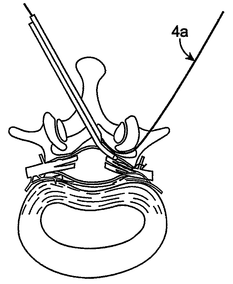
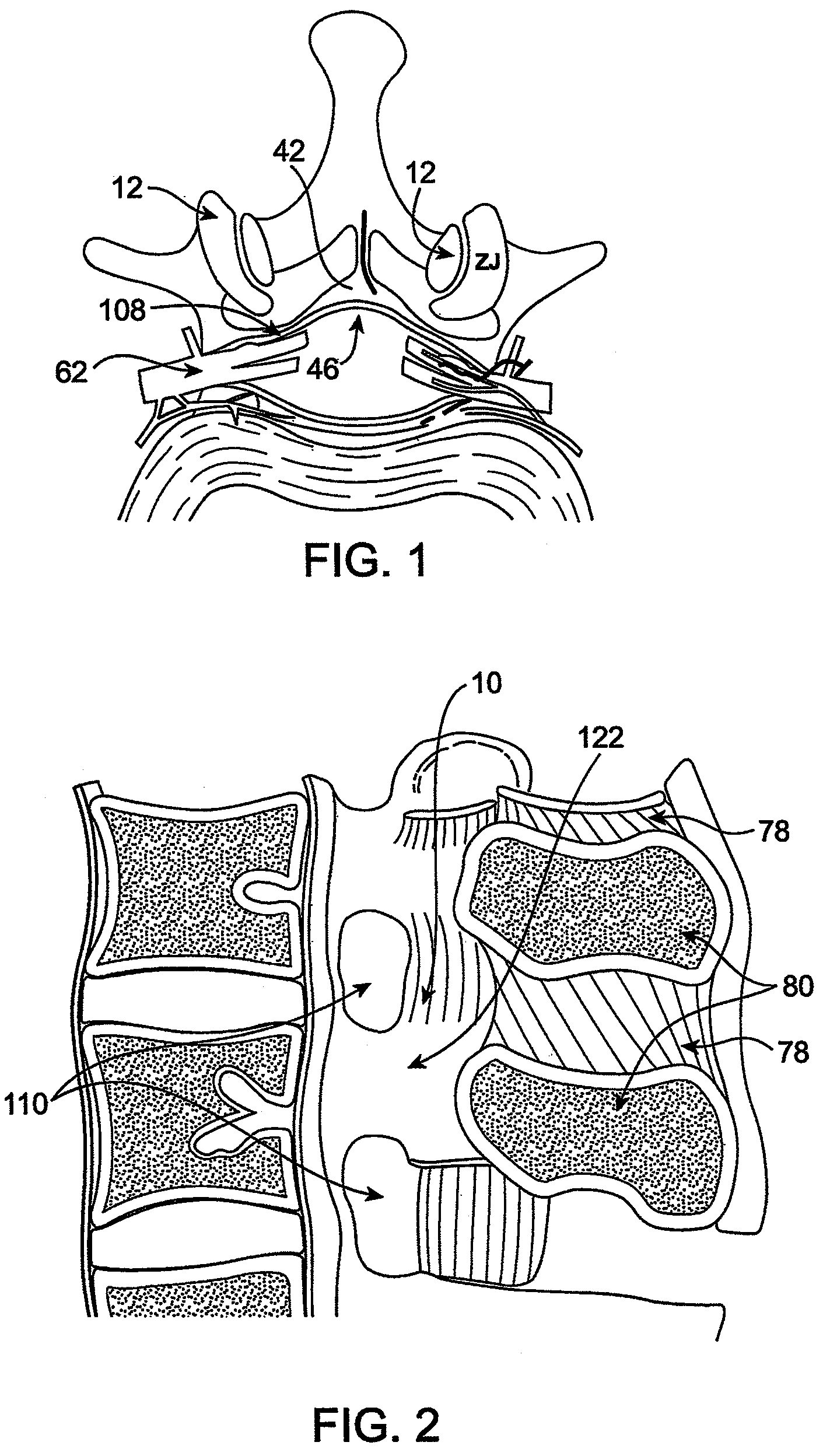
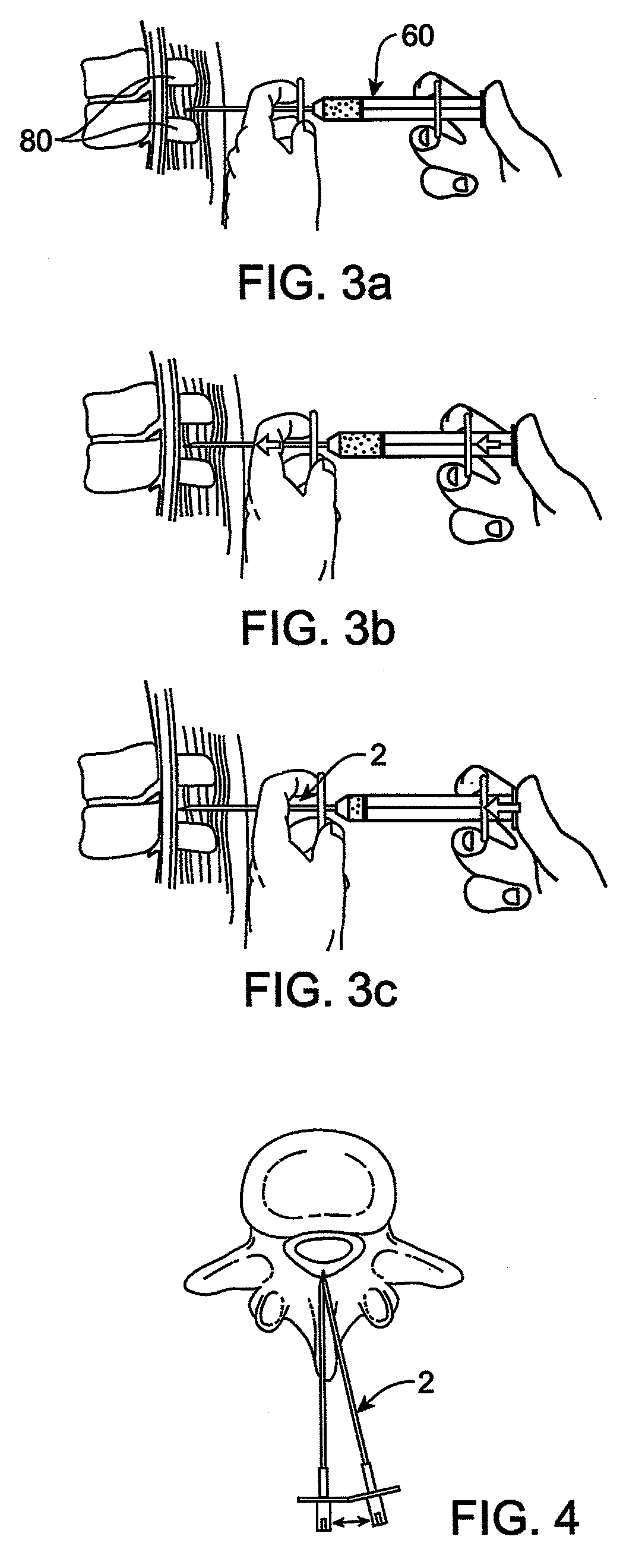
[0156]The present invention relates to methods and apparatus for the selective surgical removal or alteration of tissue that impinges upon spinal neural or vascular structures, with particular attention towards avoiding injury to the affected or adjacent neural and neurovascular structures. More particularly, a preferred embodiment of the present invention relates to methods and apparatus for lateral recess 108 and neural foraminal enlargement of the spine, in cases of neurovascular impingement, through a novel approach to selective and safe enlargement of the pathologically narrow spinal neural foramen 110, impinged lateral recess 108 and / or compromised central spinal canal. Tissues that impinge the spine's central canal, lateral recess 108, and neural foramen 110 may include, but are not limited to, ligamentum flavum 10; bone spurs or ligamentous calcifications; localized disc extrusions; enlarged facet joint complex 12, facet capsule, and superior articular processes; and scar ti...


PUM


Abstract

Methods and apparatus are provided for selective surgical removal of tissue. In one variation, tissue may be ablated, resected, removed, or otherwise remodeled by standard small endoscopic tools delivered into the epidural space through an epidural needle. The sharp tip of the needle in the epidural space, can be converted to a blunt tipped instrument for further safe advancement. The current invention includes specific tools that enable safe tissue modification in the epidural space, including a barrier that separates the area where tissue modification will take place from adjacent vulnerable neural and vascular structures. A nerve stimulator may be provided to reduce a risk of inadvertent neural abrasion.

Description

CROSS REFERENCE TO RELATED APPLICATIONS[0001]This application is a divisional of U.S. patent application Ser. No. 11 / 250,332, filed Oct. 15, 2005 entitled “DEVICES AND METHODS FOR SELECTIVE SURGICAL REMOVAL OF TISSUE” which claims the benefit under 35 U.S.C. 119 of U.S. Provisional Patent Application No. 60 / 619,306 filed Oct. 15, 2004, entitled “METHODS AND APPARATUS FOR THE TREATMENT OF TISSUE IMPINGEMENT IN THE SPINE” and U.S. Provisional Patent Application No. 60 / 622,865 filed Oct. 28, 2004, entitled “METHODS AND APPARATUS FOR SELECTIVE SURGICAL REMOVAL OF TISSUE.”INCORPORATION BY REFERENCE[0002]All publications and patent applications mentioned in this specification are herein incorporated by reference to the same extent as if each individual publication or patent application was specifically and individually indicated to be incorporated by reference.FIELD OF THE INVENTION[0003]The present invention relates to methods and apparatus for selective surgical removal of tissue, such ...

Claims


Application Information

Patent Timeline
Patent Type & Authority Patents(United States)
IPC IPC(8): A61B5/04A61B10/00A61B5/05A61N1/00A61F5/04A61B17/00A61B17/56
CPCA61B17/1671A61B17/320016A61B17/32002A61B17/32053A61B17/320758A61B17/3401A61B17/3403A61B17/3421A61B17/3496A61B18/1487A61B19/40A61B17/29A61N1/36017A61B18/1477A61B2017/00261A61B2017/00287A61B2017/003A61B2017/00867A61B2017/320004A61B2017/320044A61B2017/32006A61B2018/1407A61B2018/1425A61B2019/461A61B2019/481A61F2/0045A61N1/0551A61B90/04A61B2090/061A61B2090/08021
Inventor BLEICH, JEFFERY L.
Owner MIS IP HLDG LLC
Why Eureka
Try Eureka