Epstein-Barr virus-negative NK cell line

a technology of epstein-barr virus and nk cell line, which is applied in the field of nk cell line, can solve the problems of no report describing the involvement of ebv with nk cell line, no known technique to establish ebv-negative nk cell line, and poor prognosis, and achieves high-sensitivity measurements

Active Publication Date: 2010-08-24
JUNJIRO TSUCHIYAMA
View PDF0 Cites 3 Cited by
  • Summary
  • Abstract
  • Description
  • Claims
  • Application Information

AI Technical Summary

Benefits of technology

[0052]In addition, since the NK cell lines of the present invention are EBV negative, they can be used for the isolation and expansion of EBV. The presence of EBV in a clinical specimen can be detected, for example, by inoculating the clinical specimen, such as blood, with the NK cell lines of the present invention and confirming the infection of EBV. EBV infection in adults who have never been infected can cause infectious mono

Problems solved by technology

Primary nasal angiocentric NK lymphomas are generally resistant to various anti-cancer agents and often have a bad prognosis.
However, there

Method used


Image

  • Epstein-Barr virus-negative NK cell line
  • Epstein-Barr virus-negative NK cell line
  • Epstein-Barr virus-negative NK cell line

Examples


example 1

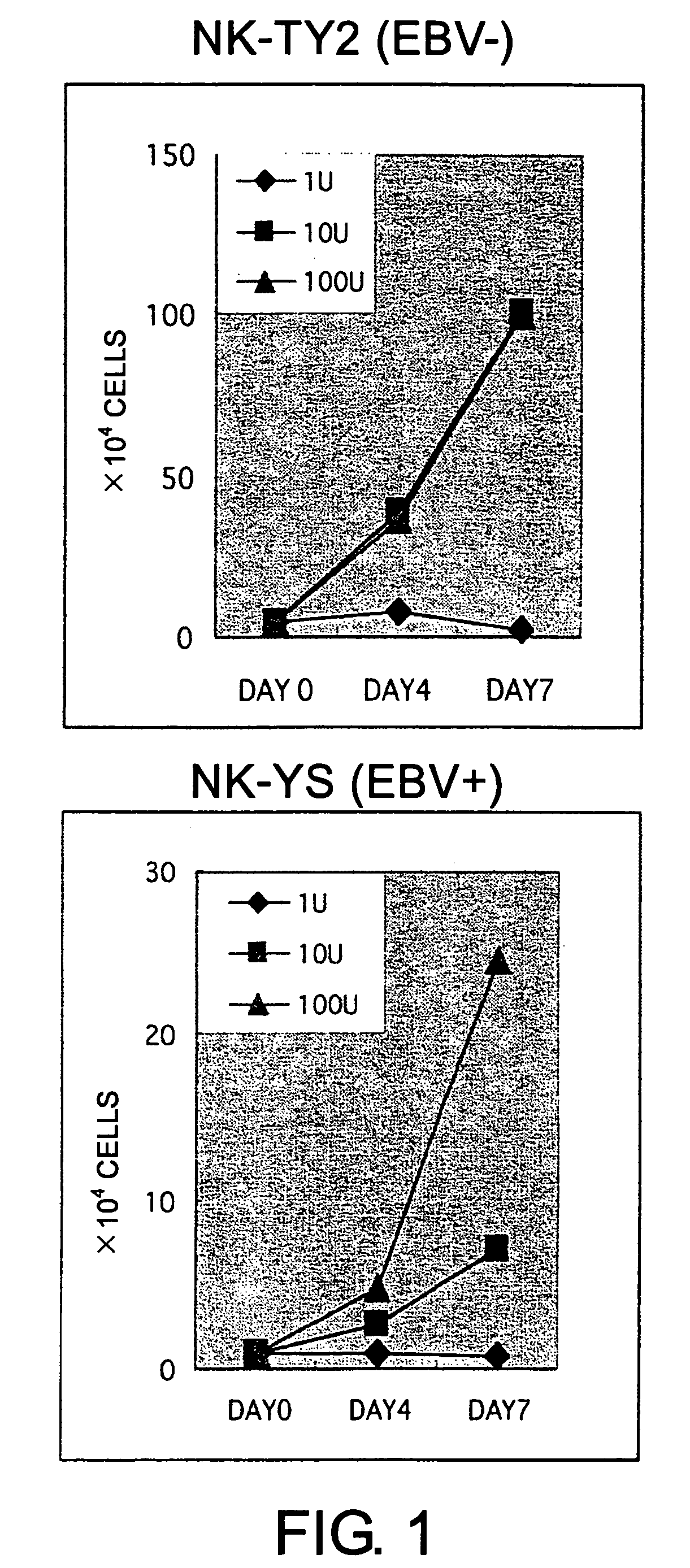
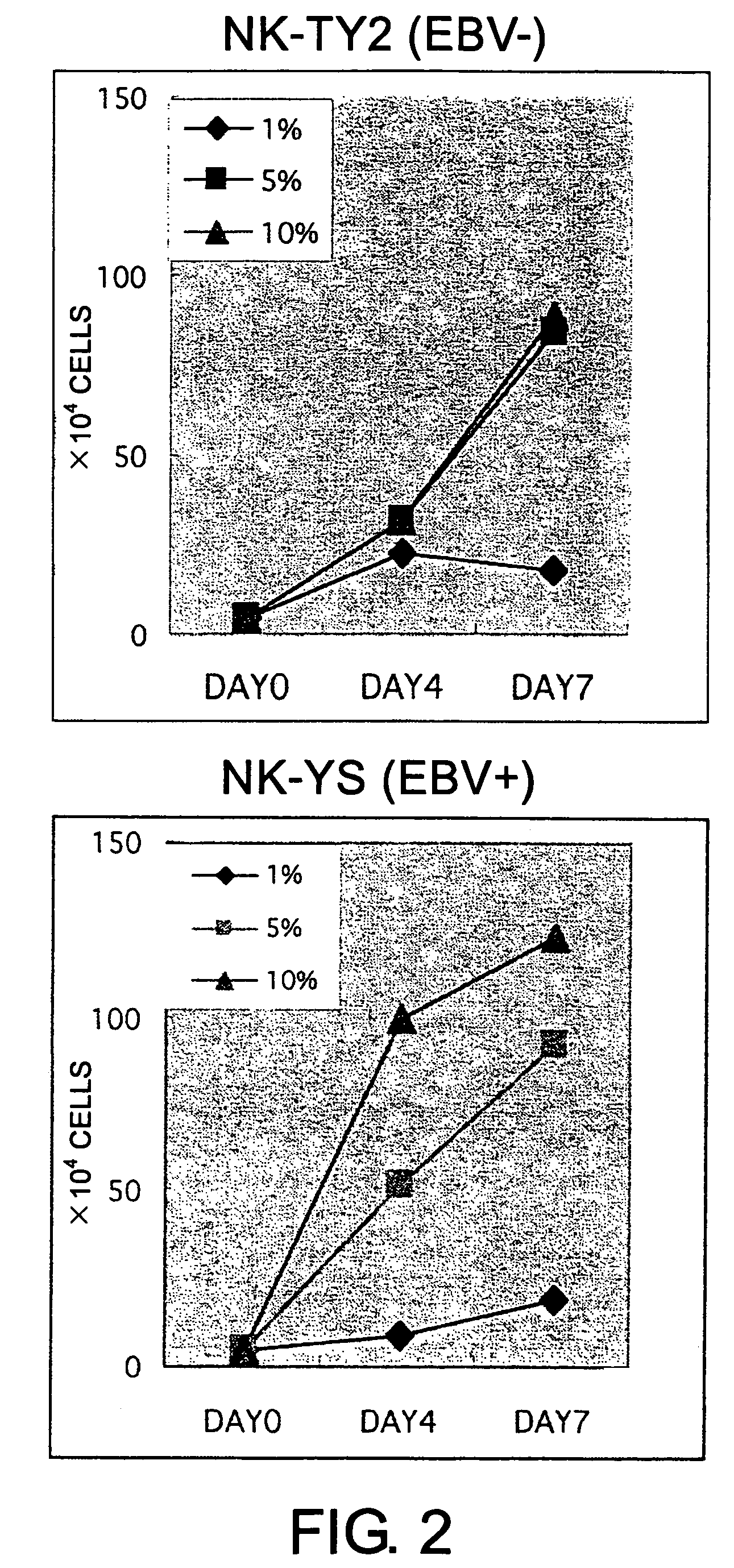
Establishment of Cell Line NK-TY2

[0090]Mononuclear cells were isolated by the Ficoll-Hypaque method from peripheral blood of a consenting patient after the crisis of leukemia. The patient was a 45 year-old female diagnosed as having primary nasal angiocentric NK lymphoma. The peripheral blood of the patient comprised CD2+, CD3−, CD7+, and CD56+ heteromorphic lymphocytes and the patient was EBV positive.

[0091]The isolated mononuclear cells were suspended in Iscove's modified Dulbecco's medium (IMDM; GIBCO, Grand Island, N.Y.) supplemented with 10% fetal bovine serum (FBS; Sanko Junyaku Co., Tokyo, Japan). More than 90% of the cells of this cell preparation were CD56-positive. The mononuclear cells (1×10⁶ cells) were inoculated into 5 mL of IMDM supplemented with 10% FBS containing 100 units of rhIL2 (Shionogi and Co., Osaka, Japan) in a 25 cm2-culture flask (Falcon 3013; Becton Dickinson, Oxnard, Calif.). This flask was maintained under a humidified atmosphere with 5% CO2 at 37° C. H...

example 2

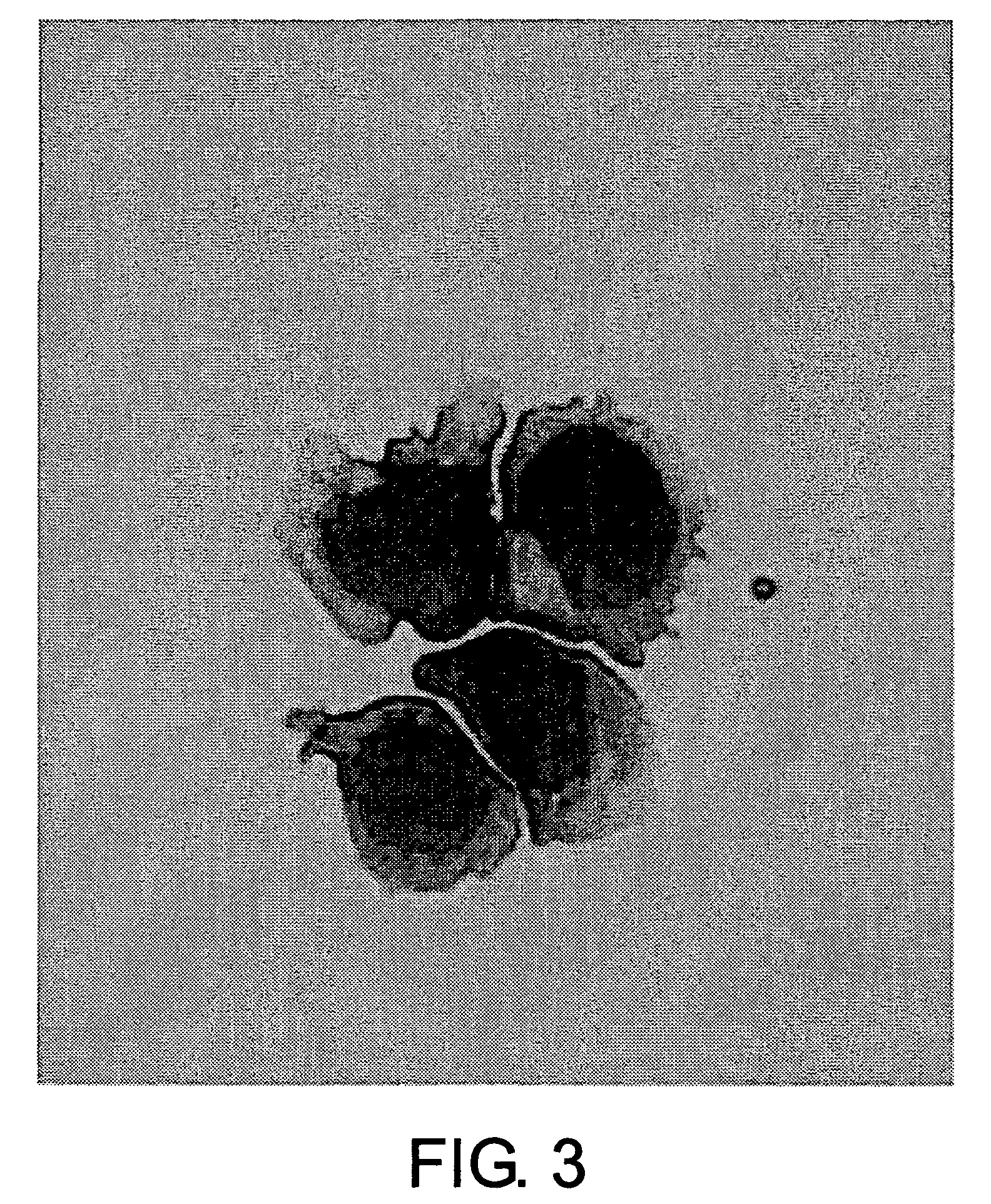
Morphological Evaluation

[0093]Cytocentrifuge smears of the peripheral blood mononuclear cells and NK-TY2 cells were stained with May-Grünwald-Giemsa for observation under a light microscope.

[0094]The result of evaluation showed that NK-YS cells had no azurophilic granules. The cells had large nuclei with coarse chromatin and conspicuous nucleoli, and abundant basophilic cytoplasm (FIG. 3). NK-TY2 cells were negative for peroxidase staining (data not shown).

example 3

Flow Cytometric Analysis (Cell Marker)

[0095]NK-TY2 cells were analyzed by single-color immunofluorescence with a flow cytometer (FACScan; Becton Dickinson and Co., Mountain View, Calif.) for the expression of surface markers. Fluorescein isothiocyanate (FITC)- or phycoerythrin (PE)-conjugated antibodies used in the experiment are shown below:

[0096]Leu5b (CD2), Leu4 (CD3), Leu3a (CD4), Leu1 (CD5), Leu9 (CD7), Leu2a (CD8), CALLA (CD10), LFA1α (CD11a), Leu15 (CD11b), LeuM5 (CD11c), LeuM7 (CD13), LeuM3 (CD14), LeuM1 (CD15), Leu11 (CD16), Leu12 (CD19), Leu16 (CD20), CR2 (CD21), IL2R (CD25), LeuM9 (CD33), HPCA1 (CD34), HLe1 (CD45), Leu19 (CD56), Leu7 (CD57), TCR α / β, and SmIg (κ+λ) from Becton-Dickinson;

OKT6 (CD1) from Ortho Diagnostic Systems (Raritan, N.J.);

B1 (CD21) from Coulter Immunology (Hialeah, Fla.);

LFA3 (CD58), Fas (CD95), HP-3B1 (kp43; CD94), EB6 (CD158a), GL183 (CD158b), FES172 (CD158c), Z27.3.7 (CD159), 191B8 (NKRP1A; CD161), and Z199 (NKG2A) from Immunotech (Marseilles, Fra...


PUM

PropertyMeasurementUnit
Timeaaaaaaaaaa

Abstract

The present invention provides Epstein-Barr virus (EBV)-negative NK cell lines. The NK cell lines of the present invention are useful for screening factors associated with the proliferation and expression functions of NK cells and to discover factors produced by the NK cells. In addition, the cell lines are immortalized despite the fact that they are EBV-negative. Thus, unknown mechanisms of oncogenesis may be elucidated through an understanding of the mechanisms underlying the immortalization of the cell lines of the present invention.

Description

TECHNICAL FIELD[0001]The present invention relates to an NK cell line.BACKGROUND ART[0002]A natural killer cell (hereinafter abbreviated as “NK cell”) is activated by interferon and interleukin-2 (hereinafter abbreviated as “IL2”), and the activated cell destroys tumor cells and such. While the biological defense mechanism dependent on T cells and B cells functions as a result of immune response, the NK cell-dependent defense mechanism works via the activation by cytokines. Namely, the complicated process of immune response is not required for the defensive function of NK cells. Therefore, NK cells have been believed to play an important role at the forefront of the biological defense mechanisms.[0003]Thus, the biological defense mechanisms may be controlled by modifying the activation and proliferation of NK cells. However, many obscurities regarding the function of NK cells remain to be clarified.[0004]On the other hand, NK cells are lymphocytes and form lymphomas upon tumorigenic...

Claims


Application Information

Patent Timeline
IPC IPC(8): C12N15/85C12Q1/70C12N7/00C12P21/00C12N5/0783G01N33/50
CPCC12N5/0646G01N33/5047C12N2503/00C12N2503/02C12N2510/04A61P35/00A61P37/02A61P43/00
Inventor TSUCHIYAMA, JUNJIROYOSHINO, TADASHIHARADA, MINE
Owner JUNJIRO TSUCHIYAMA
Why Eureka
Try Eureka