Monitoring QRS complex to identify left ventricular dysfunction

a technology of qrs complex and monitoring device, applied in the field of medical devices, can solve the problems of lvd fatigue, more severe lvd, end-organ failure, complex cause-effect interrelationship, etc., and achieve the effect of detecting more quickly

Active Publication Date: 2011-04-19
MEDTRONIC INC
View PDF28 Cites 3 Cited by
  • Summary
  • Abstract
  • Description
  • Claims
  • Application Information

AI Technical Summary

Benefits of technology

[0006]The invention is directed to techniques for cardiac monitoring. In particular, an implantable medical device identifies onset and/or progression of left ventricular dysfunction (LVD). The implantable medical device receives a signal that represents electr

Problems solved by technology

However, such disorders often have complicated cause-and-effect interrelationships, and LVD in any particular patient is most often caused by the progression of two or more such disorders.
Moreover, some disorders, such as dilated cardiomyopathy, become progressively worse in an attempt to overcome LVD, which ultimately

Method used


Image

  • Monitoring QRS complex to identify left ventricular dysfunction
  • Monitoring QRS complex to identify left ventricular dysfunction
  • Monitoring QRS complex to identify left ventricular dysfunction

Examples


Embodiment Construction

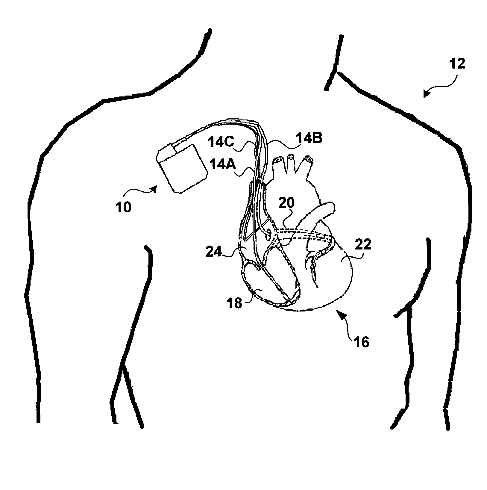
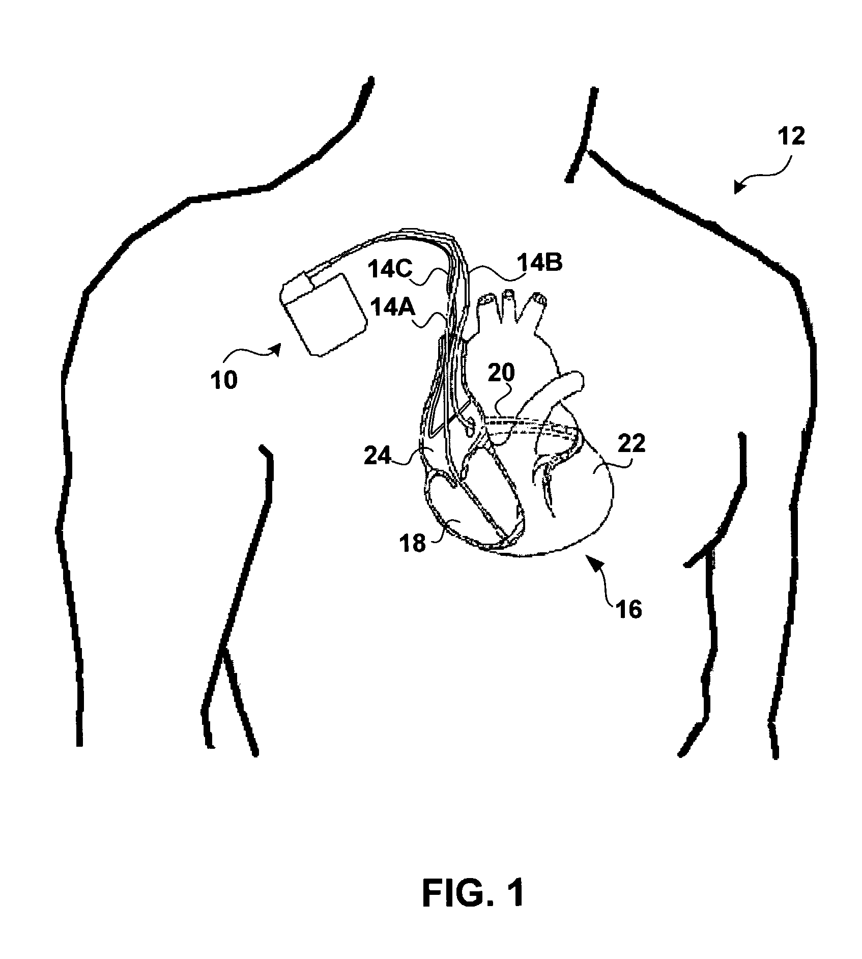
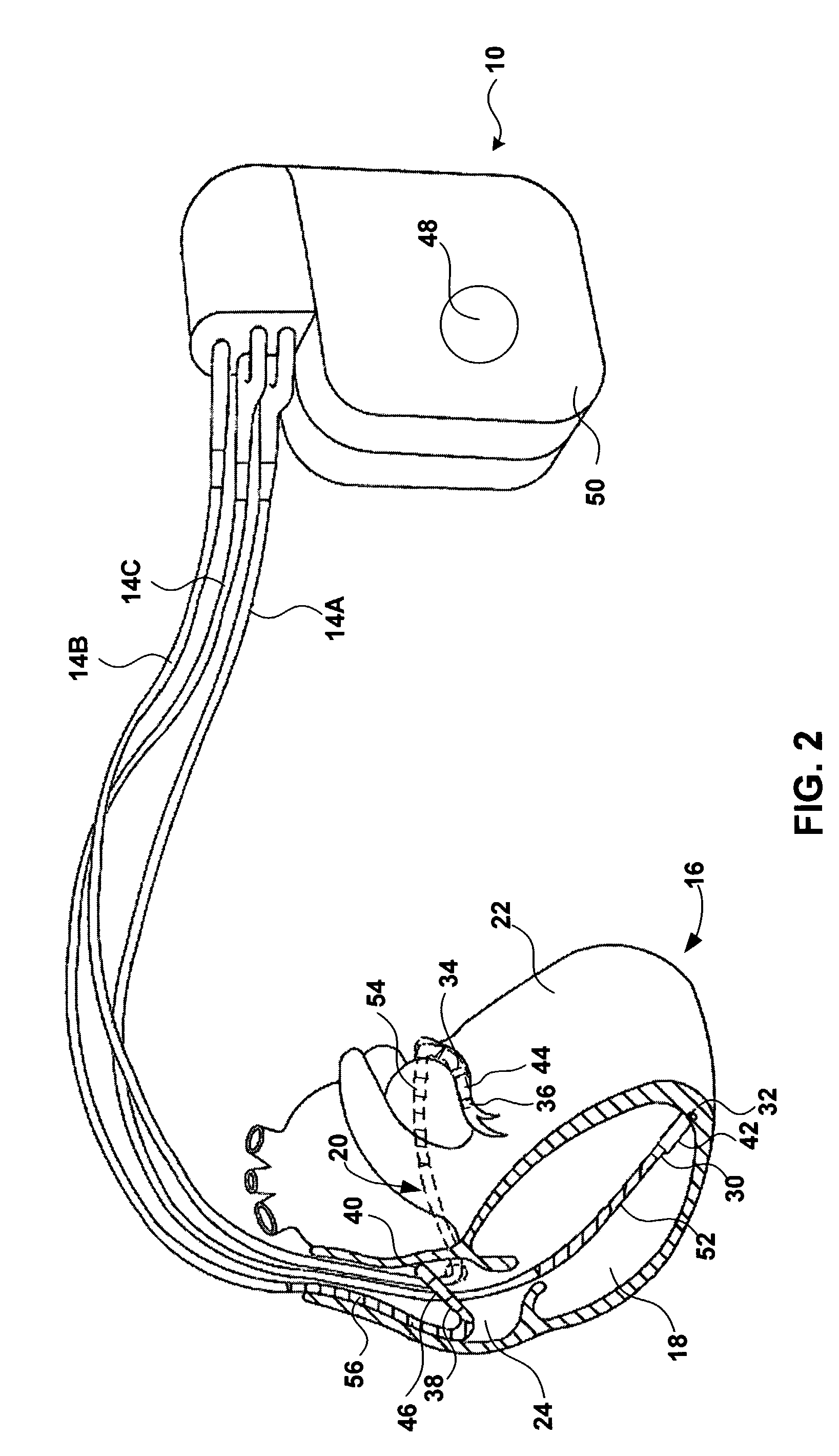
[0020]FIG. 1 is a conceptual diagram illustrating an exemplary implantable medical device (IMD) 10 implanted in a patient 12. IMD 10 may, as shown in FIG. 1, take the form of a multi-chamber cardiac pacemaker. In the exemplary embodiment illustrated in FIG. 1, IMD 10 is coupled to leads 14A, 14B and 14C (collectively “leads 14”) that extend into the heart 16 of patient 12.

[0021]More particularly, right ventricular (RV) lead 14A may extend through one or more veins (not shown), the superior vena cava (not shown), and right atrium 24, and into right ventricle 18. Left ventricular (LV) coronary sinus lead 14B may extend through the veins, the vena cava, right atrium 24, and into the coronary sinus 20 to a point adjacent to the free wall of left ventricle 22 of heart 16. Right atrial (RA) lead 14C extends through the veins and vena cava, and into the right atrium 24 of heart 16.

[0022]IMD 10 may sense electrical signals attendant to the depolarization and repolarization of heart 16 via e...


PUM


Abstract

An implantable medical device identifies onset and/or progression of left ventricular dysfunction (LVD). The implantable medical device receives a signal that represents electrical activity within a heart, e.g., an electrogram signal, and digitally processes the signal to assess left ventricular function. In particular, the implantable medical device measures at least one characteristic of QRS complexes within the signal, and assesses left ventricular function based on measurements. The implantable medical device may, for example, measure the width of the QRS complexes and/or the amplitude of the R-waves within the QRS complexes. The implantable medical device may alert a patient or clinician of the onset or progression of LVD, and may control delivery of therapies, such as rate-responsive pacing and cardiac resynchronization pacing, based on the measurements. The implantable medical device may also control delivery of a drug by an implanted drug delivery device, e.g., drug pump, based on the measurements.

Description

FIELD OF THE INVENTION[0001]The invention relates to medical devices and, more particularly, to implantable medical devices used for monitoring cardiac function.BACKGROUND OF THE INVENTION[0002]Left ventricular dysfunction (LVD) is a symptom of a variety of cardiac disorders. For example, LVD may be caused by dilated cardiomyopathy or ischemia. LVD may also be caused by left bundle branch block (LBBB), which is a conduction disorder that may cause ventricular dysynchrony.[0003]In any particular patient, a single cardiac disorder may cause LVD. However, such disorders often have complicated cause-and-effect interrelationships, and LVD in any particular patient is most often caused by the progression of two or more such disorders. Moreover, some disorders, such as dilated cardiomyopathy, become progressively worse in an attempt to overcome LVD, which ultimately leads to even more severe LVD.[0004]LVD refers to the ineffectiveness of a heart, and more particularly the left ventricle of...

Claims


Application Information

Patent Timeline
IPC IPC(8): A61N1/362A61B5/366
CPCA61B5/0472A61N1/3627A61B5/4839A61B5/366
Inventor BOUTE, WILLEMVAN HOVE, JOS W
Owner MEDTRONIC INC
Why Eureka
Try Eureka