Methods of inducing apoptosis in hyperproliferative cells

a technology of hyperproliferative cells and apoptosis, which is applied in the direction of peptide/protein ingredients, drug compositions, peptide sources, etc., can solve the problems of insufficient resectability, radioisotope therapy can induce extensive damage to normal tissues, tumors cannot be treated with radiotherapy, etc., and achieve the effect of increasing the level of kchap protein

Inactive Publication Date: 2012-05-29
CASE WESTERN RESERVE UNIV
View PDF1 Cites 0 Cited by
  • Summary
  • Abstract
  • Description
  • Claims
  • Application Information

AI Technical Summary

Benefits of technology

[0013]In another aspect, the present invention provides a method of inhibiting cell cycling cancer cells that cont

Problems solved by technology

Unfortunately, some tumors cannot be treated with radiotherapy.
Moreover, irradiation and radioisotope therapy can induce extensive damage of normal tissues.
Although most tumors are operable, they not fully resectable.
This leads to a recurrence of the cancer close to the initial site of occurrence.
Cancer chemotherapeutic agents, even though widespread in use, have proved to be of limited effect in treating most cancer types.
Although there have been some notable successes in the treatment of some specific tumor types (e.g., childhood leukemias) with conventi

Method used


Image

  • Methods of inducing apoptosis in hyperproliferative cells
  • Methods of inducing apoptosis in hyperproliferative cells
  • Methods of inducing apoptosis in hyperproliferative cells

Examples


example 1

KChAP Overexpression of in Prostate Cancer Cells

KChAP increases K+ Efflux in LNCaP Cells

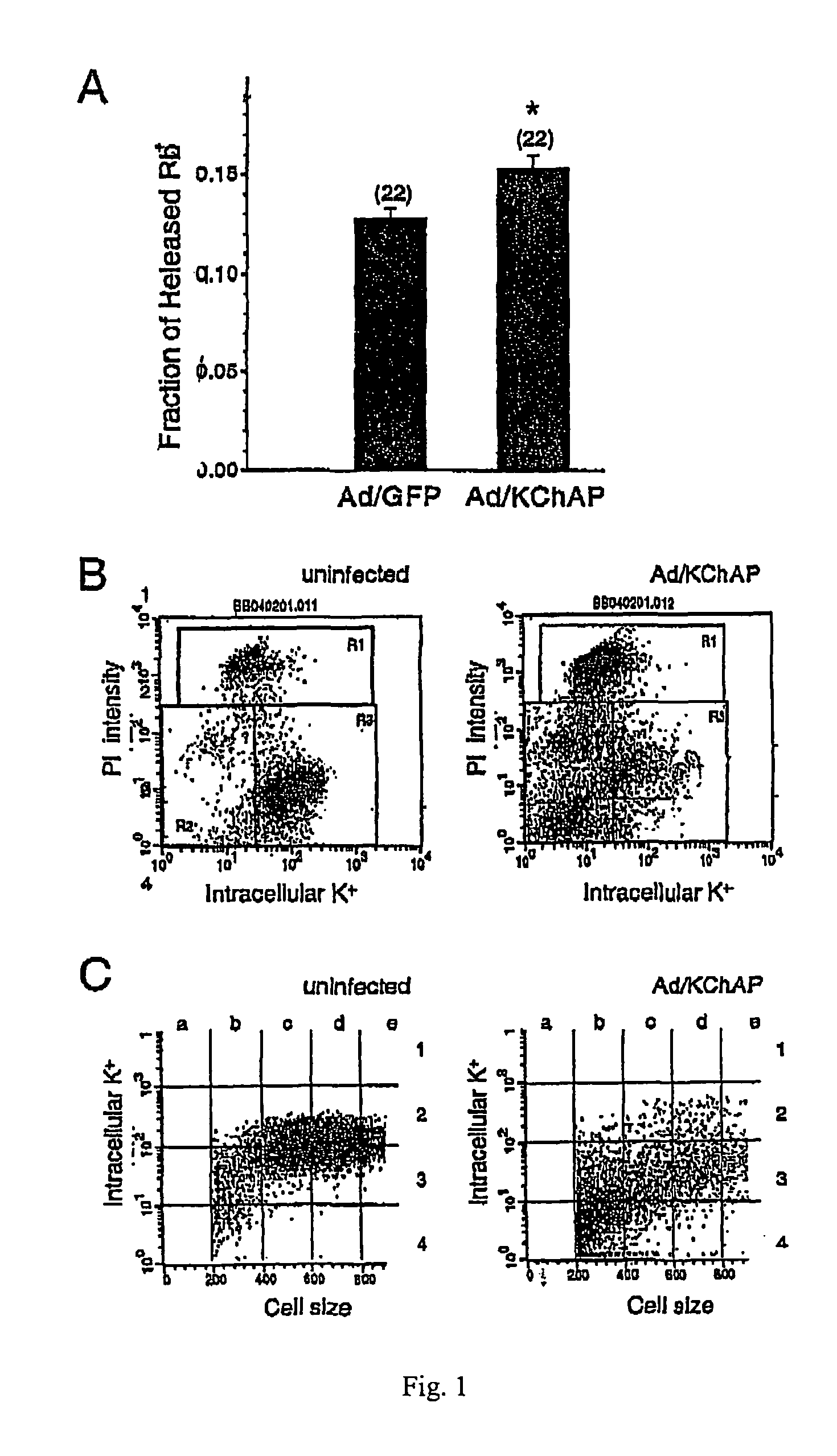
[0105]To examine the effect of increased intracellular levels of KChAP on K+ flux, LNCaP cells were infected with either Ad / GFP or Ad / KChAP (moi of 100) for 24 hours after which they were loaded with the potassium surrogate rubidium (Rb+) and assayed for Rb+ release by flame atomic absorption spectroscopy. LNCaP are a model cell line for prostate cancer cells which comprise native or wild-type p53 protein. As shown in FIG. 1A, KChAP—overexpressing cells showed a significant increase (about 20%) in the fraction of Rb+ released compared to cells infected with a GFP virus. In a 10 minute period, about 12.5% of the loaded Rb+ was released from Ad / GFP infected cells whereas over 15% was released from cells infected with Ad / KChAP.

[0106]The relative amount of K+ in Ad / KChAP-infected cells was measured at later times after infection using flow cytometry with the potassium sensitive dye, PBFI. LNCaP cells...

example 2

Inducing Apoptosis in Prostate Cancer Cells Comprising a Mutated p53 Protein by Increasing Intracellular Levels of KChAP

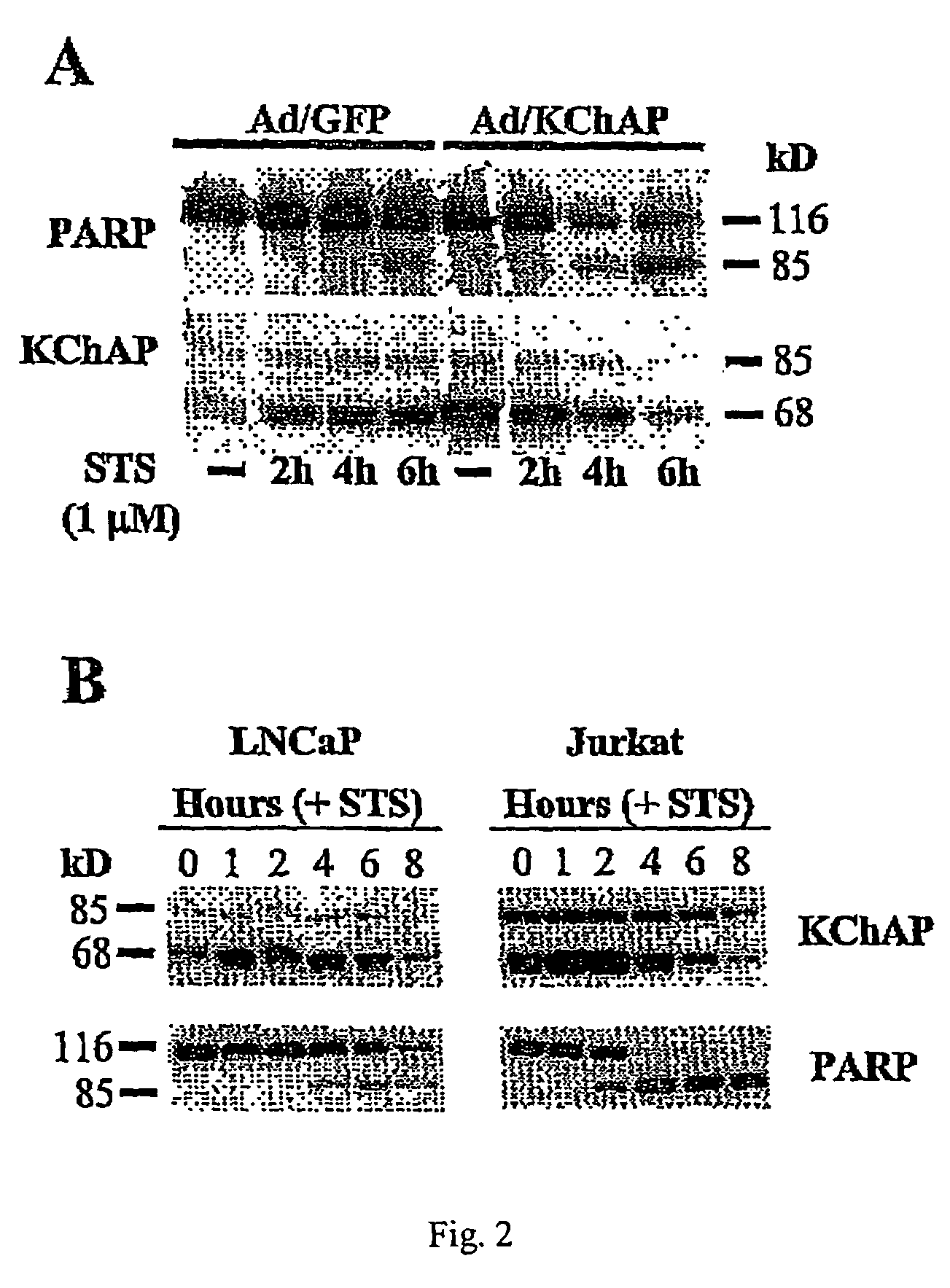
[0116]To determine whether wild-type p53 is essential for KChAP effects, we tested the effects of in a cell line with mutant p53. The prostate cancer cell line, Du145, has p53 with several point mutations rendering it nonfunctional as a transcription factor (ref). Du145 cells were infected with Ad / GFP or Ad / KCHAP at two different m.o.i. (200 and 400) and lysates prepared 72 hours after infection. Greater than 95% of the cells were infected in these experiments as determined by GFP fluorescence and most of the infected cells were floating by day 3 (data not shown). Western blotting shows significant PARP cleavage in Du145 cells infected with Ad / KChAP compared to control, Ad / GFP infected cells (FIG. 7). Steady-state p53 levels are already high in Du145 cells as is often seen when p53 is mutated, and those levels do not increase with KChAP overexpression. The phosphor...

example 3

Inhibiting In Vivo Growth of Subcutaneous Implants of Human Prostate Cancer Cells by Increasing Intracellular Levels of KChAP

[0117]We have shown that KCHAP is a potent inducer of apoptosis in cell lines with diverse p53 status. To assess its potential usefulness as an anticancer agent, we created subcutaneous tumors in nude mice by injecting either Du145 or LNCaP cells into the flank area. Du145 cells, mixed with matrigel, formed well established tumors in the flanks of nude mice in about two weeks. Once tumors were established, Ad / KChAP was injected directly into the tumors every 48-72 hours for a total of 9 injections over a period of 19 days. Two batches of control tumors were injected with either PBS or Ad / GFP. As shown in FIG. 8A, injection of Ad / KChAP significantly suppressed the growth of Du145 tumors compared with Ad / GFP or PBS treatments. In the animals treated with Ad / KChAP, the mean tumor volume was 81 mm3 after 19 days (n=8). In contrast, the mean tumor volume reached 49...


PUM

PropertyMeasurementUnit
Levelaaaaaaaaaa
Hyperproliferativeaaaaaaaaaa

Abstract

Methods of inducing apoptosis in hyperproliferative cells, particularly cancer cells are provided. Such method involves increasing the levels of a potassium channel modulatory protein in the cell. Examples of such proteins are native KChAP protein, a biologically active variant of native KChAP protein, or a biologically active KChAP-related protein (collectively referred to hereinafter as “KChAP protein”). In one embodiment, the cells are contacted with the KChAP protein under conditions permitting uptake of the protein by the cells. In another embodiment, the cells are contacted with (i) a nucleic acid encoding the KChAP protein, and (ii) a promoter active in the cancer cell, wherein the promoter is operably linked to the region encoding the KChAP protein, under conditions permitting the uptake of the nucleic acid by the cancer cell. Methods of detecting cancerous cells in a biological sample selected from the group consisting of a colorectal tissue sample or brain tissue sample are also provided. Such method comprises assaying for the presence of elevated levels of KChAP mRNA or KChAP protein in the sample.

Description

CROSS-REFERENCE TO RELATED APPLICATIONS[0001]This application is a continuation of the commonly assigned, U.S. patent application Ser. No. 10 / 000,778, filed on Oct. 31, 2001 now abandoned.[0002]This invention was made, at least in part, with government support under National Institutes of Health Grant No. HL36930, HL55404, HL61642, HL60759, DK54178. The U.S. government has certain rights in the invention.FIELD OF THE INVENTION[0003]This invention relates to methods of inducing apoptosis in hyperproliferative cells. More particularly, this invention relates to methods of inducing apoptosis in cancer cells by increasing levels of a potassium channel modulatory protein in such cells.BACKGROUND[0004]One-third of all individuals in the United States will develop cancer (American Cancer Society Yearly Outlook for 1990). Cancer is second only to cardiac disease as a cause of death in this country (American Cancer Society Yearly Outlook for 1990). Currently, cancer therapy employs a variety...

Claims


Application Information

Patent Timeline
IPC IPC(8): A01N63/00A01N65/00A61K48/00C12N15/00A61K38/00C07K14/47
CPCC07K14/4702A61K38/00
Inventor BROWN, ARTHUR M.WIBLE, BARBARA A.
Owner CASE WESTERN RESERVE UNIV
Why Eureka
Try Eureka