One step entry pedicular preparation device and disc access system

a preparation device and a technology of disc access, applied in the field of spine surgery, can solve the problems of major drawbacks of current guide pin systems, and achieve the effects of reducing the size of the guide pin, maximizing safety, and optimal performan

Active Publication Date: 2012-08-07
LIFE SPINE INC
View PDF10 Cites 78 Cited by
  • Summary
  • Abstract
  • Description
  • Claims
  • Application Information

AI Technical Summary

Benefits of technology

[0024]The system and method of the invention, illustrates a variety of structures and techniques to enable a staged location and entry into the pedicle for providing insertive, progressively larger threaded fixation and superior surgical control a distance from the spine. The PediDart™ pedicle dart system offers a one step preparation for pedicle screw insertion in the safest and most accurate way possible with current imaging technology which may include x-ray or infrared, or RF navigational guidance. This allows for faster complex spine surgery procedures, decreased cost, decreased anesthesia time and complications such as surgery time dependent post operative infections. The device and method can be utilized manually or power driven and can be used open, minimal open or percutaneous surgical approaches.
[0025]The advantages of the PediDart™ pedicle dart system includes:(1) The larger than usual guide pin (of about 2 millimeters in diameter) allowing for a sharper and more stable trocar point to precisely dock and hold to the entry point without slippage.(2) The larger guide pin resists bending and consequently binding which is a major drawback to current guide pin systems.(3) The limited exposure length of the guide pin not only resists bending, but minimizes excessive penetration in the spine and beyond which can result in vascular or bowel injury.(4) The tip end of the PediDart™ pedicle dart system begins with a cutting thread to quickly capture the cancellous bone in the case of the bony pedicle.(5) The PediDart™ pedicle dart systems fluted shaped advances smoothly down the inner pedicle and gradually expands the cancellous inner bone so that sequential tapping is not necessary.(6) The fluted shape can also safely expand immature pedicular bone such as with adolescent idiopathic scoliosis surgery allowing placement of the largest pedicle screw for the best stability.(7) The non-cutting threads at the larger end of the flute compresses and strengthens the cancellous pedicular bone to give better purchase to the pedicle screw.(8) The smaller diameter of the entry end of the PediDart™ pedicle dart system preserves the bone in the vertebral body for best pedicle screw purchase especially important for the osteopathic spine.(9) The cone shaped channel created by the fluted PediDart™ pedicle dart system allows maximum contact to the entire length of the pedicle screw.(10) The cone shaped channel created by the PediDart™ pedicle dart system allows for accurate advancement of the pedicle screw.(11) The smooth part of the shank gives greater length to the PediDart™ pedicle dart system when used as a temporary implant to compress or distract a motion segment such as for the placement of an intervertebral or disc space implant.(12) The sloped buttress at the tail end of the PediDart™ pedicle dart system keeps the system from excessive penetration.(13) The sloped buttress at the tail end provides for a “flared” entry point for the pedicle screw.(14) The inner slot and lock mechanism provides the most powerful way to rotate the PediDart™ pedicle dart system into and out of the spine.
[0026](15) The “hockey stick” shaped external slot provides an additional way to remove the PediDart™ pedicle dart system when it is deeply inserted in a large patient, and particularly where some other object or tissues blocks rearward motion of the pedicle dart.(16) The tapered inner hollow structure facilitates proper engagement between the pedicle dart and the manipulator / insertion tool.(17) The inner spring of the PediDart™ pedicle dart system mates to the circular slot of the insertion tool / manipulator capturing the pedicle dart but yet still allows for disengagement.(18) The handle of the insertion tool / manipulator is designed for the best tactile feel and control for the surgical practitioner.(19) The modular end of the insertion tool / manipulator can adapt for navigational points, impacting tool or power attachment.(20) The length of the insertion tool / manipulator keeps the surgeon's hands away from the x-ray beam.(21) The PediDart™ pedicle dart system can serve as a temporary implant, a permanent implant or a structure for a stand alone screw and rod system. It can also be adapted for intervertebral disc access (“Disc PediDart™).

Problems solved by technology

(1) The larger than usual guide pin (of about 2 millimeters in diameter) allowing for a sharper and more stable trocar point to precisely dock and hold to the entry point without slippage.
(2) The larger guide pin resists bending and consequently binding which is a major drawback to current guide pin systems.

Method used


Image

  • One step entry pedicular preparation device and disc access system
  • One step entry pedicular preparation device and disc access system
  • One step entry pedicular preparation device and disc access system

Examples


Embodiment Construction

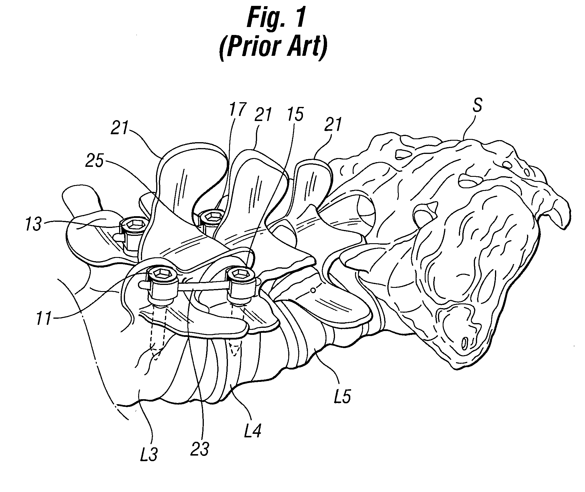
[0103]A detailed description of the preferred embodiment will be best begun by examining a perspective view of the lower lumbar vertebrae L3, L4, L5 and sacrum S shown in perspective in FIG. 1. In operative procedures in which work is to be done between two adjacent vertebrae, those two vertebrae must be fixed and held apart so that the space between those vertebrae is maintained. Traditionally this has been done by using conventional pedicle screws 11, 13, 15 and 17 seen in FIG. 1. The conventional pedicle screws 11, 13, 15 and 17 engage significant bone mass by placement through the pedicle structure of each vertebra into which they are placed. In the three dimensional view of FIG. 1, the conventional pedicle screws 11, 13, 15 and 17 seem to have an open placement, during an actual surgical procedure on a patient, the pedicle screws 11, 13, 15 and 17 must be located through and despite significant layers of muscular tissue.

[0104]As previously discussed, dissection of the muscles s...


PUM


Abstract

A one step entry pedicular preparation device works well with Minimal Invasive Spine Surgery (MISS) to facilitating such approach. A related intervertebral disc access system and pedicle screw compatible with the systems is also illustrated. The systems include a manipulator having a bar handle and main body with main barrel bore a pedicle dart having a proximal tip end and a second distal open end selectably attachable to the manipulator using a variety of interconnect configurations, both of which work in conjunction with a guide pin. The pedicle darts can be made of any material, disposable or re-usable and can be utilized for a variety of purposes, including bone structure formation for faster conventional pedicle screw insertion with precision and vertebra fixation.

Description

[0001]This is a continuation-in-part of U.S. patent application Ser. No. 12 / 009,496 filed Jan. 17, 2008 now U.S. Pat. No. 8,075,579.FIELD OF THE INVENTION[0002]The present invention relates to improvements in the field of spine surgery and more particular to a system for the ability to perform spine surgery through a small opening, the location, placement, and insertion of temporary pedicular implants called PediDart™ pedicle darts which are applicable to both MIS and open surgical technique and which allows for more exact and rapid placement with correct angular insertion of fixation members into the spinal pedicle, which will facilitate one of the most difficult phases of spine surgery. The result is increased safety of placement and insertion of the pedicle screws, avoiding potential devastating complications to the adjacent neurovascular structures as well as less time spent on the operating table. The same basic design of the PediDart™ pedicle dart system with minor but importa...

Claims


Application Information

Patent Timeline
Patent Type & Authority Patents(United States)
IPC IPC(8): A61B17/58
CPCA61B17/7001A61B17/7082A61B17/8897A61B17/861A61B17/863A61B18/1477A61B17/864A61B2019/5483A61B17/8635A61B2090/3983
Inventor HAMADA, JAMES S.
Owner LIFE SPINE INC
Why Eureka
Try Eureka