Noninvasive measurements in a human body

a human body and non-invasive technology, applied in the field of medical devices, can solve the problems of uncertainty in path length, computation-intensive tomographic devices and algorithms, and localization of probed volume, and achieve the effect of facilitating non-invasive measurements

Inactive Publication Date: 2013-04-16
OR NIM MEDICAL
View PDF34 Cites 19 Cited by
  • Summary
  • Abstract
  • Description
  • Claims
  • Application Information

AI Technical Summary

Benefits of technology

[0006]The present invention utilizes the principles of ultrasound tagging of light. According to the present invention, tagging of light with acoustic radiation is used to enable distinguishing between the optical response of a region of interest (e.g., cerebral tissue, blood vessel) and regions outside the region of interest; and / or to significantly improve oximetry and pulse oximetry based measurements.
[0014]Preferably, a body part (e.g., a human head) containing a region of interest (e.g., cerebral tissue) is illuminated with light (e.g., of at least two different wavelengths) and is irradiated with acoustic radiation, in a manner to ensure optimal operating condition for measurements. This optimal operating condition is such that the illuminating light and acoustic radiation overlap within the region of interest and thus light scattered from the region of interest is “tagged” by acoustic radiation (the light is modulated by the frequency of the acoustic radiation) while substantially do not overlap in a region outside the region of interest. Moreover, the optimal operating condition is such as to ensure that detected light includes a portion of light scattered by the region of interest and tagged by acoustic radiation, and a portion of untagged light scattered by regions outside the region of interest. This allows for distinguishing between the light responses of the region of interest and its surroundings (e.g., cerebral and extracranial tissues; a vascular cavity and surrounding tissues; or a blood pool and surrounding tissues).
[0028]a control unit connectable to the optical unit and to the acoustic unit, the control unit being preprogrammed to operate the acoustic unit with at least two different operating conditions to vary at least one characteristic of acoustic radiation, the control unit being responsive to the measured data and preprogrammed to process and analyze the measured data to extract therefrom a data portion associated with the light response of said certain region, thereby enabling determination of a property of a tissue component in said certain region based on a relation between the measured data corresponding to the at least two different operating conditions of the acoustic unit.
[0029]According to another broad aspect of the invention, there is provided a method for use for non-invasive measurements in a human body, the method comprising: applying acoustic radiation to a certain illuminated region in the body, with at least two different conditions of the applied radiation achievable by varying at least one characteristic of the acoustic radiation; detecting light scattered from the body part and generating measured data indicative of detected photons tagged and untagged by the acoustic radiation; analyzing the measured data to extract therefrom a data portion corresponding to the tagged photons and being therefore associated with a light response of said certain region, to thereby enable determination of tissue properties of said certain region based on a relation between the measured data portions corresponding to the at least two different operating conditions.

Problems solved by technology

However, near infrared spectroscopy (NIRS) suffers from several drawbacks associated for example with the fact that differential scattering of two different wavelengths used in the measurements result in an uncertainty in the path length that each wavelength passes; an inherent inability to localize the probed volume which requires computation-intensive tomographic devices and algorithms to resolve; analysis of the detected signal depends on a model being used to characterize the tissue structure being probed.
This impedes the use of NIRS in real time medical settings.

Method used


Image

  • Noninvasive measurements in a human body
  • Noninvasive measurements in a human body
  • Noninvasive measurements in a human body

Examples


Embodiment Construction

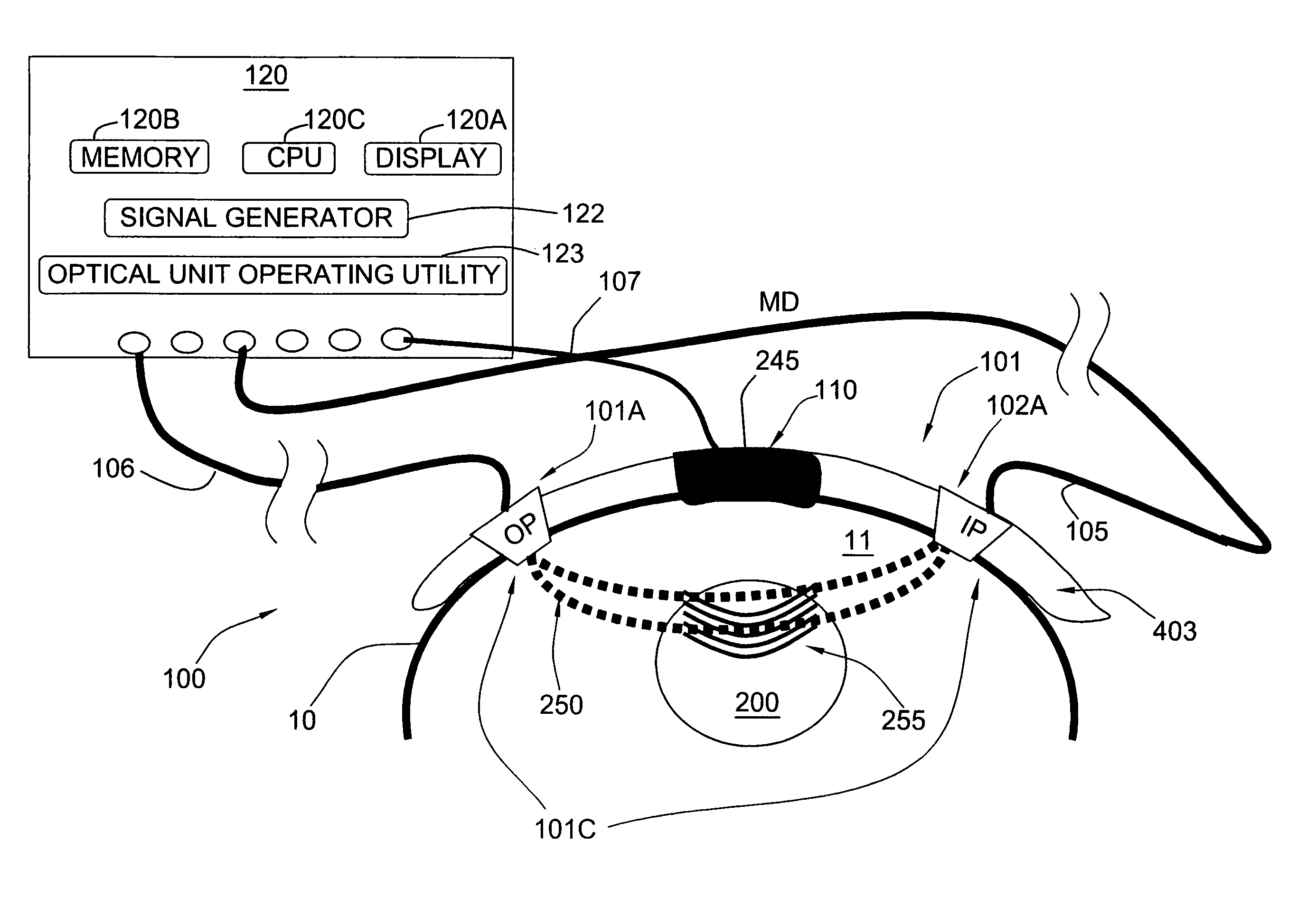
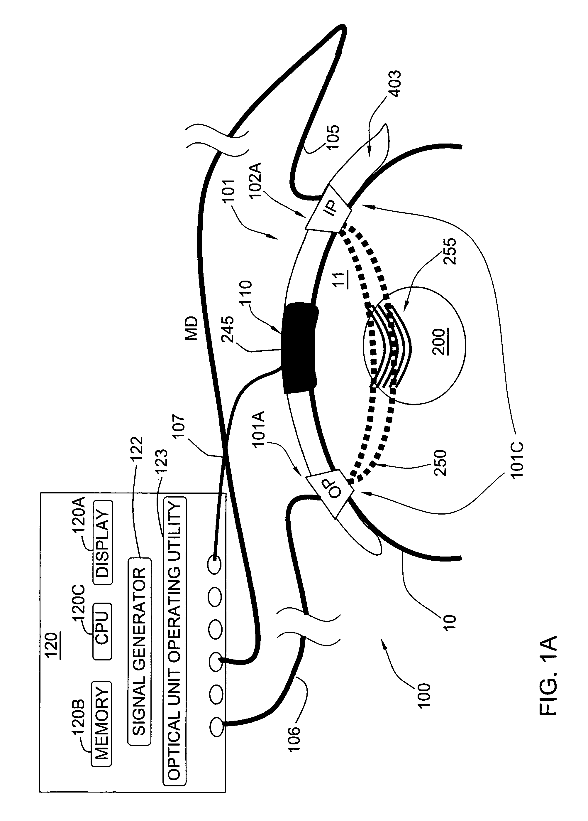
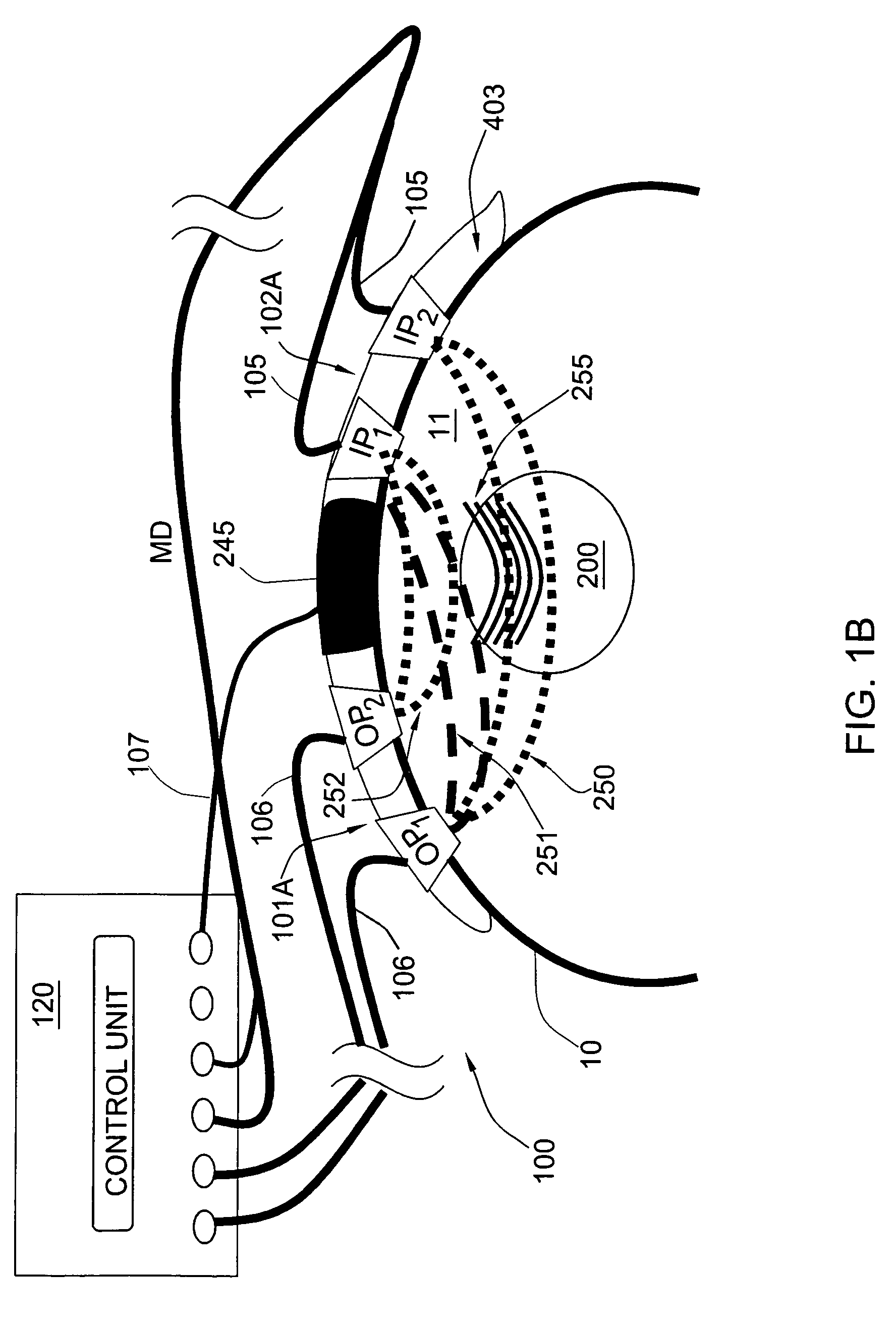
[0042]Reference is made to FIGS. 1A-1C illustrating schematically three specific but not limiting examples of a measurement system, generally designated 100, configured and operable according to the invention for non-invasive measurements of one or more parameters (properties of tissue components) of a region of interest 200 in a human or animal body. This may be an oxygen saturation level, or various other parameters such as the concentration of an analyte in the patient's blood, or the perfusion of an analyte / metabolite in tissues. To facilitate understanding, the same reference numbers are used for identifying components that are common in all the examples of the invention.

[0043]The system 100 includes a measurement unit 101 and a control unit 120. The measurement unit 101 includes: an optical unit 101C formed by an illumination assembly 101A and a light detection assembly 102A; and an acoustic unit formed by a transducer arrangement 110. The control unit 120 is configured to con...


PUM


Abstract

A measurement system and method are presented for use for non-invasive measurements in a human body. Acoustic radiation is applied to a certain illuminated region in the body, with at least two different conditions of the applied radiation achievable by varying at least one characteristic of the acoustic radiation. Light scattered from the body part is detected, and measured data indicative of detected photons tagged and untagged by the acoustic radiation is generated. The measured data is analyzed to extract therefrom a data portion corresponding to the tagged photons and being therefore associated with a light response of said certain region, thereby enabling determination of tissue properties of said certain region based on a relation between the measured data portions corresponding to the at least two different operating conditions.

Description

FIELD OF THE INVENTION[0001]This invention is generally in the field of medical devices, and relates to a method and system for noninvasive measurements in a human body. The invention is particularly useful for monitoring such parameters of the human body as oxygen saturation and / or concentration of analyte(s) in blood.BACKGROUND OF THE INVENTION[0002]Monitoring of the oxygenation level of a tissue region is required in order to determine if the tissue is viable or necrotic. For example, measuring the oxygen saturation level allows for determining whether a patient that experienced a stroke event should undergo a therapeutic procedure, whether a certain procedure is not necessary or whether performing certain procedures entails a high risk. Such measurements are also imperative for determining the efficiency of a treatment.[0003]Near infrared light has been used to non-invasively probe the patient's brain based on different absorption characteristics of oxygenated and deoxygenated h...

Claims


Application Information

Patent Timeline
Patent Type & Authority Patents(United States)
IPC IPC(8): A61B5/05
CPCA61B5/14546A61B5/14553A61B5/7475A61B8/4227G01S15/8968A61B5/1455A61B8/0808A61B8/06A61B8/0816A61B2562/043A61B2562/0233A61B8/4494
Inventor BALBERG, MICHALPERY-SHECHTER, REVITALOLSHANSKY, MICHAL
Owner OR NIM MEDICAL
Why Eureka
Try Eureka