

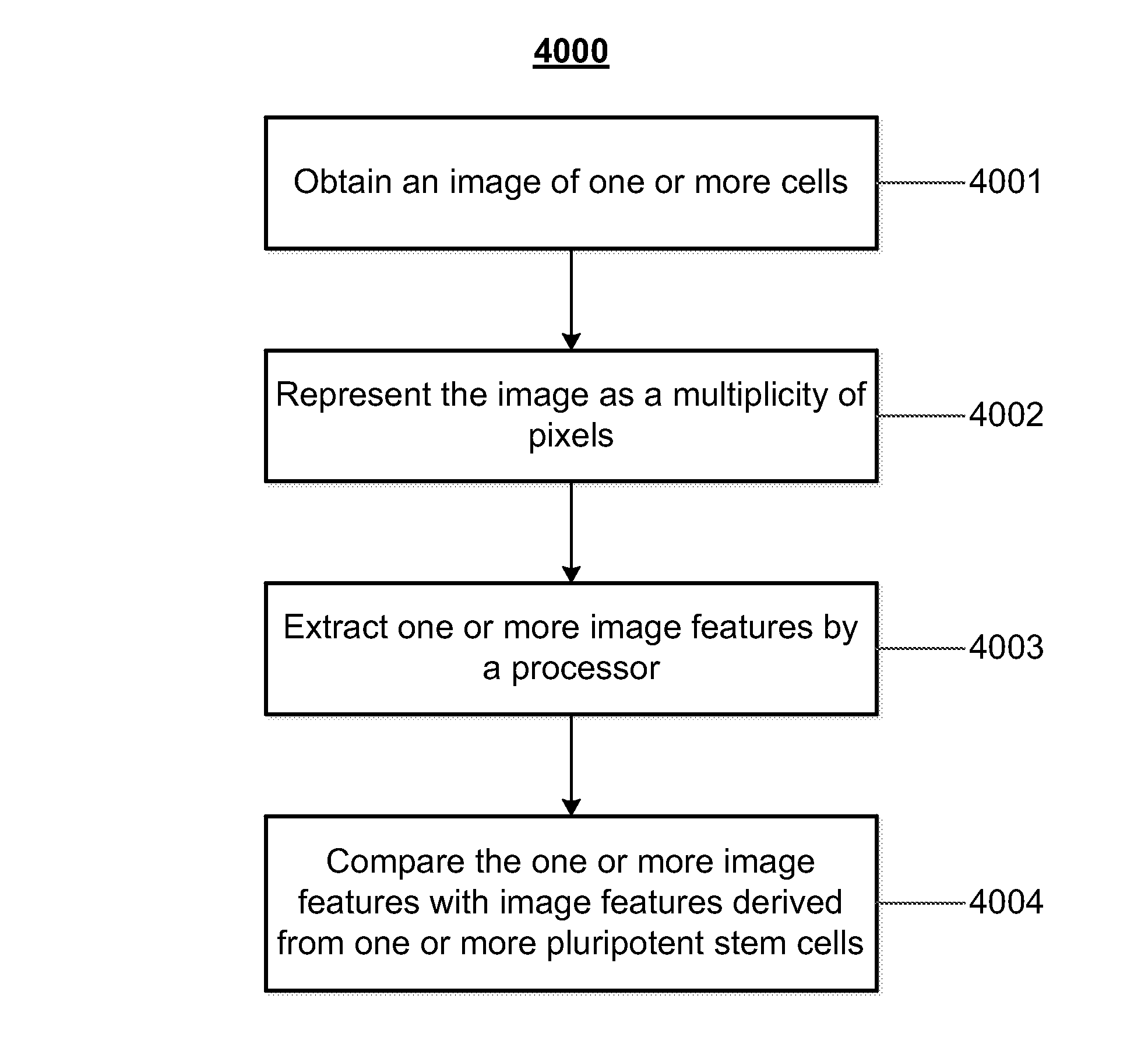
Mathematical image analysis based cell reprogramming with applications for epigenetic and non-epigenetic base induced pluripotent stem cell derivation
a technology of epigenetic and non-epigenetic bases, applied in the field of mathematical image analysis based cell reprogramming with applications for epigenetic and non-epigenetic base induced pluripotent stem cell derivation, can solve the problems of inability to scale up for the large quantity of cells expected in a therapeutic or commercial setting, limited methods for human hesc classification, time-consuming, etc., and achieve the effect of reducing the activity of one or more epigeneti
- Summary
- Abstract
- Description
- Claims
- Application Information
AI Technical Summary
Benefits of technology
Problems solved by technology
Method used
Image

Examples
example 1
Molecular Characterization and Validation of Reprogramming
[0145]To validate non-invasive measures of reprogramming, an immunostaining approach is used for a panel of nuclear and cytoplasmic factors characteristic of pluripotency including Oct4, Nanog, Sox2, FGF5R, HNF3b, Fox D,3 and Rex1. Markers of later differentiation stages also differ in pluripotent cells including Gata 6, Bracyury, and AFP (not shown). Additional markers useful for discriminating early pre and post implantation pluripotent lineages have been identified for mouse ESCs (Tesar et al., 2007). Preliminary studies have found these markers in pluripotent hESCs, but not in neuronally differentiated cells. However, the karyotypically normal hESC line H7 grown in different media all have positive immunostaining, but quantitation with automated microscopy reveals very different profiles. The pluripotency panel of H7s on mouse embryo fibroblast feeders in DSR DMEM with knockout Serum Replacer media are much lower than in ...
example 2
Epigenetic State of Pluripotent Stem Cells Compared to Somatic Cells
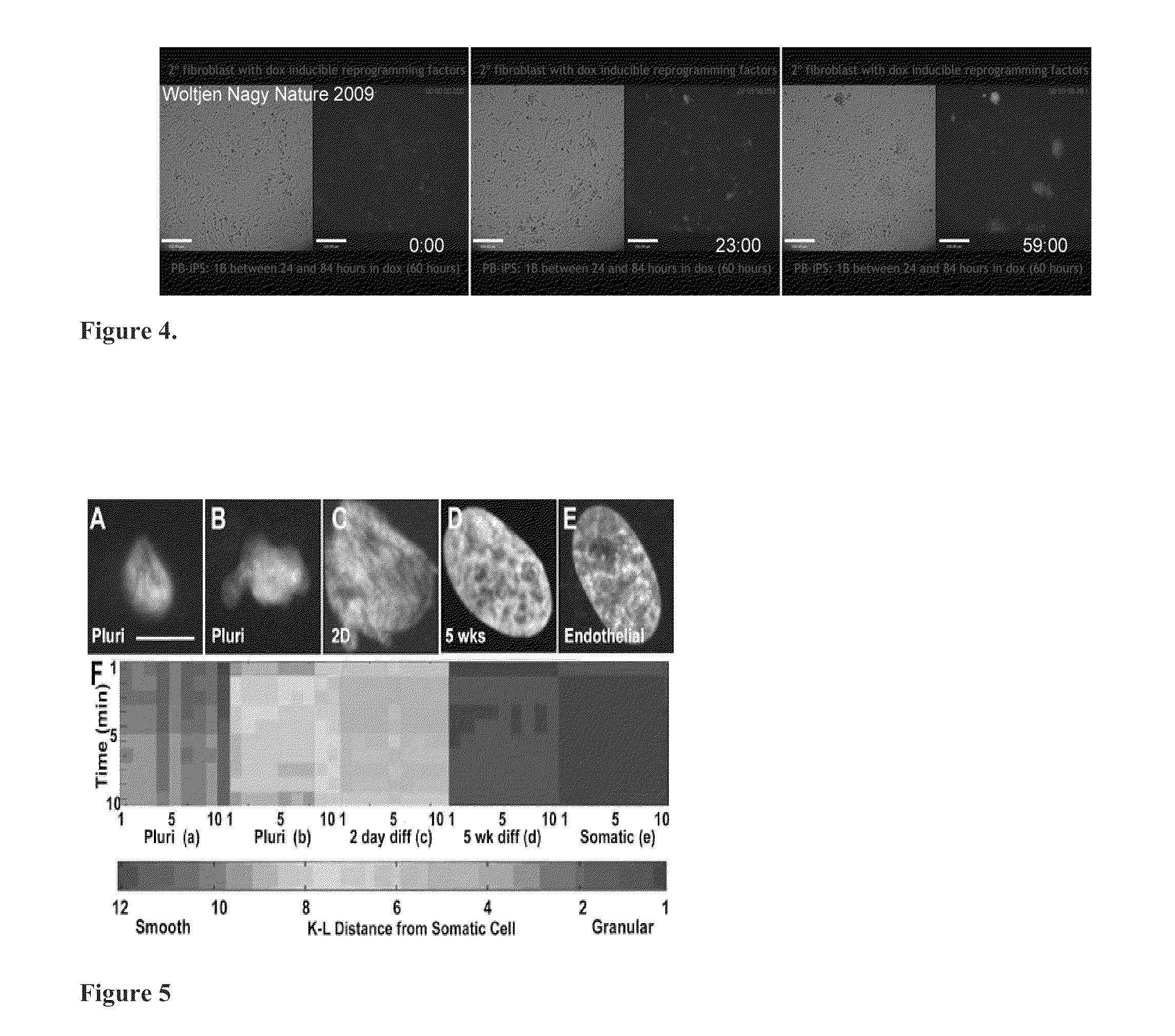
[0146]Different markers of heterochromatin (H3K9me3 [in green] and H3K27me3 [in red], FIG. 24, A-C) have different distributions in pluripotent cells but are more colocalized (yellow) in neurally differentiated cells and in somatic cells. Conversely, markers of euchromatin (H3K9ac [in green], H3K4me2 [in red], FIG. 24d-24e) are more colocalized in pluripotent cells. Euchromatin and heterochromatin markers are spatially distinct (FIG. 24g-24i) at all stages.
[0147]Pluiripotent stem cells show different organization of histone posttranslational markers than differentiated cells. Different markers of heterochromatin (H3K9me3 [in green] and H3K27me3 [in red], FIG. 24a-24c) have different distributions in pluripotent cells but are more colocalized (yellow) in neurally differentiated cells and in somatic cells. Conversely, markers of euchromatin (H3K9ac [in green], H3K4me2 [in red], FIG. 24d-24e) are more colocalized in pl...
example 3
Establishing Inducible HDAC1, 2 Knockdown Lines of hESCs & Inhibition of Differentiation
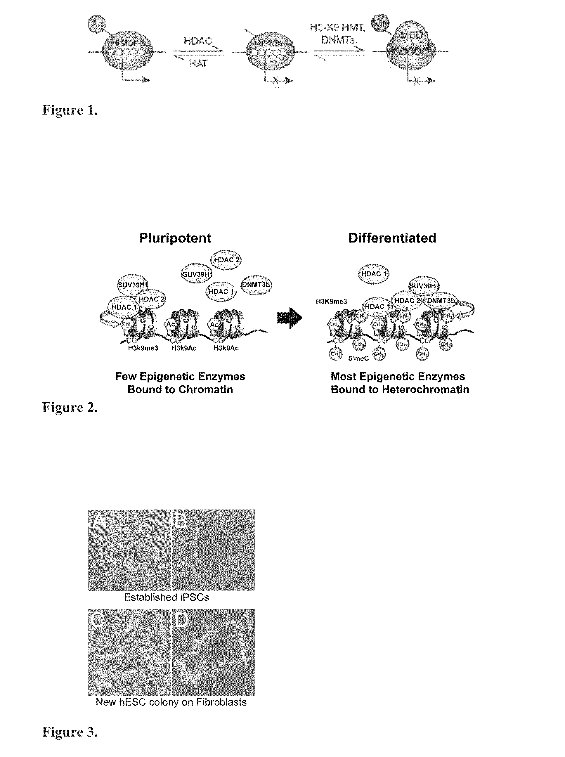
[0149]Pluripotent cells express several epigenetic enzymes, including DNMT 1, 3a, 3b, and HDAC 1 and 2 (FIG. 28a-28d). In some cases, despite high enzyme levels, enzyme activity is low in pluripotent cells, which reflects by low methylation of DNA (FIG. 28e) or deacetylation of histone H2BK5 (FIG. 280. The exception are those cells at the colony periphery that have initiated differentiation. Notably, HDAC levels do not change during differentiation on feeders.
[0150]HDAC 1 and 2 levels in hESCs were modified with tet-inducible lentivirus for HDAC1 & 2 knockdown and a nonsilencing control (FIG. 29-30). Several lines of stably transformed hESC have been established and initial testing of the effects of knockdown show an inhibition in the rate of differentiation in doxycyclin-induced HDAC 1 & 2 knockdown lines when challenged to differentiate with BMP4 (FIG. 30). HDAC KD fibroblasts will also be esta...
PUM
 Abstract
Description
Claims
Application Information