Mucoadhesive drug delivery devices and methods of making and using thereof

a technology of adhesives and drug delivery devices, which is applied in the direction of powder delivery, peptide/protein ingredients, macromolecular non-active ingredients, etc., to achieve the effect of reducing pain caused by wounds and/or lesions and enhancing drug absorption

Active Publication Date: 2013-09-10
PETVIVO HLDG INC
View PDF137 Cites 64 Cited by
  • Summary
  • Abstract
  • Description
  • Claims
  • Application Information

AI Technical Summary

Benefits of technology

[0013]In one embodiment of the present invention it has been found that mucoadhesive devices including ovalbumin have produced excellent mucoadhesive results. Such embodiment generally comprise ovalbumin combined with one or more mucoadhesive agents (e.g. glycerol), one or more pharmacologically active agents and water. These embodiments have been shown to provide excellent adhesion (e.g. strength and contact duration) and have provided excellent transmucosal delivery of the pharmacologically active agents.
[0016]The mucosa is characterized by an outermost layer of stratified squamous epithelium. Below this layer lies a basement membrane (lamina propria) followed by the submucosa as the innermost layer. The oral epithelium is similar to stratified squamous epithelia found in the rest of the body (e.g., skin) in that it has a mitotically active basal cell layer, advancing through a number of differentiating intermediate layers to the superficial layers, where cells are shed from the surface of the epithelium. The epithelium of the buccal mucosa is about 40-50 cell layers thick, while that of the sublingual mucosa is somewhat thinner. The turnover rate for the buccal epithelium is estimated to be at 5-6 days, which is representative of the entire oral mucosa. The mucosae of tissues subject to mechanical stress, that is the gingiva and hard palate, are keratinized. The mucosa of the soft palate, the sublingual, and the buccal regions are not keratinized. The keratinized epithelia contain neutral lipids like ceramides and acylceramides that serve as a physical barrier, to make the tissue impermeable to water and microorganisms. In contrast, the non-keratinized epithelia, such as the floor of the mouth and the buccal epithelia, do not contain acylceramides and only trace amounts of ceramide. The non-keratinized epithelia have been found to be considerably more permeable to water than keratinized epithelia, thereby enhancing drug absorption.

Problems solved by technology

The dynamic nature of oral mucosal and its wide range of variable health and tissue characteristics make this a challenging endeavor for the medical and dental community.

Method used


Image

  • Mucoadhesive drug delivery devices and methods of making and using thereof
  • Mucoadhesive drug delivery devices and methods of making and using thereof
  • Mucoadhesive drug delivery devices and methods of making and using thereof

Examples


example i

Mucoadhesive Composition

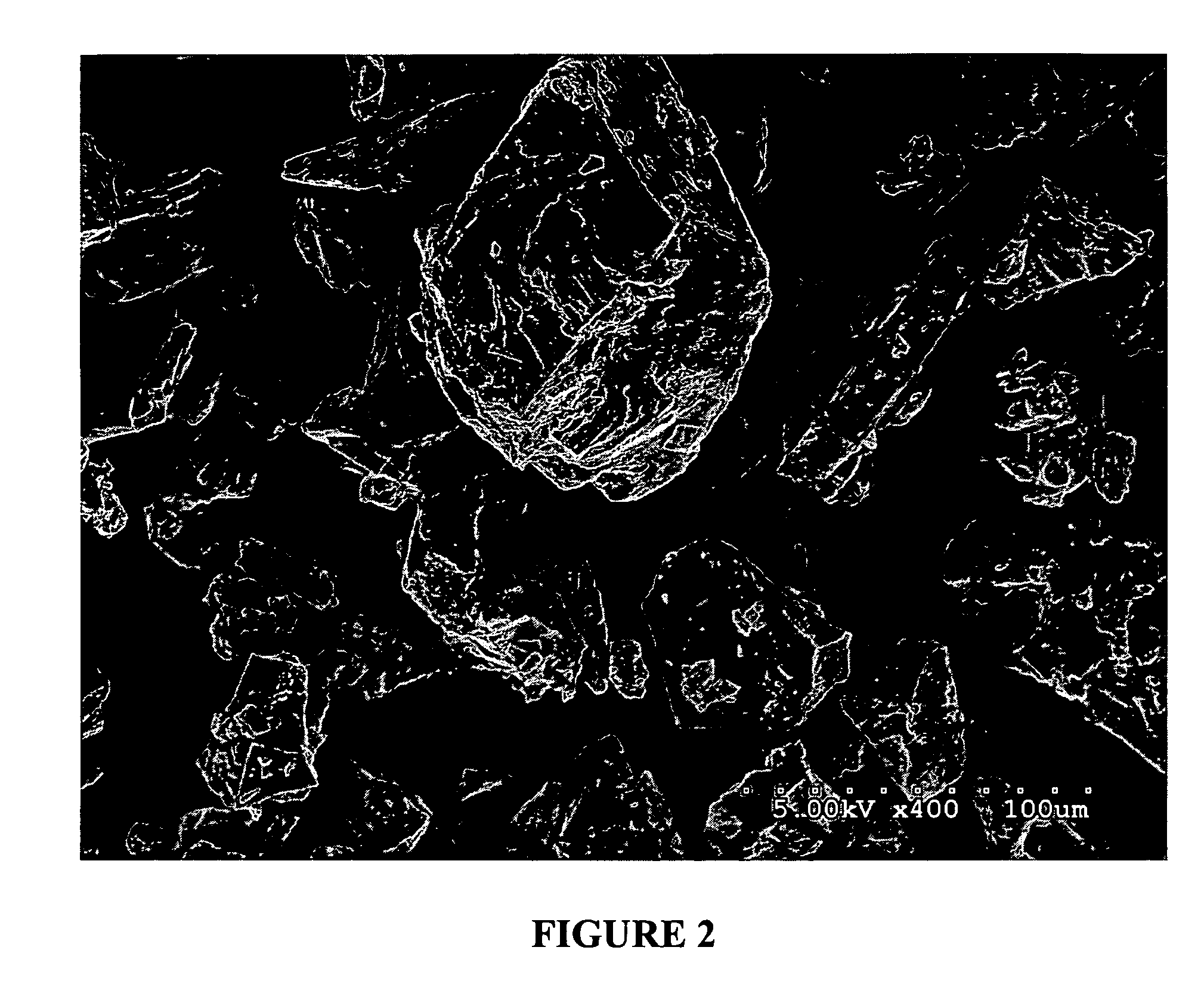
[0178]Powdered egg albumin (Alfachem) (50 gs) is added to a stirred (magnetic stirrer) solution of glycerol (15 gs) in saline (0.9%) (187.5 mls) in a 500 ml beaker. After all the albumin has been added, and mostly dispersed, the solution is shaken at the slow settting for 30 minutes on a platform shaker (Eberbach). (note: an active drug may be added to the glycerol / saline solution before the albumin is added). The resulting solution / dispersion was divided into two-250 ml beakers up to the 125 ml mark (approximately one-half full). The two beakers were covered with foil and placed in a freezer at −20° C. overnight. The frozen solutions were next placed onto a freeze-drier platform previously held at −30° C. and left to equilibrate for one hour. Cooling of the platform was then discontinued when a vacuum of 200 millitor was obtained. The condenser was then turned off. Full vacuum was then continued for 48 hours or until the platform temperature reached ambient ...

example ii

In Vitro Bioadhesion Tests on Mucosal Tissue

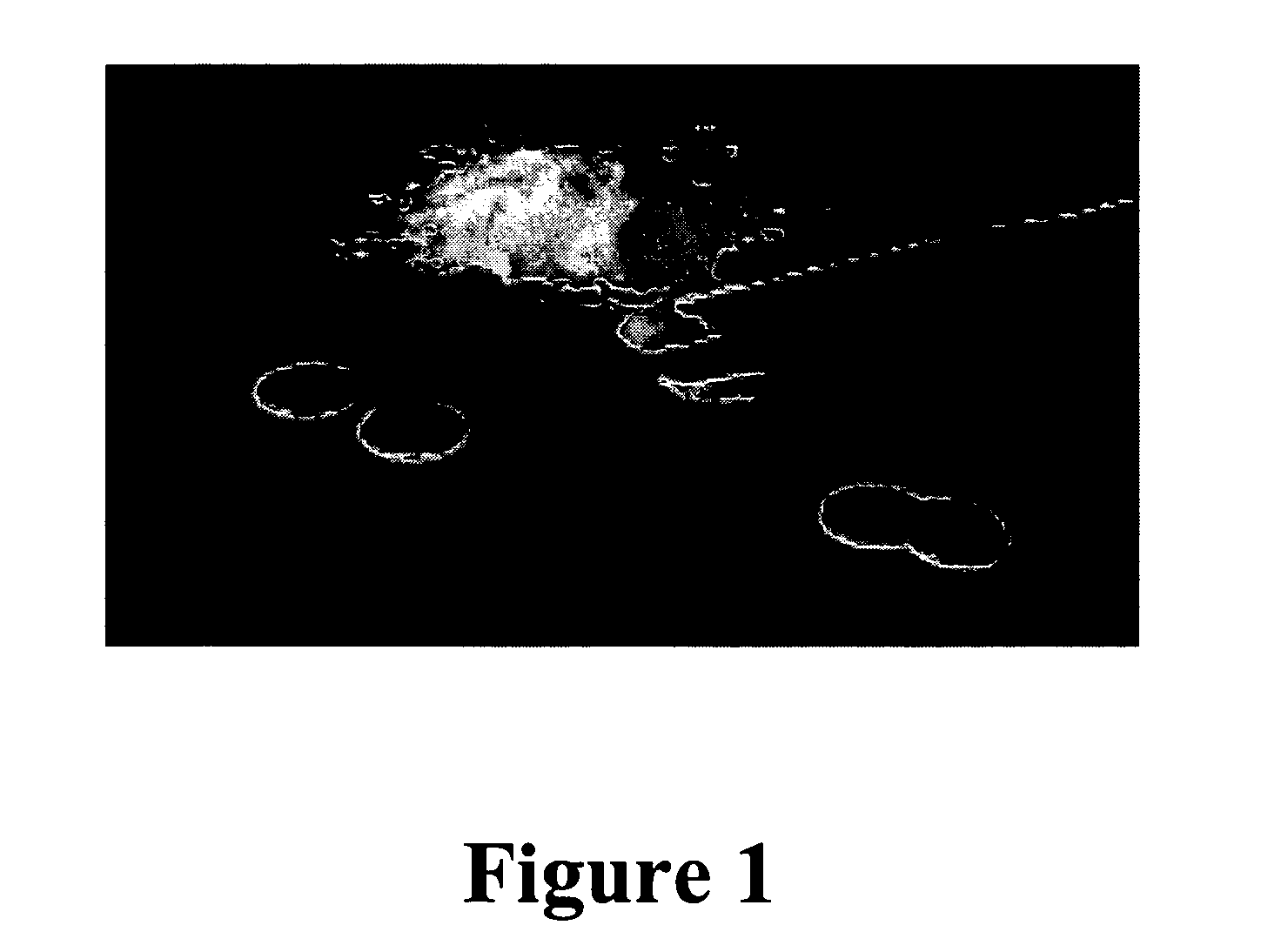
[0179]Mucoadhesive devices including ovalbumin, glycerol and water (no drug) were made into particles utilizing the methods described above. 150 mg of the particles were compressed into 1 cm diameter wafers at 1375 psi for 3 min (MAD1, MAD 2 and MAD3). The wafers were then tested for mucoadhesive attachment by adhering them to the bottom of a glass beaker with double sided Scotch®, 3M tape and pressing the bottom of the beaker to fresh wetted mucosal membrane from pig intestine that was cleaned for usage in sausage casing. The bottom of the beaker was pressed to the mucosal membrane for 30 seconds at approximately 10 psi. The beaker was then placed on a Satorius electronic balance and the membrane pulled slowly in an upward motion at a 45° angle away from the wafer and beaker until completely detached. The measurement taken was the detachment force, which is the difference between the starting weight of the beaker, wafer and membrane and t...

example iii

In Vitro Bioadhesion Tests on Mucosal Tissue

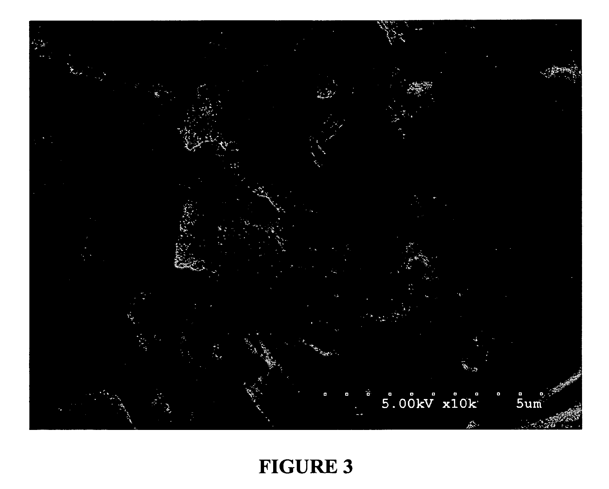
[0181]Mucoadhesive devices including ovalbumin, glycerol and water were made into wafers and were tested for mucoadhesive attachment to fresh mucosal membrane from pig intestine by Christer Nyström, Ph.D., of the University of Uppsala, Uppsala, Sweden. A TA-HDi texture analyser (Stable Micro Systems, Haslemere, UK) with a 5 kg load cell was used. The pig intestine was cut into approximately 2 cm2 pieces and placed in a tissue holder. The mucoadhesive devices were attached to the upper probe using double-sided tape (Scotch, 3M). After spreading 3 ml of buffer [Krebs-Ringer Bicarbonate] with a pipette onto mucosa, the studied material was brought into contact with mucosa under a force of 0.5 N over 30 seconds. The probe was then raised at a constant speed of 0.1 mm / s and the detachment force was recorded as a function of displacement. The detachment force was measured at a sampling rate of 25 measurements / second throughout the measuring cycl...


PUM

PropertyMeasurementUnit
thicknessaaaaaaaaaa
thicknessaaaaaaaaaa
thicknessaaaaaaaaaa

Abstract

The present invention relates to mucoadhesive drug delivery devices and their methods of preparation and use. More specifically the present invention relates to mucoadhesive drug delivery devices comprising one or more biocompatible purified proteins combined with one or more biocompatible solvents and one or more mucoadhesive agents. The mucoadhesive drug delivery devices may also include one or more pharmacologically active agents. The drug delivery devices of the present invention adhere to mucosal tissue, thereby providing a vehicle for delivery of the pharmacologically active agent(s) through such tissue.

Description

FIELD OF THE INVENTION[0001]The present invention relates to mucoadhesive drug delivery devices and their methods of preparation and use. More specifically the present invention relates to mucoadhesive drug delivery devices comprising one or more biocompatible purified proteins combined with one or more biocompatible solvents and one or more mucoadhesive agents. The mucoadhesive drug delivery devices may also include one or more pharmacologically active agents. The drug delivery devices of the present invention adhere to mucosal tissue, thereby providing a vehicle for delivery of the pharmacologically active agent(s) through such tissue.BACKGROUND OF THE INVENTION[0002]The localized and systemic delivery of pharmacologically active agents for treatment of body tissues, diseases, and wounds requires that for proper absorption of the particular pharmaceutical component, the drug delivery device be maintained at the site of administration for an effective period of time. Given the tend...

Claims


Application Information

Patent Timeline
Patent Type & Authority Patents(United States)
IPC IPC(8): A61K39/08A61K38/00A61K31/557A61K48/00A61K9/20A61K39/12A61F13/00A61KA61K9/00
CPCA61K9/006A61K9/19A61K31/557A61K38/363A61K47/42A61K38/39A61K38/57A61K47/10A61K38/38
Inventor MASTERS, DAVID B.BERG, ERIC P.
Owner PETVIVO HLDG INC
Why Eureka
Try Eureka