
Wireless endoscopic camera

a camera and wireless technology, applied in the field of reliable, high-performance wireless endoscopic camera systems, can solve the problems of increased risk of contamination of camera head during surgery, device connection to camera head, and difficulty for surgeons to operate, so as to increase the battery life of the camera head, improve the signal fidelity, and ensure the effect of reliable operation

Active Publication Date: 2013-10-01
STRYKER CORP
View PDF49 Cites 55 Cited by
  • Summary
  • Abstract
  • Description
  • Claims
  • Application Information

AI Technical Summary

Benefits of technology

The system provides reliable, high-performance wireless transmission of video data with improved fidelity and reduced interference, maintaining image quality and enabling flexible operation within the sterile field, while ensuring continuous operation through backup power and connectivity.

Problems solved by technology

Unfortunately, the presence of the wired connection between the camera head and monitor lead to various complications.
First, the presence of a wired connection on the camera head makes it difficult for the surgeon to operate since the wired connection often interferes with free movement of the endoscope.
In addition, a camera head utilizing a wired connection poses a greater risk of contamination during surgery.
However, the devices that connect to the camera head, i.e., video monitor, video recorder, etc., cannot be sterilized, and thus must be maintained outside the sterile field.
The wired connection subsequently complicates the maintaining of a sterile field since a physical link exists between the sterile camera head and the non-sterile monitor.
This, however, leads to various new problems.
Wireless communications are frequently subject to various types of electromagnetic interference, resulting in the camera heads being unreliable.
Disruption of the wireless signal due to obstruction can also be a problem.
During a procedure, a surgeon may frequently change their hold on the camera head or endoscope, resulting in the antenna of the camera head to be covered over or blocked.
Consequently, the position of the camera head can change frequently, thereby increasing the chance that the wireless signal path may become obstructed by an object in the room or even by the surgeon's body.
Additionally, the wireless connection between the camera head and monitor can be limited to a relatively low rate of data transfer, thereby restricting the transfer of the more bandwidth intensive high-fidelity digital video signal.
Limitations in the image compression schemes typically utilized by existing endoscopic cameras also tend to decrease the reliability of the wireless connection as well as impose limitations on the quality of the video.

Method used


Image

  • Wireless endoscopic camera
  • Wireless endoscopic camera
  • Wireless endoscopic camera

Examples


Embodiment Construction

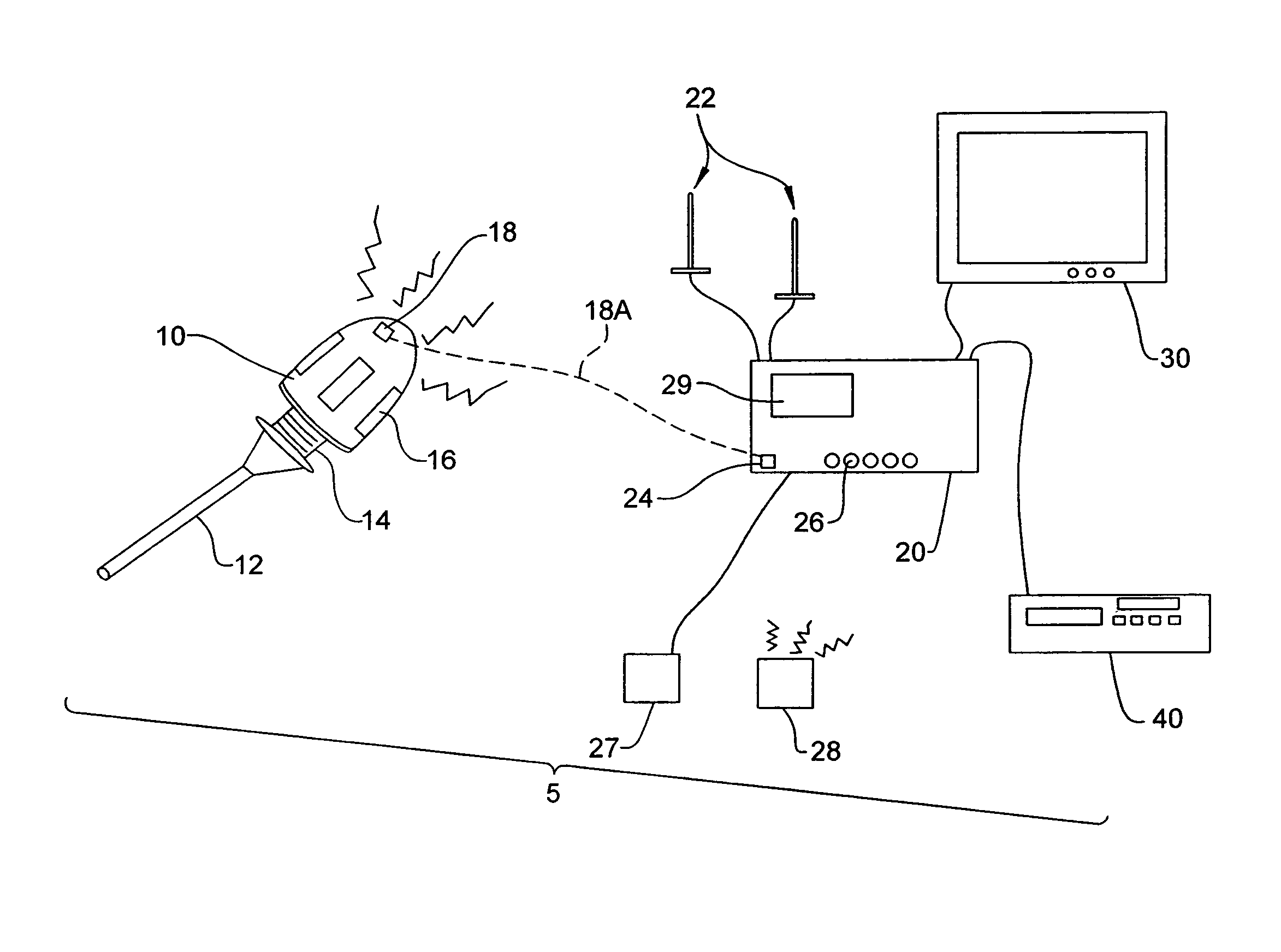
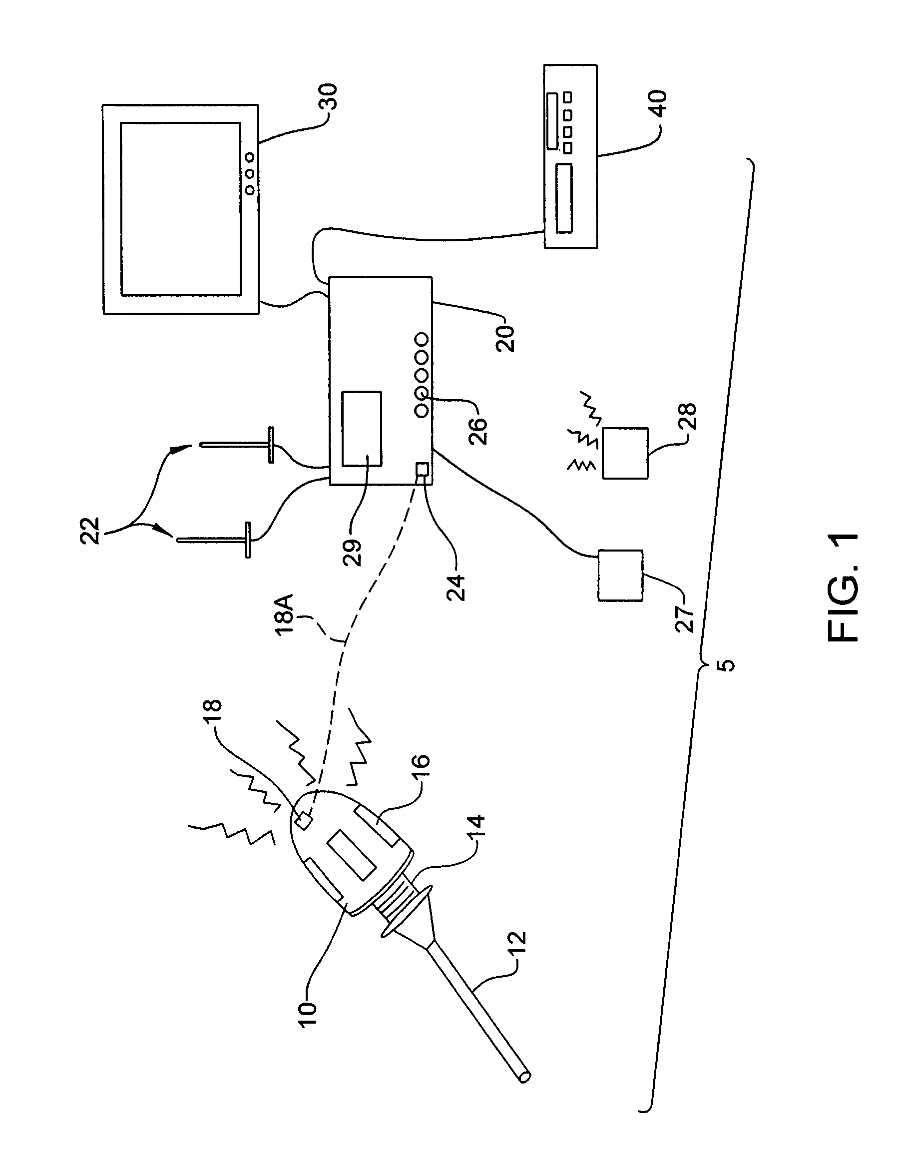
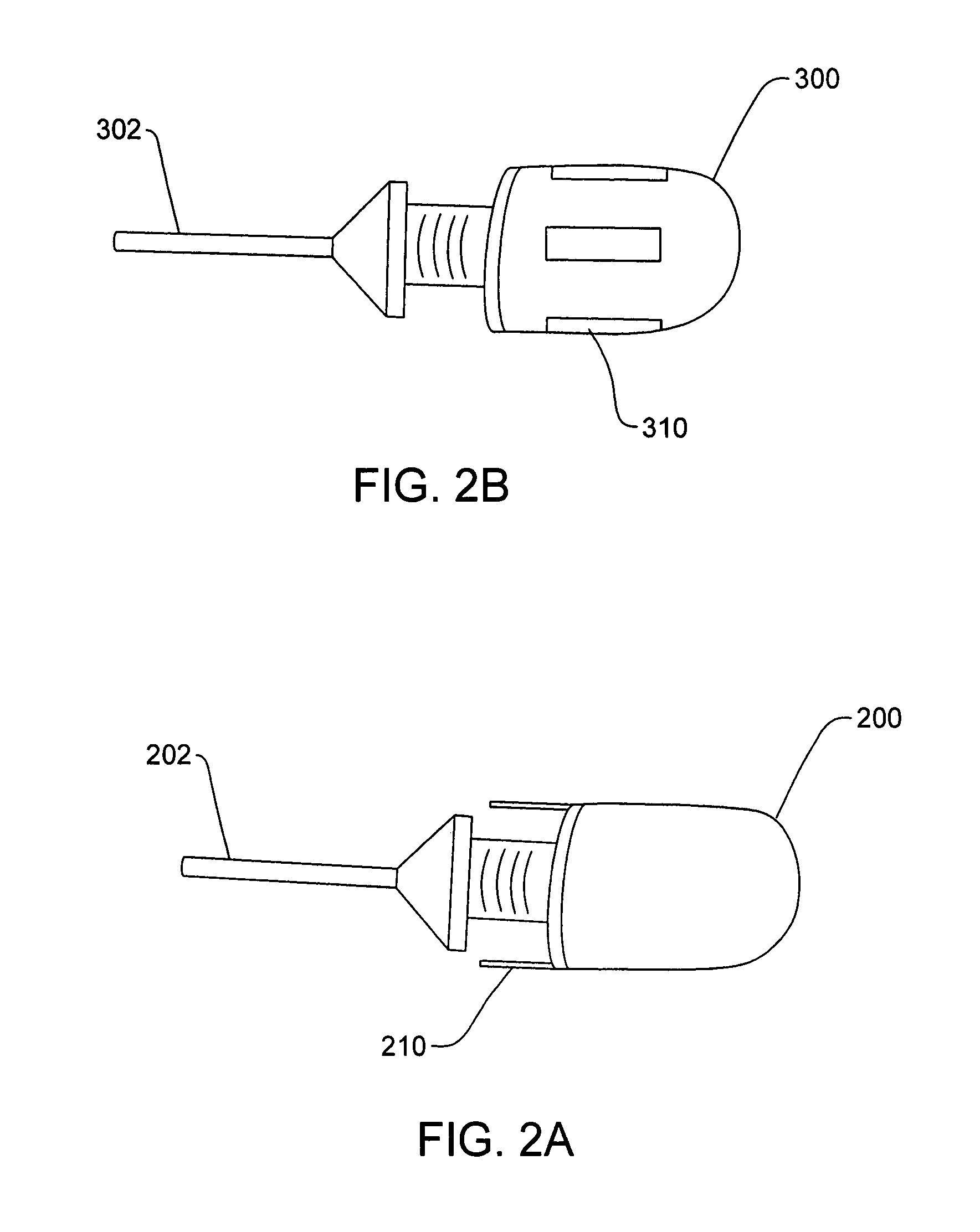
[0026]FIG. 1 depicts a wireless endoscopic camera system 5 according to one embodiment of the present invention. A wireless camera head 10 detachably mounts to an endoscope 12 by a connector 14. Contained within camera head 10 is a battery powering the electronics for the camera itself as well as the electronics making up the wireless transmitter system. Incorporated within or mounted upon the camera head 10 are one or more antennas 16 for directing the wireless signal to a receiver. The camera head 10 can also include one or more interfaces 18, such as a jack, for receiving a wired connection capable of providing power to the camera head 10 and / or for the transfer of video image data.

[0027]Located outside the sterile field is a control unit 20 that subsequently receives and processes the wireless video signal transmitted by the camera head 10. Associated with the control unit 20 are one or more antennas 22 for intercepting and conveying the wireless video signal to the control unit...


PUM


Abstract

A system and method for wirelessly transmitting a video image signal from an endoscopic camera to a receiver or control unit for storage and / or display on a video monitor. Use of a frame-specific, variable compression algorithm capable of progressively encoding a data stream provides for a better performing and higher quality wireless endoscopic camera system capable of generating images at varying resolutions. Use of a short-range, high-performance wireless technology, such as Ultrawideband (UWB), improves the performance capabilities of the system, while minimizing power consumption and extending battery life. Implementations of error correcting codes, as well as the use of multiple transmitting and receiving antennas, further improve the fidelity of the wireless communication.

Description

CROSS REFERENCE TO RELATED APPLICATION[0001]This application claims the benefit of U.S. Provisional Application No. 60 / 859,413, filed Nov. 16, 2006, the disclosure of which is incorporated herein by reference in its entirety.FIELD OF THE INVENTION[0002]The present invention relates to a reliable, high-performance wireless endoscopic camera system and corresponding method for wirelessly transmitting images from an endoscopic camera head to a control unit.BACKGROUND OF THE INVENTION[0003]Endoscopy is a technology that allows minimally-invasive viewing of internal features of the body of a patient. In medicine, endoscopy allows acquisition of high-quality images of internal features of a human body without the need for invasive surgery. The basic tool of endoscopy is the endoscope, which is inserted into the patient's body to be viewed. Some endoscopic procedures involve the use of a flexible scope, as in the medical field of gastroenterology, for example. Other medical procedures, suc...

Claims


Application Information

Patent Timeline
Patent Type & Authority Patents(United States)
IPC IPC(8): A61B1/04A61B1/06A61B1/00
CPCA61B1/00036A61B1/00105A61B1/041H04N7/185H04N19/60A61B1/00016H04N23/555H04N23/56A61B1/045A61B1/044A61B1/00009
Inventor COVER, REIDMCCARTHY, EMMETNAMBAKAM, VASUDEV
Owner STRYKER CORP
Learn More