Erythrocytes differentiated in vitro from nanofiber expanded CD133+ cells

a nanofiber and erythrocyte technology, applied in the field of erythrocyte differentiation in vitro from nanofiber expanded cd133+ cells, can solve the problems of significant ramifications of treatment limitations, insufficient treatment for a significant number of patients who remain incompletely revascularized, and morbidity and mortality, so as to reduce the need for surgical or pharmaceutical intervention, delay or eliminate the effect of treatment limitations

Inactive Publication Date: 2014-03-11
ARTERIOCYTE +1
View PDF7 Cites 2 Cited by
  • Summary
  • Abstract
  • Description
  • Claims
  • Application Information

AI Technical Summary

Benefits of technology

[0014]In certain aspects the invention provides a method further comprising administering a composition comprising said CD133+ cells to a subject in need thereof. In certain embodiments, said subject in need thereof is suffering from a condition selected from the group consisting of: ischemia, diabetes, a wound in need of healing, a need for tissue or organ replacement, a need for dialysis, need for blood transfusion, a blood condition, blood disease and blood loss. In certain embodiments, the CD133+ cells are autologous to said subject. In certain embodiments, the CD133+ cells are allogeneic to said subject. In certain embodiments, the CD133+ cells are administered by infusion into an artery. In certain embodiments, the composition comprises a matrix in which the CD133+ cells are embedded. In certain embodiments, the matrix comprises polyethylene glycol (PEG), collagen, fibrin, fibronectin, gelatin, poly-lysine, laminin, heparan sulfate proteoglycan, entactin, elastin, nidogen, hyaluronin, or a combination thereof. In certain embodiments, the fibrin matrix is polymerized from a solution that contains from about 50 mg / ml to about 400 mg / ml fibrinogen and from about 250 units / mL to about 2000 units / mL thrombin. In certain embodiments, the CD133+ cells are held in a solution comprising buffered saline for 6-36 hours prior to administering to the subject. In certain embodiments, the composition further comprises soluble human fibronectin, hyaluronan, type I collagen, fibrin, gelatin, poly-lysine, laminin, heparan sulfate proteoglycan, entactin, elastin, nidogen, or a combination thereof. In certain embodiments, the method further comprises administering to the subject a cytokine, chemokine or growth factor. In certain embodiments, the growth factor is PDGF or VEGF. In certain embodiments, the method further comprises pre-incubating said CD133+ cells with a cytokine, chemokine, or growth factor before administering said CD133+ cells to the subject. In certain embodiments, the growth factor is PDFG or VEGF. In certain embodiments, the method further comprising directing differentiation in any of the ways described above of said CD133+ cells before administering said CD133+ cells to the subject. In certain embodiments, the method further comprises administering to the subject an anticoagulant. In certain embodiments, at least 10% of cells in the composition are CD133+ cells. In certain embodiments, the CD133+ cells are isolated from umbilical cord blood, bone marrow or peripheral blood or combinations thereof. In certain embodiments, the CD133+ cells are enriched from umbilical cord blood, bone marrow or peripheral blood or combinations thereof. In certain embodiments, the composition comprises CD133+ enriched at least 2-fold over bone marrow mononuclear cells. In certain embodiments, the CD133+ cells are recultured for further expansion prior to administering to the subject. In certain embodiments, administering the composition to the subject consists of a single administration of the composition to the subject. In certain embodiments, administering the composition to the subject consists of multiple administrations of the composition to the subject. In certain embodiments, administering the composition reduces, delays or eliminates the need for surgical or pharmaceutical intervention. In certain embodiments, administering the composition increases by at least 25% the likelihood that the subject will survive over a one-year period following treatment.

Problems solved by technology

Atherosclerotic cardiovascular disease is a leading cause of morbidity and mortality in the industrialized western hemisphere.
Although catheter-based revascularization or surgery-based treatment approaches have been successful in restoring blood flow to ischemic myocardium in the majority of cases, the treatments are inadequate for a significant number of patients who remain incompletely revascularized.
The ramifications of treatment limitations may be significant in patients who have large areas of ischemic, but viable myocardium jeopardized by the impaired perfusion supplied by vessels that are poor targets for conventional revascularization techniques.
Treatment alternatives, including mechanical approaches such as percutaneous transluminal myocardial revascularization, and the like, have not produced encouraging results.
Gene therapy using adenoviral vectors to augment cytokine production and, therefore, promote angiogenesis has shown promise, but this therapy has limitations and has not yet emerged as the optimal treatment for these patients.
Although percutaneous transluminal angioplasty and aorto-bifemoral bypass procedures are associated with acceptable morbidity and mortality risk and are usually initially successful, these interventions have not been shown to be effective long-term.
Moreover, administration of cytokines to mobilize sufficient patient-derived responsive cells may worsen cardiovascular pathophysiology secondary to leukocytosis and / or activation of pro-coagulant processes.
Clinical use of autologous patient-derived sources of stem cells is advantageous to avoid potential adverse allogeneic immune reactivity; however, the disadvantages include the need to subject the patient to stem cell collection at a time of active vascular disease.
Advanced medical support requirements for military personnel have become an ever growing challenge with the advent of smaller more widespread deployments in the war on terrorism.
Moreover, cellular hemoglobin-based blood substitutes are not suitable due to their short circulatory life span of less than 48 hours, compared to 42-120 days for human red blood cells.
Clinical grade media-only based culturing systems are inefficient due to the requirement for multiple media changes, and have demonstrated difficulty maintaining the starting populations in undifferentiated states.
In addition, co-culture methods have not been shown to effectively produce clinical grade stem cells.23-27 However, the failure of erythroid development, which normally culminates in mature enucleated reticulocytes and red blood cells in vitro or ex vivo is articulated to the need for cell-cell interactions.
Although the work of Giarratana et al. is a significant milestone in red blood cell engineering, its practical implications are limited due to the constraints in obtaining enough erythrocyte progenitor cells using the co-culture techniques.
In addition, the complex engineering needed to implement the three-step protocol would make each unit of blood produced prohibitively expensive.

Method used


Image

  • Erythrocytes differentiated in vitro from nanofiber expanded CD133+ cells
  • Erythrocytes differentiated in vitro from nanofiber expanded CD133+ cells
  • Erythrocytes differentiated in vitro from nanofiber expanded CD133+ cells

Examples


example 1

[0129]Using biofunctional nanofiber scaffold that can at least partially mimic the bone marrow (BM) stem cell niche for efficient expansion of human umbilical cord blood (UCB) derived hemangioblasts, we transfected expanded (˜225 fold) stem cell population with pro-angiogenic growth factors, and evaluated the neovascularization potential. Flow cytometric analysis revealed that expanded cells retained their progenitor stem cell characteristics. These expanded cells express higher levels of CXCR4 and LFA-1 molecules, are directly linked to cellular homing and adhesion as compared to freshly selected UCB selected cells. Functional analysis revealed that these cells can uptake efficiently AcLDL, migrate in a transwell membrane plate, and can differentiate into endothelial or smooth muscle phenotype. To evaluate neovascularization potential in vivo, hind limb ischemic model was generated and tested with modified stem cell therapy. Expanded cells transfected with angiogenic factors effici...

example 2

Isolation of Fresh Umbilical Cord Blood Derived Stem Cells and Ex Vivo Expansion

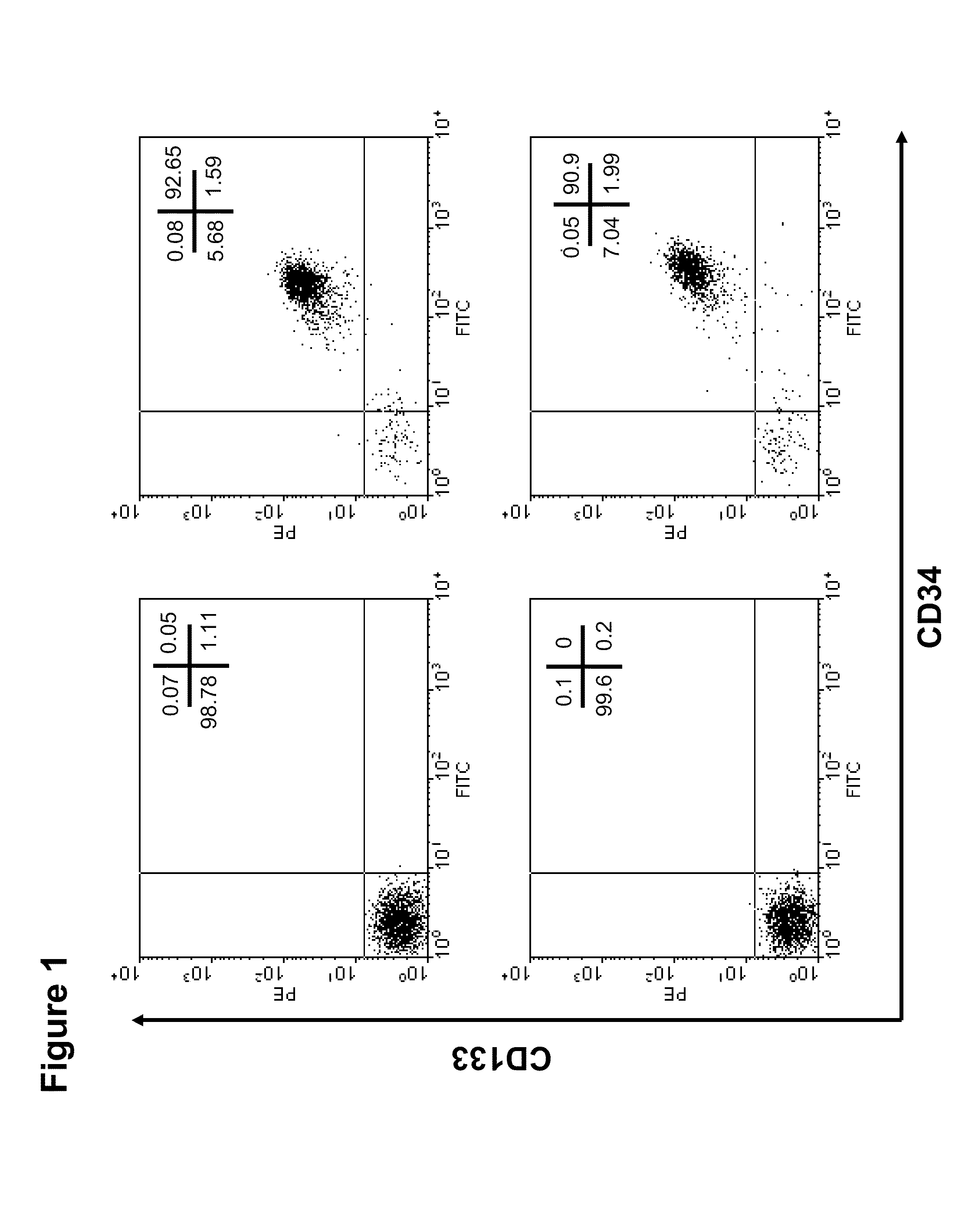
[0167]In general, via regular processing of human umbilical cord blood samples (60-80 ml sample volume) we are able to isolate 5×10⁵ to 1×10⁶ CD133+ cells using a magnetic bead-conjugated immunopurification system (AutoMACS, Miltenyi Biotec Inc.) Representative flow cytometric analysis revealed that UCB-derived CD133+ cells isolated by this method were above 90% pure (FIG. 1). However, it would be preferable to obtain a larger number of cells than this purification method yields, especially for applications such as ex vivo characterization. Applicants describe herein a way to significantly expand these cells ex vivo without major changes in their phenotype. We demonstrate here our ability to successfully expand an initial population of 20,000 cells to a quantity of at least 4.5 million (225-fold amplification) cells over 10 days of culture using a 24-well nanofiber coated plate. Even at day 5, a 20 to 25...

example 3

Phenotypic Characteristics of the Expanded Stem Cells

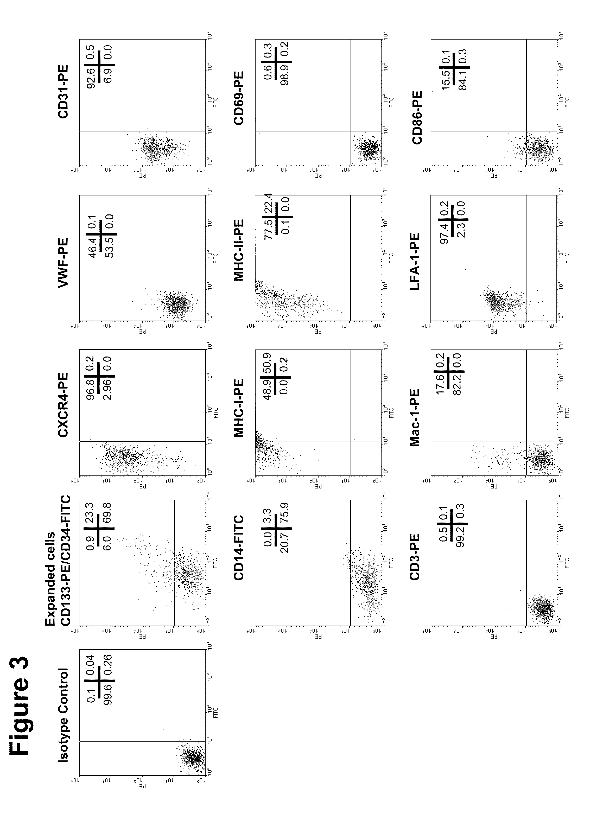
[0168]To examine phenotype of these expanded cells, flow cytometric analysis was performed using several relevant antibodies (FIGS. 3, 23 and 25). Almost 24% of the total cells retain their CD133 positivity and 93% of the cells retain their CD34 positivity. A remarkable increase in expression of two important pro-migratory and pro-adhesive molecules (CXCR4 and LFA-1, respectively) was observed in this analysis. Mild to moderate expression of other myeloid markers were observed such as CD14, CD86, vWF, CD31 or Mac-1. These expanded cells do not express any activation markers (CD69) or pan T cell markers (CD3) molecules on their surface, indicating that these expanded cells retain their stem cell characteristics. However, these cells highly express MHC class-I or Class-II molecules on their surface.


PUM

PropertyMeasurementUnit
concentrationsaaaaaaaaaa
lengthaaaaaaaaaa

Abstract

The invention provides, among other things, methods and systems for expanding CD133+ cells. The invention further provides methods and systems for increasing the blood flow to an ischemic tissue in a subject in need thereof, such as to ischemic myocardium. The invention further provides methods and systems for directing differentiation of expanded CD133+ cells. The invention further provides methods and systems for treating a subject with differentiated cells in a subject in need thereof.

Description

RELATED APPLICATIONS[0001]This continuation-in-part application claims the benefit of the filing date of U.S. application Ser. No. 11 / 975,492, filed Oct. 18, 2007, which itself claims priority to U.S. Application No. 60 / 852,598, filed Oct. 18, 2006, and this continuation-in-part application claims the benefit of the filing date of U.S. Provisional Patent Applications 61 / 010,917, filed Jan. 10, 2008 and 61 / 190,544, filed Aug. 28, 2008. The entire teachings of all of these application are incorporated by reference herein.STATEMENT REGARDING FEDERALLY SPONSORED RESEARCH OR DEVELOPMENT[0002]This invention was made with government support under R01HL083008-01 awarded by NIH / NIBIB. The government has certain rights in the invention.BACKGROUND[0003]Atherosclerotic cardiovascular disease is a leading cause of morbidity and mortality in the industrialized western hemisphere. Coronary artery disease, the pathologic process of arterial luminal narrowing by atherosclerotic plaque resulting in o...

Claims


Application Information

Patent Timeline
Patent Type & Authority Patents(United States)
IPC IPC(8): C12N5/078C12N5/00C12N5/071C12N5/0789A61K35/12
CPCC12N5/0641B82Y30/00C12N2500/44C12N2506/11C12N2501/145B82Y5/00C12N5/0691C12N2500/46C12N2501/23C12N2501/14C12N2500/25A61K2035/124C12N2510/02C12N5/0075C12N2533/30C12N2501/125C12N5/0647C12N5/069
Inventor SAKTHIVEL, RAMASAMYBROWN, DONALD J.MAO, HAI-QUANDOUAY, LUCPOMPILI, VINCENT J.MCINTOSH, KEVINDAS, HIRANMOYZHAO, YUKANG
Owner ARTERIOCYTE
Why Eureka
Try Eureka