
Method of infiltrating enamel lesions

a technology of enamel lesions and infiltration methods, which is applied in the direction of impression caps, instruments, prostheses, etc., can solve the problems of difficult cleaning of approximal surfaces, difficult remineralization of approximal lesions that have reached dentin, and difficult remineralization of approximal lesions, etc., to achieve the effect of preventing and/or treating carious lesions

Active Publication Date: 2014-04-01
CHARITE UNIVS MEDIZIN BERLIN
View PDF12 Cites 1 Cited by
  • Summary
  • Abstract
  • Description
  • Claims
  • Application Information

AI Technical Summary

Benefits of technology

This approach allows for deeper resin penetration into enamel lesions, effectively inhibiting caries progression and providing mechanical stabilization of the fragile enamel structure, potentially delaying or preventing the need for invasive treatments.

Problems solved by technology

While smooth surfaces of the tooth are more susceptible for improved cleaning strategies, approximal surfaces are particularly difficult to clean.
Nevertheless, remineralization in approximal lesions that have reached the dentin seems to be hardly achievable, since several clinical studies showed that from this threshold a visible cavitation of the lesion is established in most cases (Rugg-Gunn A J. Approximal carious lesions.
Therefore, if a cavitation occurs even at such an early stage of the caries process, a remineralization seems very unlikely under clinical conditions.
In approximal carious lesions which are difficult to reach, the ratio of carious and intact substance being removed is particularly unfavorable.
Moreover, the connection between an inserted filling and the endogenous tooth material is susceptible for carious lesions itself, and renewal of fillings due to the ageing process leads to further removal of sound tooth material.
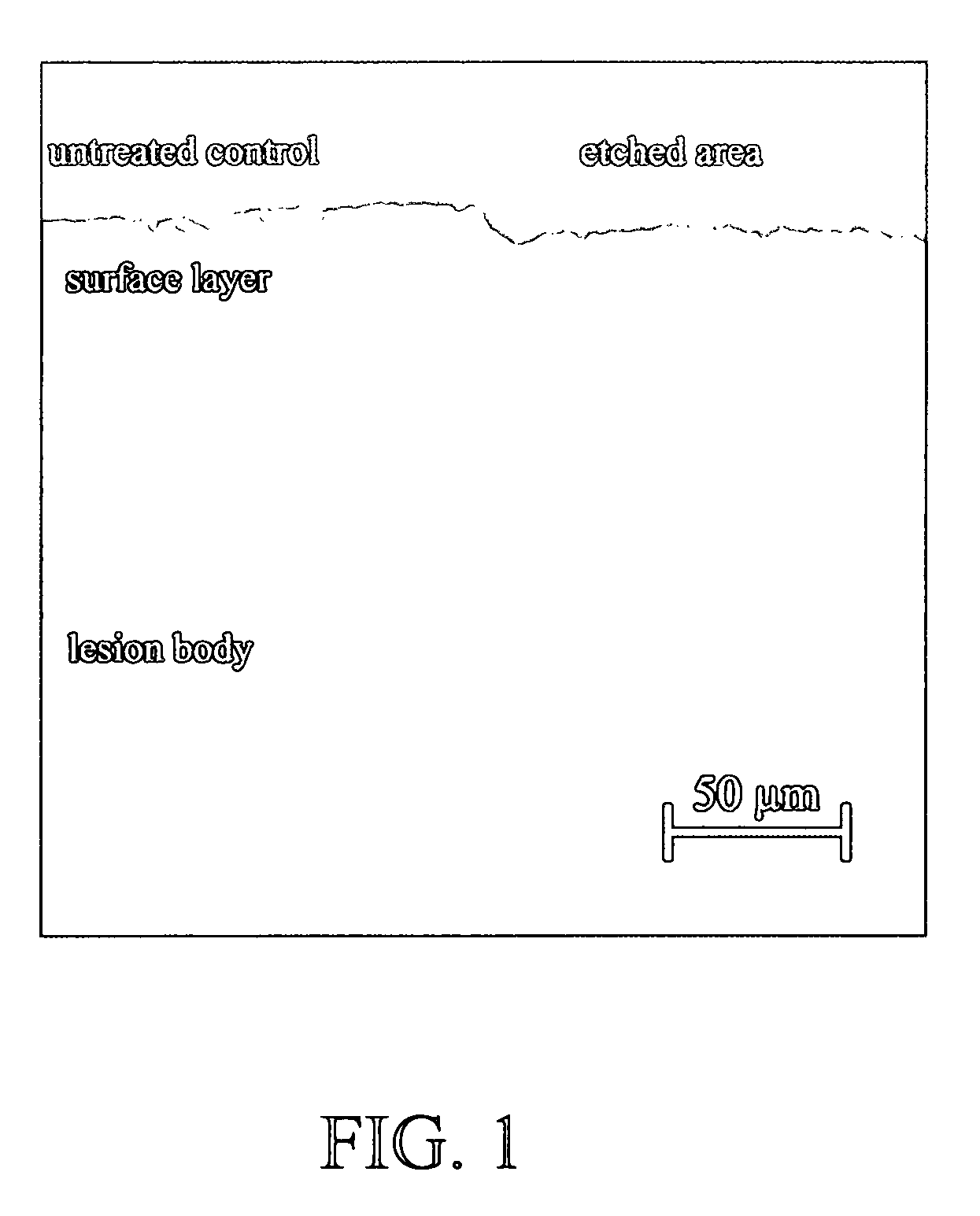
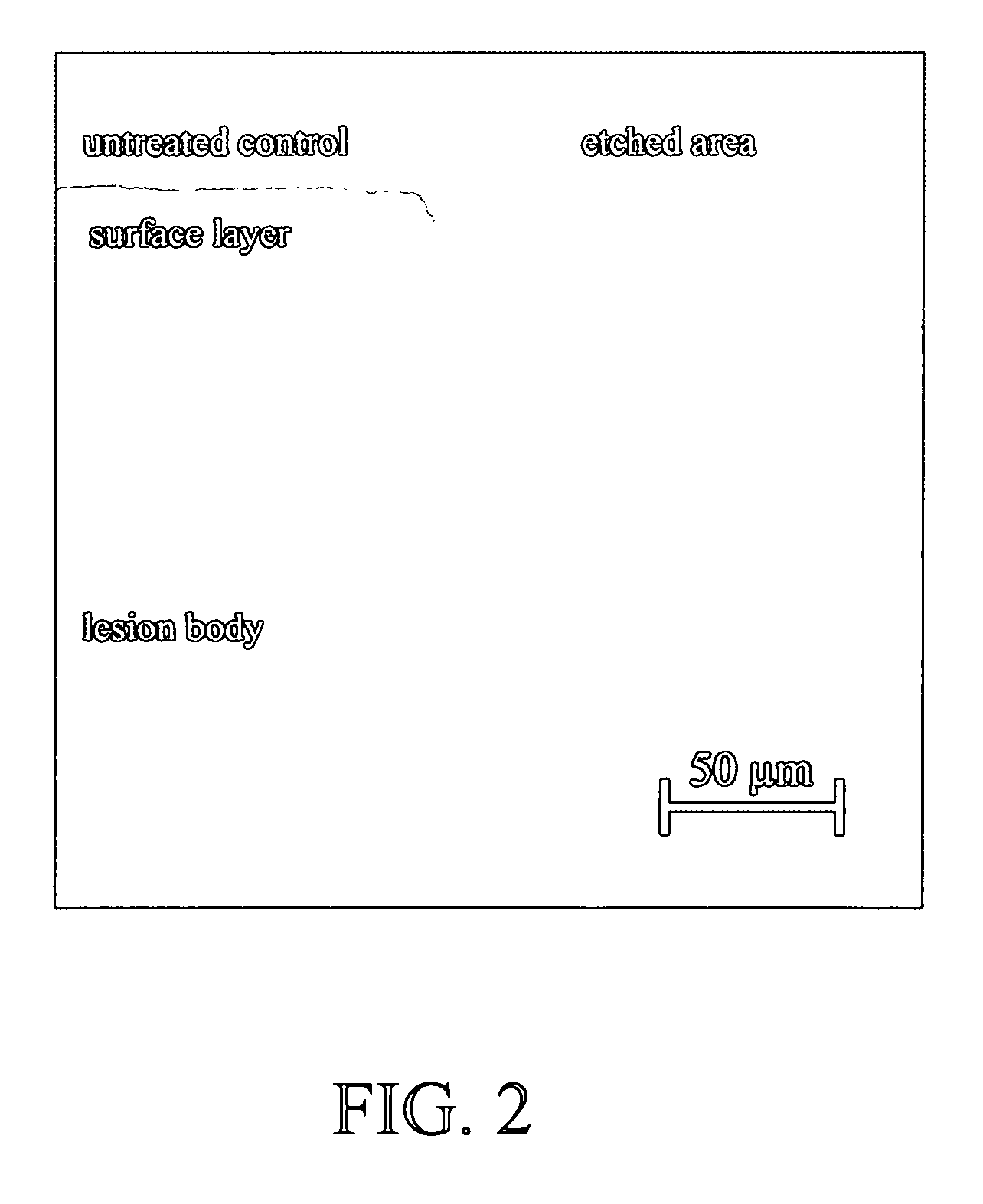
However, one of the problems in sealing natural enamel lesions is that “pseudo-intact surface layers” have higher mineral contents compared to carious bodies of lesion.
In the end, the surface layer may be superficially sealed, but the carious body may be insufficiently penetrated by the resin.
At worst, the carious process further proceeds below the “seal”.
Thus, conditioning with phosphoric acid, although demonstrated as successful in vitro, must not necessarily provide for a benefit in vivo.
Nevertheless, an in vivo study reported that the application of a conventional adhesive onto enamel lesions pre-treated with phosphoric acid gel resulted in retardation of caries progression compared to controls (Ekstrand et al.
However, patients were monitored for two years only and diagnosis was done by X-raying, a rather insensitive method for analyzing successful penetration.
Moreover, it remains unclear whether this initial success would be seen after longer periods since the rather superficial “seal” might be destroyed due to the physical load in vivo.
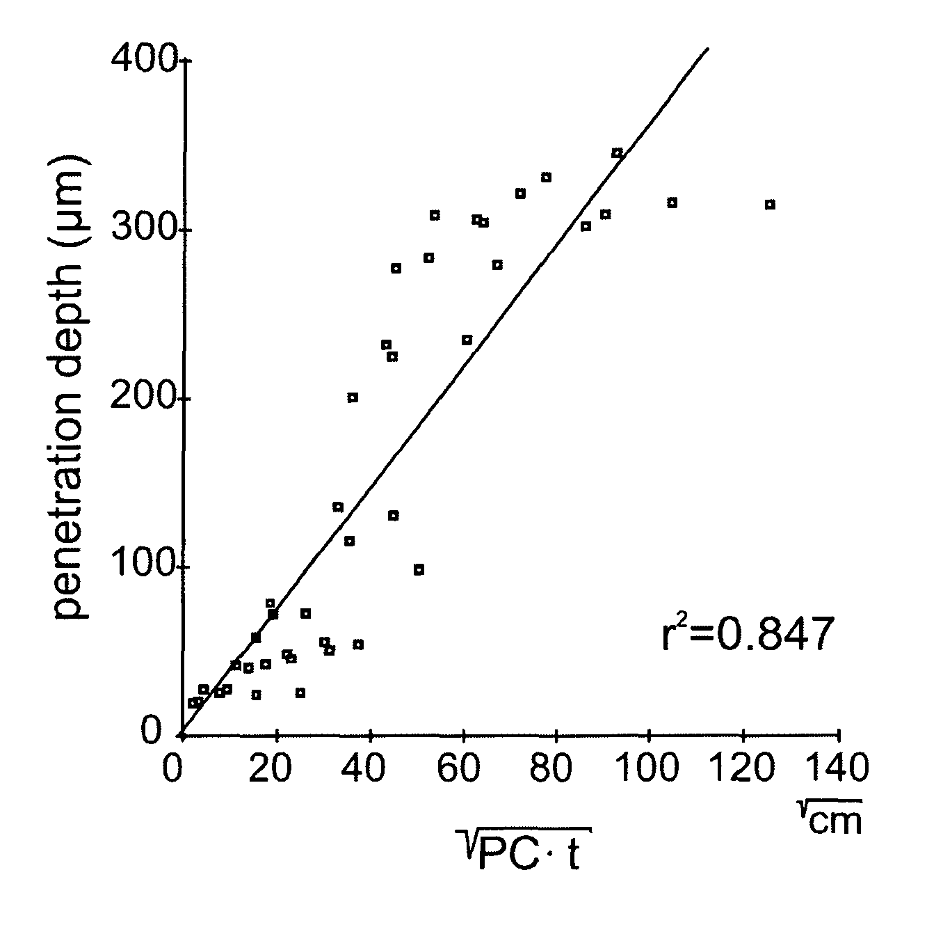
However, no study has hitherto focused on the influence of the PC on resin penetration into carious lesions.
However, application times of more than 120 seconds are hardly acceptable for use in a dentist's daily practice due to economical reasons.

Method used


Image

  • Method of infiltrating enamel lesions
  • Method of infiltrating enamel lesions
  • Method of infiltrating enamel lesions

Examples


example 1

Effect of the Pre-Treatment with a Conditioner Comprising Hydrochloric Acid

1. Material and methods

1.1 Sample Preparation

[0126]Extracted human molars and premolars, showing approximal white spots were cut across the demineralizations. One-hundred-twenty lesions confined to the outer enamel were selected. The cut surface as well as half of each lesion, thus serving as control, was covered with nail varnish. Subsequently, the lesions were etched with either phosphoric (37%) or hydrochloric (5% or 15%) acid gel for 30 to 120 seconds (n=10).

1.2 Visualization

[0127]The specimens were dried for 5 minutes in a silicone hose, closed at one end with a stopper, and separated with silicone rings. Subsequently, Spurr's resin (Spurr A R. A low-viscosity epoxy resin embedding medium for electron microscopy. J Ultrastruct Res, 1969, 26:31-43), labeled with 0.1 mmol / l of the fluorescent dye Rhodamine B Isothiocyanate (RITC), was doused over the specimens and the hose was closed with another stopper. ...

example 2

Resin Infiltration of Natural Caries Lesions after Etching with Phosphoric and Hydrochloric Acid Gels In Vitro

1. Material and Methods

[0132]Extracted human molars and premolars showing proximal white spot lesions were used in this study. After careful cleaning from soft tissues teeth were stored in 20% ethanol solution up to usage. Teeth were examined using a 20× stereo microscope (Stemi SV 11; Carl Zeiss, Oberkochen, Germany) and cavitated as well as damaged lesions were excluded.

[0133]For radiographic examination teeth were positioned in a silicone base with the buccal aspect facing to the radiographic tube (Heliodent M D; Siemens, Bensheim, Germany). To simulate cheek scatter a 15 mm wall of clear Perspex was placed between the tube and the teeth. Standardized radiographs (0.12 seconds, 60 kV, 7.5 mA) were taken from each tooth (Ektaspeed; Kodak, Stuttgart, Germany) and developed using an automatic processor (XR 24-II; Dürr Dental, Bietigheim-Bissingen, Germany). The radiographic ...

example 3

Evaluation of the PCs of Experimental Infiltrants

[0143]The aim of this investigation was the evaluation of the PCs of 66 experimental composite resins intended to infiltrate enamel lesions (infiltrants).

1. Material and Methods

[0144]A total of 66 experimental infiltrants containing two of the monomers BisGMA, UDMA, TEGDMA and HEMA in variable weight proportions each (100:0; 75:25; 50:50; 25:75; 0:100) as well as ethanol (0%, 10% or 20%) were prepared (Table 2). For each experimental resin, 10 g were mixed up in brown glass jars according to Table 2. To avoid premature polymerization, the resins were stored at 4° C. until use. To determine PCs of the experimental infiltrants, contact angles, surface tensions, and viscosities were measured.

[0145]

TABLE 2Composition (weight percent) and measured results of experimental infiltrants.Means and standard deviations (SD) are given for contact angles, surface tensions,dynamic viscosities and resulting penetration coefficients (PCs). In addition...


PUM

PropertyMeasurementUnit
wavelengthaaaaaaaaaa
wavelength maximaaaaaaaaaaa

Abstract

The present invention refers to a method of infiltrating enamel for the prevention and / or treatment of carious lesions comprising (a) exposing an enamel area to a conditioner comprising hydrochloric acid; (b) exposing the conditioned enamel area to an infiltrant; and (c) curing the infiltrant. The present invention further refers to a kit for carrying out the method of infiltrating enamel, which comprises a conditioner comprising hydrochloric acid and an infiltrant comprising at least one low viscous dental resin. The present invention also refers to a method for identifying an infiltrant by calculation of the penetration coefficient, and to an infiltrant identified by the method having a penetration coefficient of >50 cm / s or comprising a low viscous light curing resin having a penetration coefficient of >50 cm / s.

Description

CROSS-REFERENCE TO RELATED APPLICATION[0001]The present application is a continuation-in-part of U.S. application Ser. No. 11 / 040,442, filed Jan. 21, 2005, now U.S. Pat. No. 7,485,673, which is hereby incorporated by reference herein in its entirety, including any figures, tables, nucleic acid sequences, amino acid sequences, and drawings.BACKGROUND OF THE INVENTION[0002]In industrial countries, about 98% of the adult population exhibits one or more carious lesions or are already provided with fillings. Any carious lesion which eventually may lead to cavitation is initiated by demineralization of the hard tooth substance. At an early stage, referred to as “initial enamel caries”, the tooth surface remains intact without visible signs of erosion but the demineralized area below the surface becomes more and more porous.[0003]Today, the only non-operative ways to treat approximal caries are to enhance remineralization by application of fluorides and to arrest lesion progress by improve...

Claims


Application Information

Patent Timeline
Patent Type & Authority Patents(United States)
IPC IPC(8): A61L24/06A61K6/02A61K6/08A61K6/083A61K6/884
CPCA61K6/083G01N11/06G01N2013/0208A61K6/40A61K6/884A61K6/887C08L33/10C08L33/12C08L33/00A61Q11/00
Inventor MEYER-LUCKEL, HENDRIKPARIS, SEBASTIANKIELBASSA, ANDREJ M.
Owner CHARITE UNIVS MEDIZIN BERLIN
Learn More