
Vision based method for micromanipulating biological samples

a micromanipulation and biological sample technology, applied in the field of micromanipulation, automation, computer vision, microrobotics, can solve the problems of less micrometers, less micrometers thick, and more stringent requirements for microrobotic positioning, and achieve low throughput and high reproducibility

Active Publication Date: 2014-09-30
THE GOVERNING COUNCIL OF THE UNIV OF TORONTO
View PDF4 Cites 0 Cited by
  • Summary
  • Abstract
  • Description
  • Claims
  • Application Information

AI Technical Summary

Benefits of technology

The present invention is a new method for manipulating cells that solves problems with traditional manual methods such as inconsistency and fatigue. It automates the process and allows for higher reproducibility and faster research.

Problems solved by technology

Although adherent cells do not require immobilization efforts, they are highly irregular in morphology, which makes robust pattern recognition difficult and full automation challenging.
Additionally, they are only a few micrometers thick, posing more stringent requirements in microrobotic positioning.
Currently, no automated, high-throughput adherent cell micromanipulation systems are known.

Method used


Image

  • Vision based method for micromanipulating biological samples
  • Vision based method for micromanipulating biological samples
  • Vision based method for micromanipulating biological samples

Examples


example 1

[0112]A. Materials

[0113]The cells: porcine aortic endothelial cells, isolated from porcine aorta and cultured in cell medium (M199 medium, 5% calf serum, and 5% fetal bovine serum with a pH value of 7.4). Microrobotic injection was performed after 2 or 3 days of cell passage.

[0114]During system testing, fluorescent dyes (dextran, Texas Red, 70,000 MW, neutral, Invitrogen) mixed with PBS buffer.

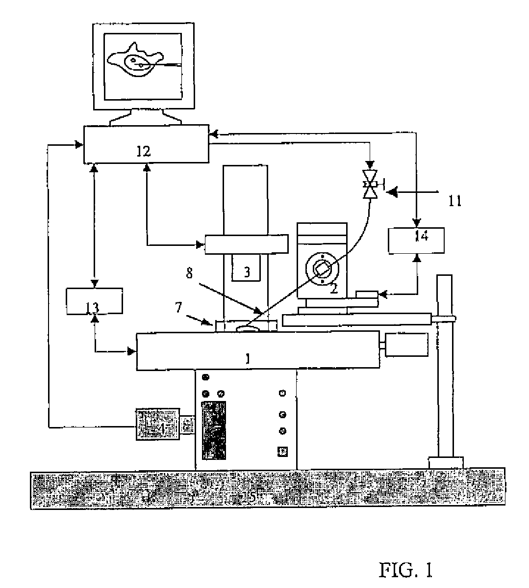
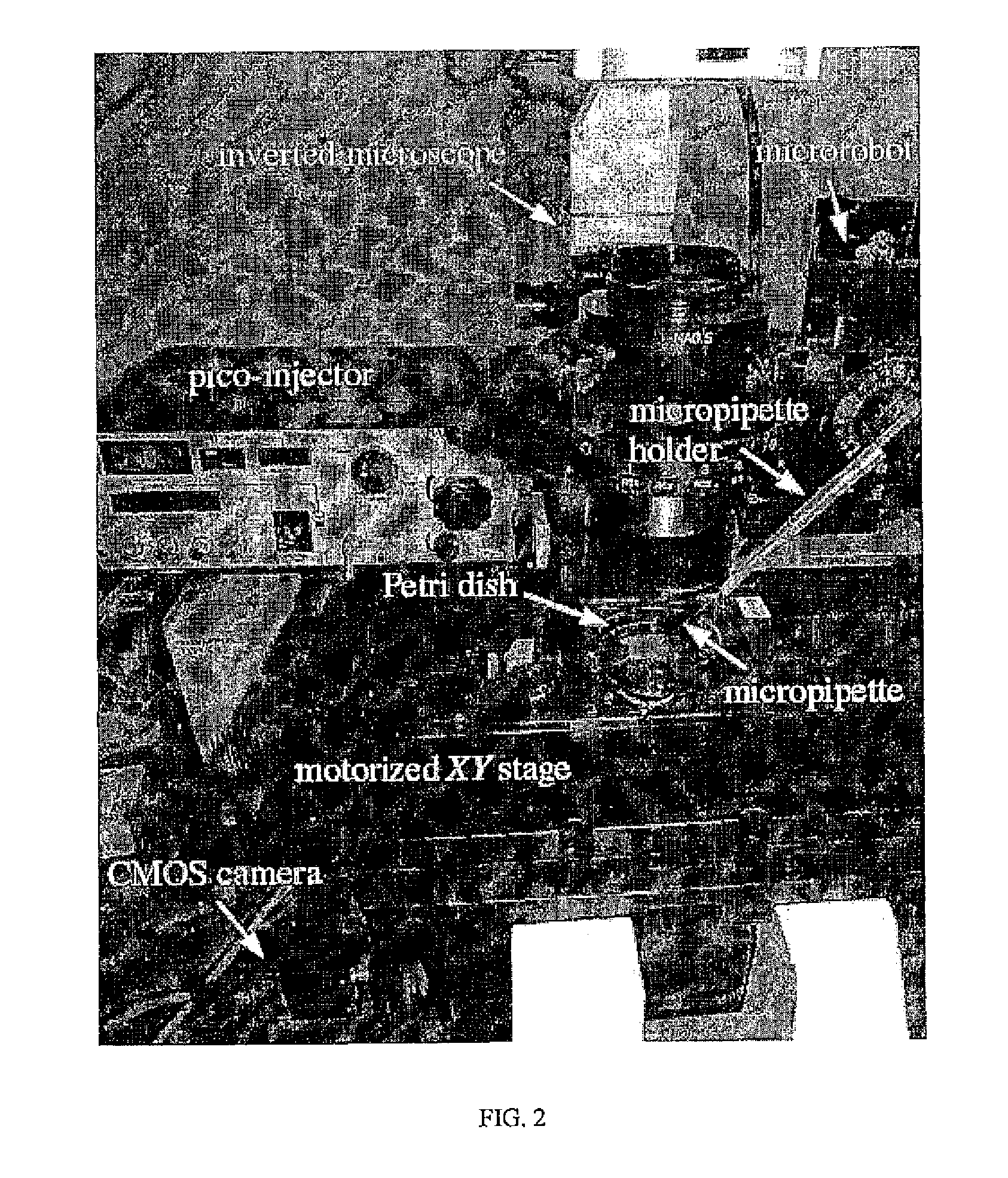
[0115]The system, used in this example is the system shown in FIG. 2, which employs a three-degrees-of-freedom microrobot (MP-285, Sutter) with a travel of 25 mm and a 0.04 μm positioning resolution along each axis. One motion control card (NI PCI-6289) is mounted on a host computer (3.0 GHz CPU, 1 GB memory) where control algorithms operate. Visual feedback is obtained through a CMOS camera (A601f, Basler) mounted on an inverted microscope (IX81, Olympus). A Polystyrene Petri dish (55 mm, Falcon) where the endothelial cells are seeded is placed on a motorized precision XYstage (ProScanII, Pri...


PUM

PropertyMeasurementUnit
outer diameteraaaaaaaaaa
heightaaaaaaaaaa
distanceaaaaaaaaaa

Abstract

A system and method for micromanipulating samples are described to perform automatic, reliable, and high-throughput sample microinjection of foreign genetic materials, proteins, and other molecules, as well as drawing genetic materials, proteins, and other molecules from the sample. The system and method overcome the problems inherent in traditional manual micromanipulation that is characterized by poor reproducibility, human fatigue, and low throughput. The present invention is particularly suited for adherent cell microinjection but can be readily extended to aspiration, isolation, and electrophysiological measurements of microorganisms, unicellular organisms, or cells.

Description

CROSS REFERENCE TO RELATED APPLICATIONS[0001]The present application is a national stage application under 35 U.S.C. 371 of International Application No. PCT / CA2008 / 000522 filed Mar. 19, 2008, the contents of which are hereby incorporated by reference into the present disclosure.FIELD OF THE INVENTION[0002]The present invention relates to micromanipulation, automation, computer vision, and microrobotics, and more specifically to a system and method for micromanipulating samples, such as microorganisms and cells.BACKGROUND OF THE INVENTION[0003]The micromanipulation of microorganisms, including unicellular and multicellular microorganisms and cells permits the insertion of foreign materials into individual cells for genetic manipulation, cellular response quantification, or intracellular structure imaging. Possessing many advantages, mechanical cell injection is highly effective for delivering macromolecules and is free from concerns about phenotype alteration.[0004]As cell injection...

Claims


Application Information

Patent Timeline
Patent Type & Authority Patents(United States)
IPC IPC(8): C12M1/36
CPCC12M23/50C12M35/02G02B21/32C12N15/89
Inventor SUN, YUWANG, WENHUI
Owner THE GOVERNING COUNCIL OF THE UNIV OF TORONTO
Learn More