Loadable polymeric particles for enhanced imaging in clinical applications and methods of preparing and using the same

a technology of enhanced imaging and polymeric particles, which is applied in the field of loading polymeric particles for enhanced imaging in clinical applications and methods of preparing and using the same, can solve the problems of most particles used in medical applications, irritation of the tissues with which they come, and initiation of adverse immune reactions, so as to minimize the agglomeration of particles formed

Active Publication Date: 2015-08-25
VARIAN MEDICAL SYSTEMS
View PDF345 Cites 4 Cited by
  • Summary
  • Abstract
  • Description
  • Claims
  • Application Information

AI Technical Summary

Benefits of technology

[0012]Microspheres of the present invention may further be provided as sized microspheres further comprising a color-coded dye incorporated into or attached to the exterior of the microspheres and contained or delivered in a similarly color-coded syringe or other transport or delivery container to further visually aid a user in providing a visual confirmation of the specific size of microspheres in use.
[0013]Also included is a method of minimizing blood flow to a tissue in a mammal comprising occluding at least a portion of a blood vessel of the mammal with at least one particle, wherein the particle comprises a poly[bis(trifluoroethoxy)phosphazene] and / or a derivative thereof.
[0019]Additionally, a method of enhanced ultrasound imaging is described herein. The method comprises administering to an ultrasound subject at least one particle comprising poly[bis(trifluoroethoxy)phosphazene] and / or a derivative thereof and a core containing one or more gas filled microbubbles to an area of the ultrasound subject to enhance echogenicity therein, and imaging the area of the subject using ultrasound.
[0022]Further, a method for minimizing agglomeration of particles formed from acrylic-based polymers is described in which the method comprises providing barium sulfate to the core and / or surface of the particles.

Problems solved by technology

Most particles used in medical applications are characterized by numerous disadvantages including irritation of the tissues with which they come in contact and initiation of adverse immune reactions.
Additionally, many of the materials used to prepare these particles may degrade relatively rapidly within the mammalian body, thereby detracting from their utility in certain procedures where long term presence of intact particles may be necessary.
Moreover, the degradation of these materials may release toxic or irritating compounds causing adverse reactions in the patients.
Some known particle types suffer from difficulties in achieving desirable suspension properties when such particles are incorporated into a delivery suspension for injection into a site in the body to be treated.
Many times, the particles settle out or tend to “float” in the solution such that they are not uniformly suspended for even delivery.
Furthermore, particles may tend to aggregate within the delivery solution and / or adhere to some part of the delivery device, making it necessary to compensate for these adhesive / attractive forces.
It can also be difficult to visualize microparticles in solution to determine their degree of suspension when using clear, transparent polymeric acrylate hydrogel beads in aqueous suspension.

Method used


Image

  • Loadable polymeric particles for enhanced imaging in clinical applications and methods of preparing and using the same
  • Loadable polymeric particles for enhanced imaging in clinical applications and methods of preparing and using the same
  • Loadable polymeric particles for enhanced imaging in clinical applications and methods of preparing and using the same

Examples


example 1

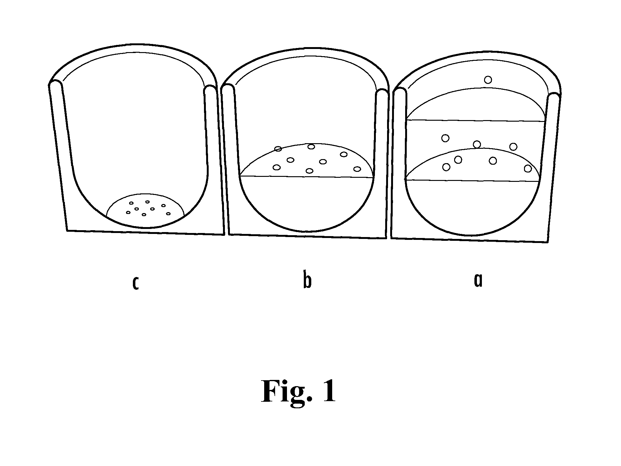
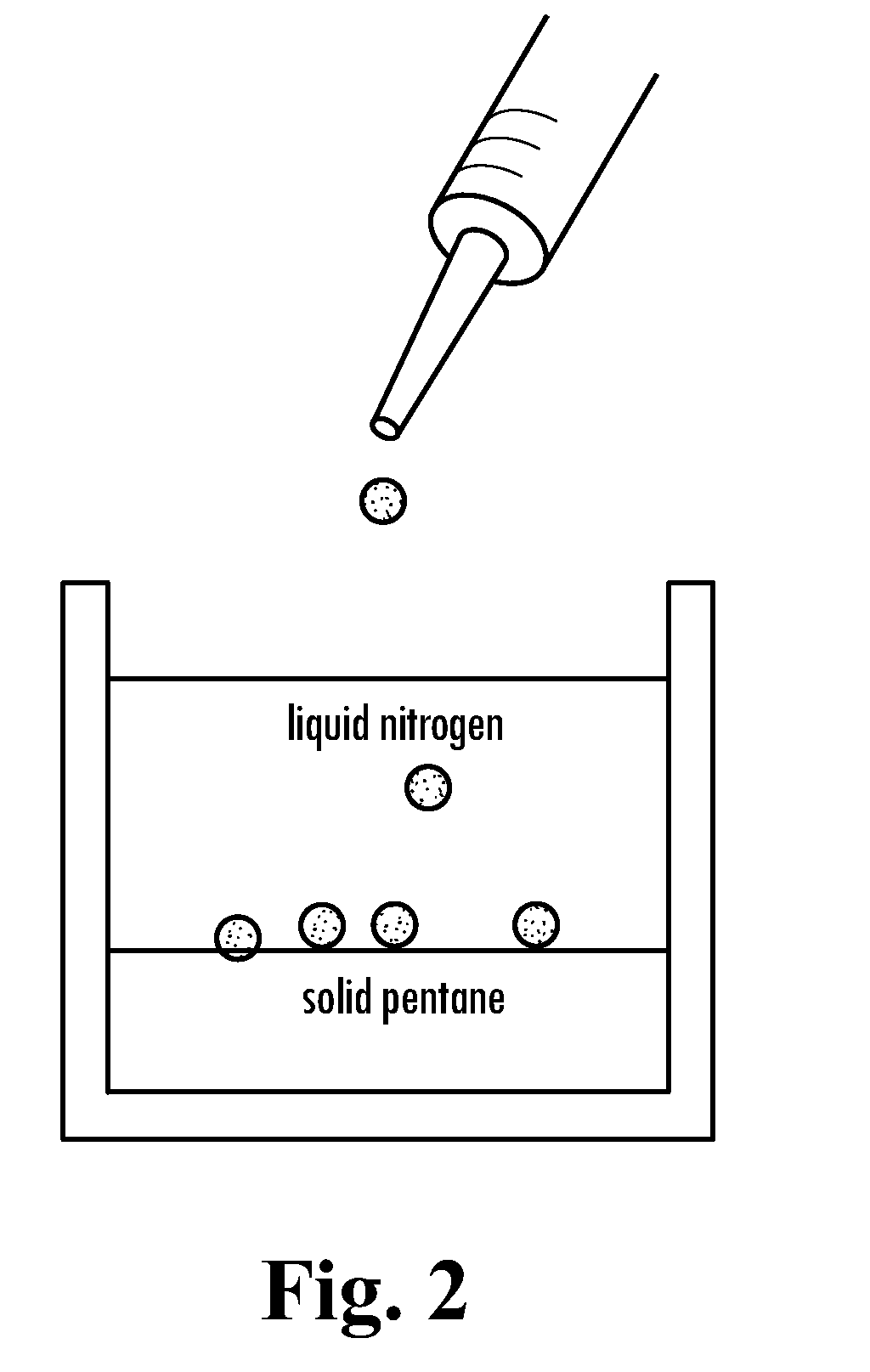
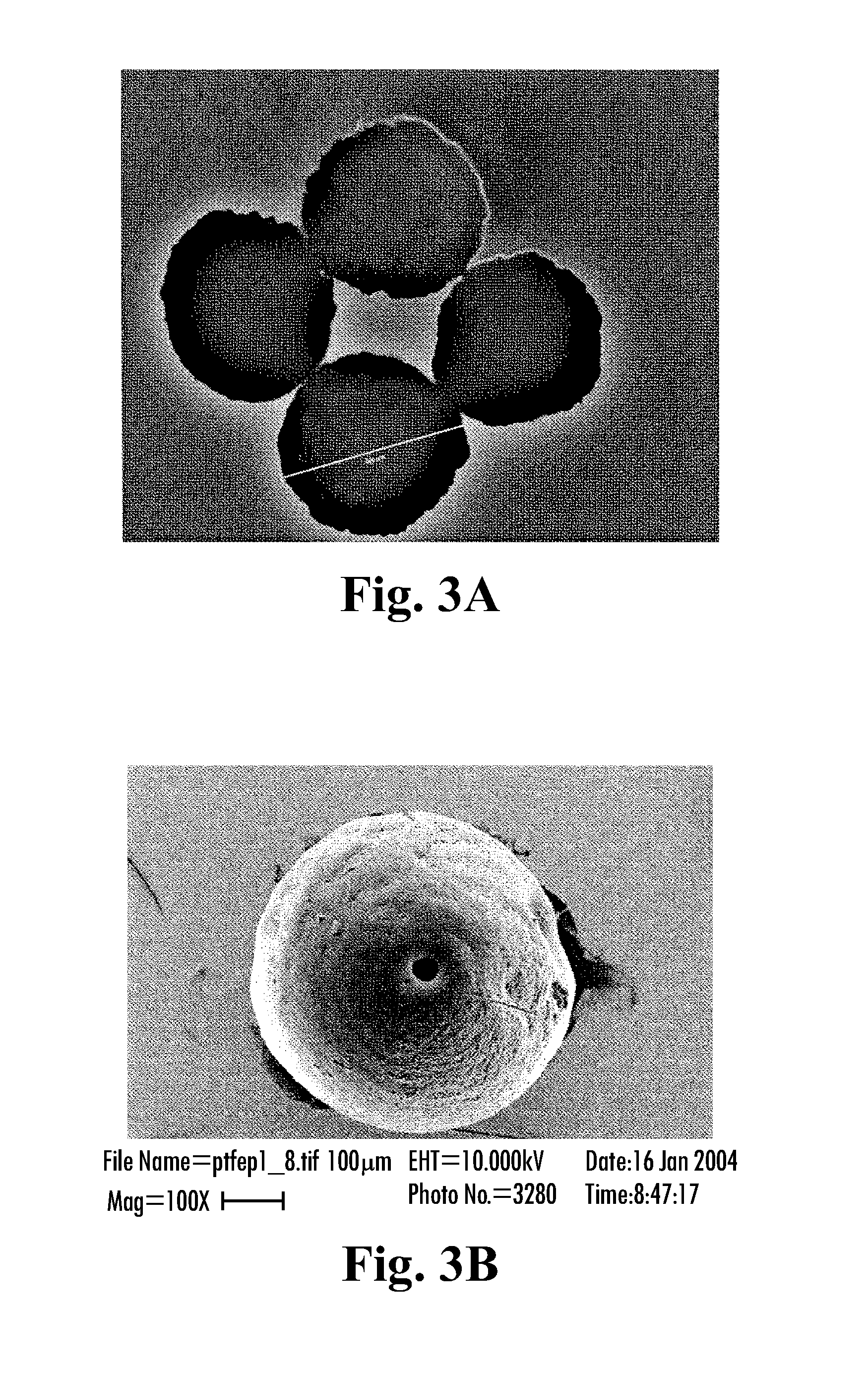
[0123]Microspheres having a diameter of approximately 500 to 600 μm were prepared. First, a polymer solution was prepared by dissolving poly[bis(trifluoroethoxy)phosphazene polymer of a molecular weight 3×10⁶ μg / mol in the polymer solvent ethyl acetate to obtain a 2% (wt / v) polymer solution. Four milliliters of this polymer solution was manually dripped into liquid nitrogen using a 5 ml syringe. This dispersion was dispensed onto a frozen layer of 150 milliliters of pentane. (See FIG. 2.) The cryoextraction was allowed to proceed for three days. Subsequently, polymeric particles were retrieved from the reaction vessel, and were air dried at 21° C.

example 2

[0124]Microspheres having a diameter of approximately 350 to 450 μm were prepared. First, a polymer solution was prepared by dissolving poly[bis(trifluoroethoxy)phosphazene polymer of a molecular weight 3×10⁶ gμmol in ethyl acetate to obtain a 1% (wt / v) polymer solution. Four milliliters of this polymer solution was manually dripped into liquid nitrogen using a 5 ml syringe. This dispersion was dispensed onto a frozen layer of 150 milliliters of pentane. (See FIG. 2.) The cryoextraction was allowed to proceed for three days. Subsequently, polymeric particles were retrieved from the reaction vessel and were air dried at 21° C.

example 3

[0125]Microspheres having a diameter of approximately 500 to 600 μm were prepared. First, a polymer solution was prepared by dissolving poly[bis(trifluoroethoxy)phosphazene polymer of a molecular weight 12×106 g / mol in methylisobutylketone to obtain a 2% (wt / v) polymer solution. Four milliliters of this polymer solution was manually dripped into liquid nitrogen using a 5 ml syringe. This dispersion was dispensed onto a frozen layer of 150 milliliters of a 1:9 (v / v) ethanol / pentane mixture (See FIG. 2.). The cryoextraction was allowed to proceed for three days. Subsequently, polymeric particles were retrieved from the reaction vessel, and dried under reduced pressure at 21° C.


PUM


Abstract

Particles are provided for use in therapeutic and / or diagnostic procedures. The particles include poly[bis(trifluoroethoxy)phosphazene] and / or a derivatives thereof which may be present throughout the particles or within an outer coating of the particles. The particles can also include a core having a hydrogel formed from an acrylic-based polymer. Such particles may be provided to a user in specific selected sizes to allow for selective embolization of certain sized blood vessels or localized treatment with an active component agent in specific clinical uses. Microspheres of the present invention may further be provided with physical and / or chemical enhancements within the particles' cores to enhance visualization of the embolized tissue using a variety of medical imaging modalities, including conventional radiography, fluoroscopy, tomography, computerized tomography, ultrasound, scintillation, magnetic resonance, or other imaging technologies.

Description

CROSS-REFERENCE TO RELATED APPLICATIONS[0001]This application is a continuation-in-part of U.S. patent application Ser. No. 11 / 257,535, filed Oct. 25, 2005, which claims the benefit of priority under 35 U.S.C. §119(e) of U.S. Provisional Patent Applications Nos. 60 / 684,307, filed May 24, 2005 and 60 / 621,729, filed Oct. 25, 2004, the entire disclosures of which are incorporated herein by reference.BACKGROUND OF THE INVENTION[0002]Small particles, including microspheres and nanospheres, have many medical uses in diagnostic and / or therapeutic procedures. In selected clinical applications, it may be advantageous to provide such microspheres and nanospheres with physical and / or chemical qualities to enhance their visual identification using various clinical imaging modalities to make the in vivo location of such microspheres apparent to a user. Such microspheres and nanospheres may allow for selective embolization of certain sized blood vessels under visual control using various clinical...

Claims


Application Information

Patent Timeline
Patent Type & Authority Patents(United States)
IPC IPC(8): A61K49/04
CPCA61K49/0419A61K49/1824A61K49/225A61K51/1244
Inventor FRITZ, ULFFRITZ, OLAFGORDY, THOMAS A.WOJCIK, RONALDBLUMMEL, JACQUESKULLER, ALEXANDER
Owner VARIAN MEDICAL SYSTEMS
Why Eureka
Try Eureka