Conduits for enhancing tissue regeneration

a tissue regeneration and tissue technology, applied in the field of tissue regeneration bundles, can solve the problems of affecting the function of the body, so as to and enhance the regeneration of the body

Active Publication Date: 2015-09-22
OSTEOCERAMICS
View PDF23 Cites 5 Cited by
  • Summary
  • Abstract
  • Description
  • Claims
  • Application Information

AI Technical Summary

Benefits of technology

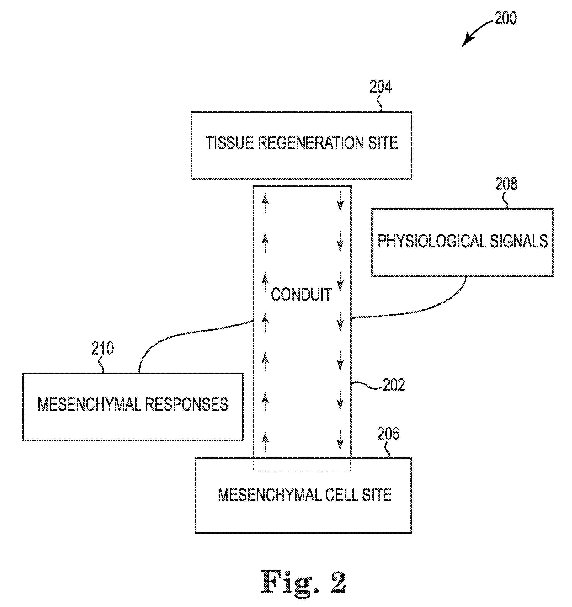
[0025]As described herein, a conduit can include one or more segments formed at least in part from an osteoceramic material. For example, a conduit can include one or more segments and, depending upon the particular application, can also include other structures contributing to the transit (e.g., delivery) of the multipotent mesenchymal cells and / or products thereof, etc., and / or contributing to the support and / or appropriate tissue regeneration of the damaged tissue. One or more segments of the conduit can enable transit of multipotent mesenchymal responses (e.g., the cells and / or their products), along with other cells, cell products, and / or blood, from one or more sources to a targeted site and / or the vicinity thereof. The conduit can provide a sheltered environment for transit of the multipotent mesenchymal responses, etc., to the target. The conduit can transit the tissue building components to and from the target site such that a geometrical construction of the conduit can control regenerated tissue structure. For example, the geometrical construction of the conduit can control bone structure and microstructure to promote and guide bone formation, bone growth, and / or bone strength to produce normally functional bone and associated tissue at the targeted site.
[0026]In some examples, the conduit can carry a fluid (e.g., blood, lymph, plasma, etc.) that serves as a transport medium for active components of wound healing and / or tissue regeneration, which can increase the kinetic rates of the wound healing and / or tissue regeneration. The conduit can define a pathway and / or orientation of structures (e.g., vascular and / or endosteal systems) that become connected from sources to targets. The conduit can serve as a barrier (e.g., with impervious walls) to excessive fluid and / or cell leakage and / or callus formation. The conduit can provide strength to withstand external forces imposed by surroundings and / or functional stresses imposed by the bodily location in which it is utilized in order to protect the associated tissue.
[0028]Provision of a conduit as disclosed herein provides for necessary cells of all types, provides a connected blood supply system to participate in wound healing, to control hematoma and callus formation, to provide for development of a new blood supply to transport the mesenchymal cell responses to the tissue growth areas adjacent to the implant, and to provide a mechanism for bone to attach to an implant. The present disclosure provides a method to allow the natural physiological processes of the patient to deliver the mesenchymal cells that form osteogenic cells to the areas of the tissue adjacent to an implant or trauma so that the cells can generate new tissue. The method is made possible during the surgical procedure by installing a conduit from the natural location of the mesenchymal cells to the areas adjacent to the site where tissue is needed for stabilization of the implant / tissue structure and to provide the strength necessary for said implant / tissue structure to support the loads necessary for normal function of that part of the body.
[0029]Using orthopedic repair as an example, the present disclosure also includes provision for a blood supply within the conduit and the areas adjacent to the implant that facilitate the normal physiology of wound healing and bone generation necessary for the load-bearing implant / bone structure to perform its function. This disclosure can extend the manufacture of mesenchymal stem cells into the conduit closer to the areas adjacent to the implant by providing the necessary conditions for that extension to occur. This disclosure also provides the geometry and composition of the implant structure to allow the natural physiological process of bone regeneration to operate by providing space to induce and / or allow the bone regeneration to occur, to control the geometry of the new bone, to provide a strong bond of bone to the implant, to allow for the physical stresses that might otherwise destroy the conduit as bone through which it passes receives the applied load, and, where appropriate, to provide a calcium phosphate environment within the conduit to the bone regeneration areas. The above accomplishments induce the bone to regenerate and / or to achieve stabilization in a much shorter time than would be possible without the conduit.

Problems solved by technology

When these massive devices explode in close proximity to personnel, they produce explosions of such force that pieces of armor may become shrapnel to cause serious local injuries, and the force of the explosion may blow the soft tissue off of the bones.
Often the injury is so severe that parts of extremities must be amputated.
This may leave insufficient bone for prosthesis attachment.
The present state of the art has not developed ways to enhance the functions supporting both soft tissue healing and bone healing needed for natural wound healing and bone regeneration.
Excessive time is often needed currently to obtain functional bone with or without medical intervention.
Often functional bone cannot be achieved.
In that case, the scaffold on which the cells are deposited is usually highly porous calcium phosphate that is too weak to support mechanical loads inherent in the tissue repair site.
Kinetic stimulants, such as DBM and growth factors, are often included, but access to mesenchymal cells is not provided.

Method used


Image

  • Conduits for enhancing tissue regeneration
  • Conduits for enhancing tissue regeneration
  • Conduits for enhancing tissue regeneration

Examples


example 1

Perthes Disease Repair

[0057]Perthes disease can occur when the femoral artery to the ball of the hip joint is destroyed or blocked. Perthes disease occurs most frequently in children and young adults, especially to highly athletic young men who are using non-steroidal anti-inflammatory medication. Alcoholic consumption may contribute. When the blood supply to the ball is interrupted, necrosis of the bone in the ball can set in. This can weaken the ball and fracturing can occur. This tends to flatten the ball. Pain is usually present and movement becomes more difficult. The contact area of the acetabular cup loses its spherical shape as it conforms to the misshapen ball and the patient has serious impairment of the function of the joint. It is possible for the ball to recover sufficient for nearly normal function in patients that are young enough to be still growing when spontaneous recovery and remodeling is complete. If the patient reaches puberty before the remodeling is complete,...

example 2

Tibia Tuberosity Advancement

[0071]FIG. 4 illustrates an example of another condition for which enhanced tissue regeneration can be implemented consistent with the present disclosure. When a canine, for example, suffers an injury that weakens, tears, or breaks an anterior cruciate ligament (ACL), the knee joint can become unstable with the tibia sliding too far forward below the femur. This is a serious injury in all mammals. The example of ACL damage illustrated in FIGS. 4 and 5 represents damage to one knee of a canine patient by way of example and not by way of limitation. That is, the aspects of utilizing conduits for enhancing tissue regeneration described herein are not limited to treatment of knee joints and / or canines, and multiple injured soft tissues and / or bones could be treated simultaneously or sequentially if needed (e.g., in contrast to the applications shown in FIGS. 3-5 in which treatment of one joint is illustrated).

[0072]FIG. 4 illustrates a lateral picture of a no...


PUM


Abstract

Apparatuses, systems, and methods for enhancing bone or soft tissue regeneration are provided. For example, a conduit, having one or more segments, can originate at a tissue regeneration site and can have a first opening to promote physiological signals to enter the conduit and transit to a second opening that penetrates a histologically rich source of multipotent mesenchymal cells, promoting the multipotent mesenchymal cells to produce tissue regeneration response products, the response products transiting through the second opening to egress at the first opening of the conduit, and promoting tissue regeneration at the tissue regeneration site. Transit of the physiological signals and the tissue regeneration response products is promoted through physical and / or chemical means, as is promotion of the anatomical functionality of the regenerated tissue. For example, a number of the segments of the conduit can be formed at least partially from an osteoceramic material.

Description

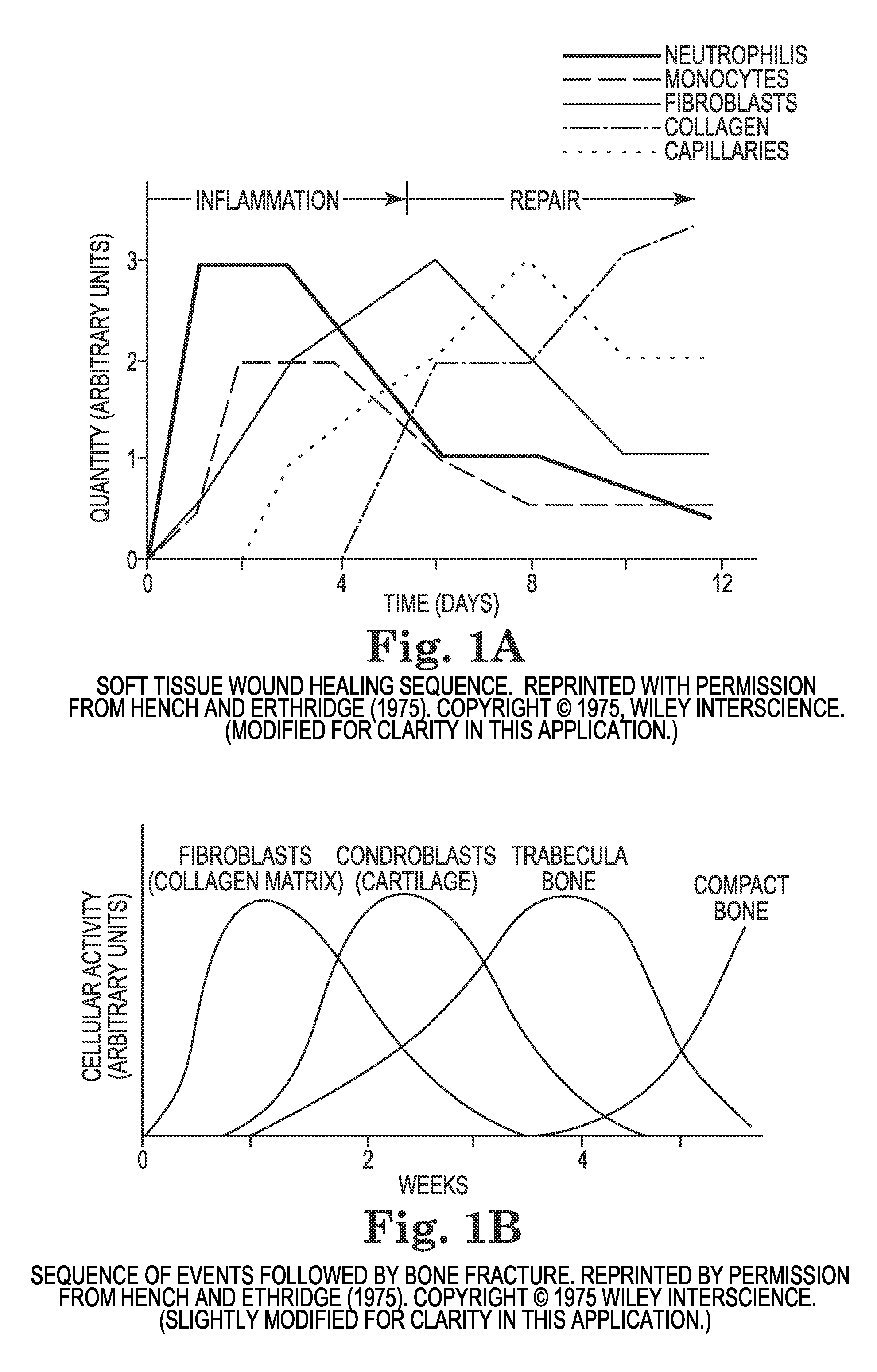
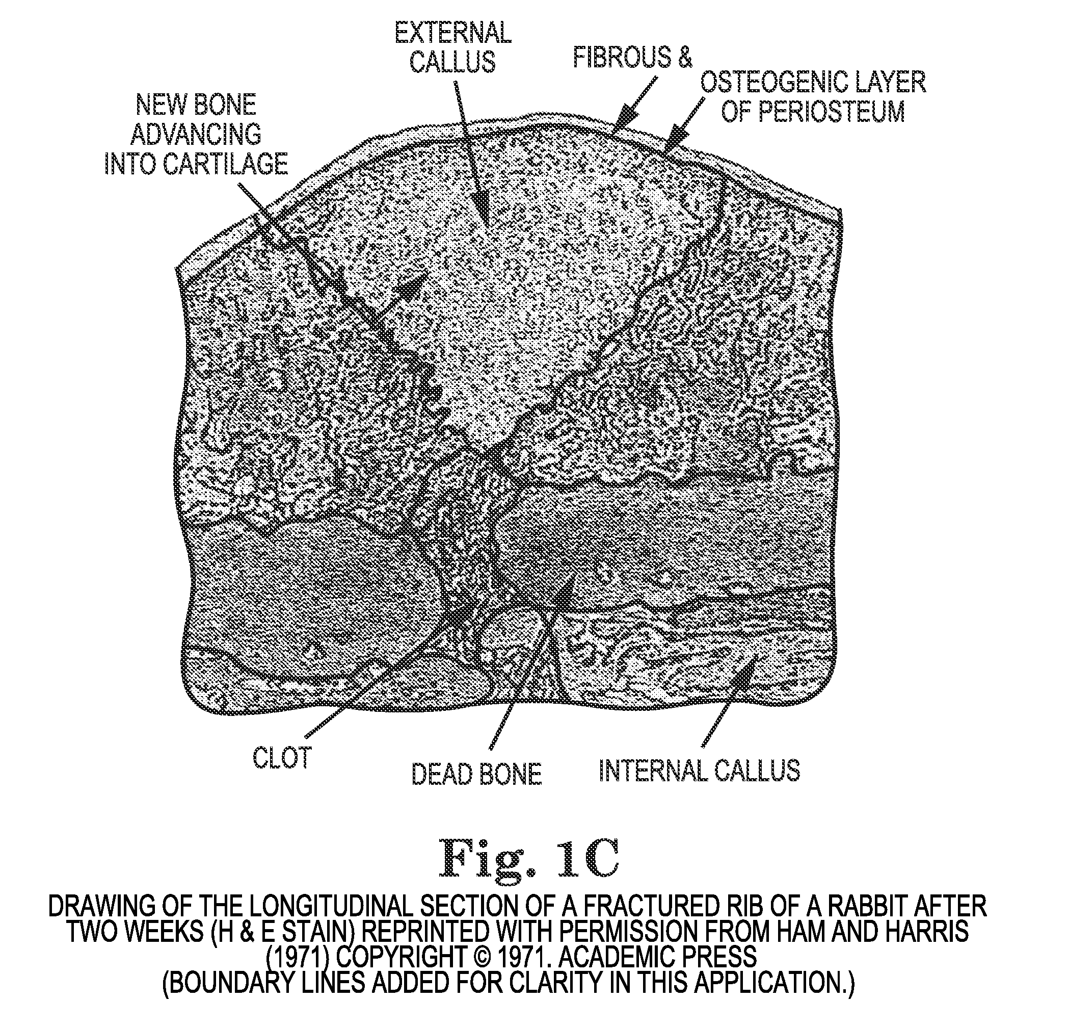
BACKGROUND[0001]During orthopedic surgery, a soft tissue wound must be made to gain access to the bones to be repaired and a bone wound is present from the original defect and from the process of treating the bone. Both types of wounds cause tissue responses that are well characterized but not well understood. Park and Lakes describe these processes in their Biomaterials book, Vol. 3, as follows, with the figure numbers being revised for consistency with the present patent application:10-1. Normal Wound-Healing Processes10.1.1. Inflammation[0002]Whenever tissues are injured or destroyed, the adjacent cells respond to repair them. An immediate response to any injury is the inflammatory reaction. Soon after injury, constriction of capillaries occurs (stopping blood leakage); then dilation. Simultaneously there is greatly increased activity in the endothelial cells lining the capillaries. The capillaries become covered by adjacent leukocytes, erythrocytes, and platelets (formed element...

Claims


Application Information

Patent Timeline
Patent Type & Authority Patents(United States)
IPC IPC(8): A61F2/02A61F2/28A61B17/56A61M27/00A61B17/74A61B17/80
CPCA61F2/28A61B17/56A61B17/74A61B17/8095A61M27/002
Inventor MCGEE, THOMAS D.
Owner OSTEOCERAMICS
Why Eureka
Try Eureka