

Aptamers that are specific for immunoglobulin-binding cell wall proteins
a technology of immunoglobulin-binding cell wall proteins and aptamers, which is applied in the field of aptamers, can solve the problems of time-consuming or expensive methods, particularly hazardous antibiotic resistance forms and especially multiresistant forms
- Summary
- Abstract
- Description
- Claims
- Application Information
AI Technical Summary
Benefits of technology
Problems solved by technology
Method used
Image

Examples
example 1
Preparation of the Target-Modified Magnetic Beads
[0244]Dynabeads® M-280 Streptavidin (Invitrogen, UK) were used in accordance with the manufacturer's instructions. 1×10⁹ magnetic beads were first washed 3× with 500 μl of PBS, pH 7.4 each time and then incubated with 510 μg of biotinylated protein A for 1 h at 21° C., while shaking (native, biotinylated protein A from Staphylococcus aureus (Sigma, P2165); dissolved in 0.1 M sodium phosphate buffer pH 7.4 to give a stock solution of 2 mg / ml). Further washing steps then followed: 3×500 μl of PBS, pH 7.4; 1×500 μl of PBS, pH 7.4+0.05% Tween20 with an incubation of 5 min at 21° C., while shaking; 1×500 μl of PBS, pH 7.4+0.05% Tween20 (without incubation); 2×500 μl of PBS, pH 7.4. The beads were separated off from the solutions each time using a Magnetic Separation Stand (Promega, Germany). The protein A-modified beads were then suspended in 500 μl of PBS, pH 7.4+0.02% sodium azide and the suspension was stored at 4° C.
example 2
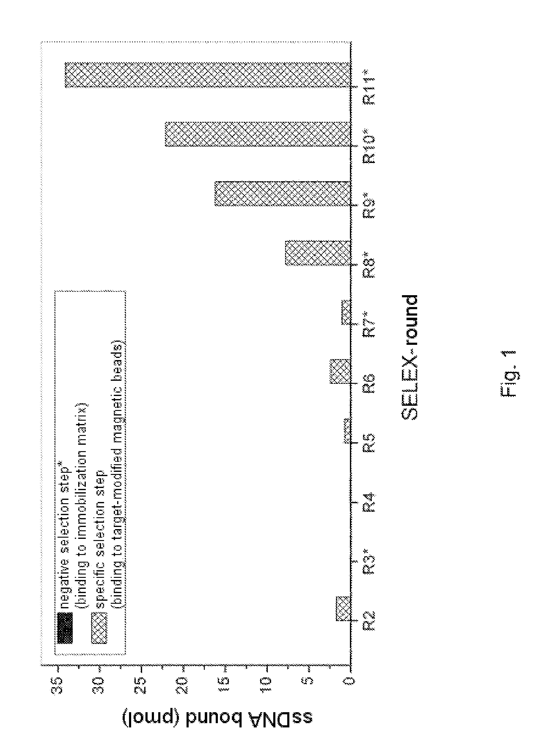
In Vitro Selection (FluMag-SELEX)
[0245]The selection of DNA aptamers for protein A was carried out using the FluMag-SELEX process. For this, biotinylated protein A was immobilized on streptavidin-functionalized magnetic beads and employed in this form as the target for the aptamer selection. A fluorescein labeling of the DNA from the second SELEX round onwards moreover rendered possible quantification thereof in the various steps of the SELEX process by means of fluorescence measurement (Wallac Victor2V Multilabel Counter; PerkinElmer, Germany; measurement conditions: excitation 485 nm / emission 535 nm, time 1 s, CW lamp energy 22500, measurement volume 100 μl / well, measurement in black 96-well microtiter plates (NUNC; Germany)).
[0246]The starting point of the selection process was a randomized DNA oligonucleotide library which was produced by means of chemical synthesis (Microsynth AG, CH): 5′-ATACCAGCTTATTCAATT-N40-ACAATCGTAATCAGTTAG-3′. N40 represents the region having variable nu...
example 3
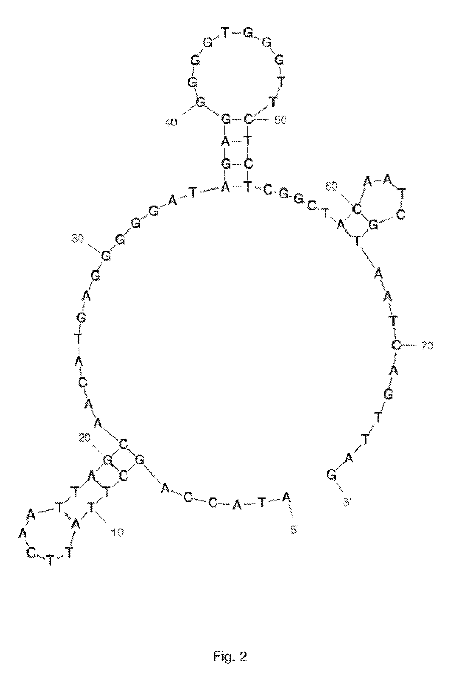
Cloning and Sequencing
[0250]In the last 11th SELEX round the oligonucleotides selected (aptamer pool) were amplified with non-modified primers in order subsequently to clone the PCR products formed directly into the vector pCR2.1-TOPO and to transform them into E. coli TOP10 cells (TOPO TA Cloning Kit; Invitrogen, UK). Positive clones were identified by means of colony PCR. Using the QIAprep 96 Turbo Miniprep Kit (Qiagen, Germany), the plasmid DNA of some of the clones was prepared and sent for sequencing of the aptamer insert (Microsynth AG, CH).
[0251]An overview of the aptamer sequences obtained from the cloning is given in Table 1. The primer sequence (sense) at the 5′ end is ATACCAGCTTATTCAATT (SEQ ID NO: 66) and the primer binding region for the antisense primer at the 3′ end is ACAATCGTAATCAGTTAG (SEQ ID NO: 67). They are in each case shown in bold. The sequences with SEQ ID NOs: 66 and 67 are a constituent of the aptamers, subject to shortened sequences, as defined in the pre...
PUM
| Property | Measurement | Unit |
|---|---|---|
| molecular weight | aaaaa | aaaaa |
| ionic strength | aaaaa | aaaaa |
| ionic strength | aaaaa | aaaaa |
Abstract
Description
Claims
Application Information