Large-volume scintillator detector for rapid real-time 3-D dose imaging of advanced radiation therapy modalities

a scintillator detector and large-volume technology, applied in the direction of luminescent dosimeters, instruments, therapy, etc., can solve the problems of difficult daily clinical use of dosimetric gels, inability to measure the time-dependent aspects of dose delivery, and time-consuming post-processing manipulation and analysis, etc., to facilitate the reconstruction process

Active Publication Date: 2017-04-18
UNIV LAVAL +1
View PDF48 Cites 5 Cited by
  • Summary
  • Abstract
  • Description
  • Claims
  • Application Information

AI Technical Summary

Benefits of technology

[0017]In some embodiments, the surface of said volumetric scintillating element may be treated to prevent light reflection at the interface between said volumetric scintillating element and any adjacent components or the surrounding medium. Particular embodiments may further include a light-guiding apparatus which directs light from the volumetric scintillating element to said means for measuring light emission. In some embodiments, the light-guiding apparatus may include fiber optics with low numerical aperture. In certain embodiments, the light-guiding apparatus may include lenses and / or mirrors.
[0029]Certain embodiments of the present invention aim to provide a three-dimensional dose detector system that can measure the three-dimensional radiation dose delivered by any type of external beam radiation therapy machine such as, but not restricted to, linear accelerators, cobalt-60 based devices, and accelerators for protons and other heavy ions. The detector can also measure the three-dimensional radiation dose delivered by radionuclide sources. Embodiments of the present invention can provide a methodology to collect the dosimetric signal in real time or near-real time with high temporal resolution, as well as a way to reconstruct a quantitative three-dimensional dose distribution from this signal. The dosimetric information thus reconstructed will be continuous (i.e. no gaps) and will possess a high spatial resolution.
[0033]The data acquisition method comprises one or more light-sensing elements such as, but not limited to, CCD cameras, CMOS cameras, photodiode arrays or photomultiplier tube arrays. In one possible embodiment, a light-field imaging device is used in order to measure both the light and the direction of the light, hence acting as both the guiding apparatus and the data acquisition mechanism. In one embodiment a combination of different types of light-sensing elements is used to provide complementary information and facilitate the reconstruction process.

Problems solved by technology

However, they usually share a delicate fabrication process; they require time-consuming post-processing manipulation and analysis; and they may suffer from various artifacts.
These characteristics make the daily clinical usage of dosimetric gels somewhat challenging.
They are not capable of measuring the time-dependent aspects of dose delivery.
The use of detector arrays is therefore not ideal because such detectors are either based on silicon diodes or air-filled ionization chambers, both of which significantly differ from water and human tissues.
Consequently detector arrays cause perturbations of the radiation fields.
Furthermore, detector arrays often suffer from intrinsic limitations.
The size of the detector elements might limit the spatial resolution of the measurements (e.g. the ionization chambers used in arrays often have a detector size of more than 5 mm).
The minimum possible spacing between detectors might also create gaps where no information can be obtained.
Even if the images contain information about the entire 3-D dose distribution, it is impossible to extract the dose delivered at a given point.

Method used


Image

  • Large-volume scintillator detector for rapid real-time 3-D dose imaging of advanced radiation therapy modalities
  • Large-volume scintillator detector for rapid real-time 3-D dose imaging of advanced radiation therapy modalities
  • Large-volume scintillator detector for rapid real-time 3-D dose imaging of advanced radiation therapy modalities

Examples


Embodiment Construction

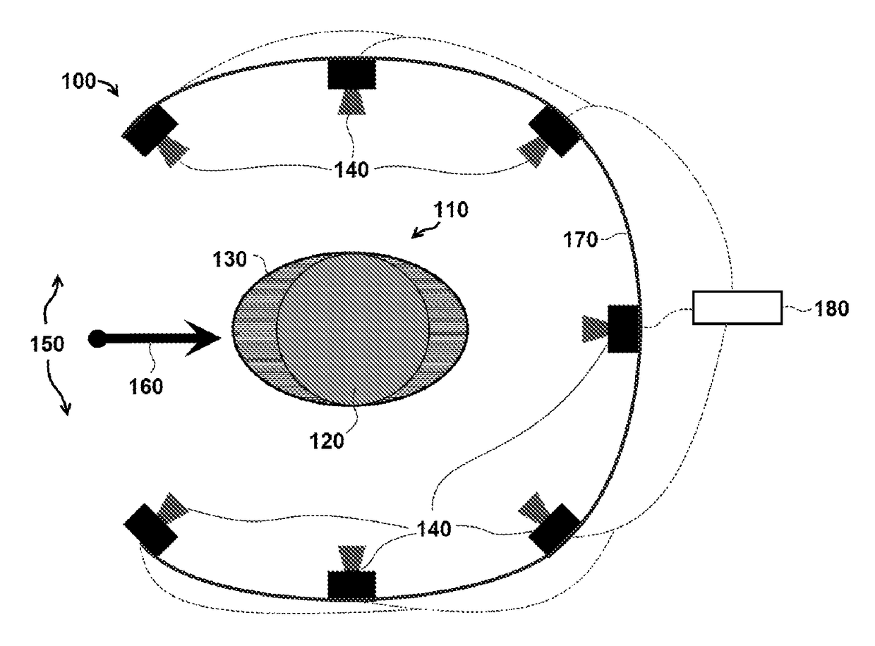
[0045]FIG. 1 is a schematic of one exemplary embodiment of a three dimensional (3-D) radiation detector system 100 for measuring radiation dose distributions intended for cancer treatment. A cylindrical plastic scintillator assembly 110 comprises an active element 120 in detector system 100, and in certain embodiments, the dimensions of cylindrical plastic scintillator assembly 110 include a height of approximately 10 to 30 cm and a radius of approximately 5 to 15 cm. In particular embodiments, active element 120 is encased in an elliptic cylinder encasing body 130, which is also called a ‘phantom’ in the terminology of the medical physics community. In specific embodiments, the external dimensions of encasing body 130 are similar in size to a specific part of a human anatomy, including for example, a human head or human torso. In particular embodiments, encasing body 130 can be made of a clear plastic or other clear material with radiation absorption properties similar to those of ...


PUM


Abstract

An apparatus and method for measuring three-dimensional radiation dose distributions with high spatial and temporal resolution using a large-volume scintillator. The scintillator converts the radiation dose distribution into a visible light distribution. The visible light is transported to one or more photo-detectors, which measure the light intensity. The light signals are processed to correct for optical artifacts, and the three-dimensional light distribution is reconstructed. The reconstructed light distribution is post-processed to convert light amplitudes to measured radiation doses. The high temporal resolution of the detector makes it possible to observe the evolution of a dynamic dose distribution as it changes over time. Integral dose distributions can be measured by summing the dose over time.

Description

CROSS-REFERENCE TO RELATED APPLICATIONS[0001]This application is a national phase application under 35 U.S.C. §371 of International Application No. PCT / US2014 / 040228, filed May 30, 2014, which claims priority to U.S. Provisional Patent Application Ser. No. 61 / 829,397, filed May 31, 2013, the contents of each of which are incorporated by reference herein.BACKGROUND OF THE INVENTION[0002]Certain embodiments of the present invention relate to the field of radiation therapy and radiation dose measurement for all types of external ionizing radiation, including photons, proton beams, heavy ion beams, and for internally-administered radioactive isotopes and x-ray sources.[0003]The properties of plastic and organic scintillators are known to be ideal for radiation dosimeters used in radiation therapy. A series of seminal papers published in the early 90s have established that plastic scintillation detectors have a unique set of advantages such as water-equivalence, dose linearity, dose rate...

Claims


Application Information

Patent Timeline
Patent Type & Authority Patents(United States)
IPC IPC(8): G01T1/00G01T1/29G01T1/20A61N5/10G01T1/10G01T1/204
CPCG01T1/10A61N5/1071G01T1/2042G01T1/29
Inventor BEDDAR, A. SAMARCHAMBAULT, LOUISROBERTSON, DANIEL
Owner UNIV LAVAL
Why Eureka
Try Eureka