Biomedical implant for use in fluid shear stress environments

a biomedical implant and environment technology, applied in the field of biomedical implants and biomedical implants, can solve the problems of delaying the clinical trial of anticoagulant therapies, affecting so as to improve the clinical trial results, promote cell survival, and promote cell adhesion

Active Publication Date: 2017-04-25
CORNELL UNIVERSITY
View PDF10 Cites 1 Cited by
  • Summary
  • Abstract
  • Description
  • Claims
  • Application Information

AI Technical Summary

Benefits of technology

[0016]One advantage of the present invention is that it provides geometric modification of surfaces of biomedical devices to promote cellular adhesion, cellular survival, cellular proliferation, and to improve biological function. In one example, the present invention provides cellular niches on the surfaces of biomedical implants to protect and enhance cell growth for applications in which cellular coatings are currently unavailable due to shear stress exposure. Fluid flows induce high shear stresses and prevent cellular coatings of biomedical devices. While the highest fluid shear stress in the body is in the aortic valve, renal filtration offers a smaller scale version of a similar phenomenon. The present invention is effective to lower the shear stresses on the majority of the implant surface, creating a cell friendly environment. Coating cardiovascular and renal filtration devices could have a great impact medically as patients would not require the current dosage of anti-coagulant drug therapies and would be able to resorb nutrient loss due to dialysis. The present invention allows for the cellular coating of biomedical devices in fluid shear exposed environments by creating a protected niche where cells can proliferate and interact biologically with their surroundings, increasing implant functionality and improving patient quality of life.
[0017]The cellular niches of the present invention also are effective to protect adhered cells from fluid derived mechanical forces while simultaneously enabling their robust secretion of soluble factors. Many implanted and extracorporeal biomedical devices are in contact with flowing blood. Receiving and providing signals through this medium is of critical importance, but shielding the sensory elements from potentially damaging shear stresses has been a continual challenge. Prominent examples include prosthetic heart valves, left-ventricular assist devices, and renal filtration systems. The present invention overcomes these limitations by creating mechanically protected niche environments where adherent cells can restore and maintain homeostasis. Further, the present invention provides micropatterned surfaces for local control of hemodynamic properties on biomedical implants.

Problems solved by technology

However, these technologies are deficient in various aspects.
While this application allows molecules or cells to be incorporated into the structure, the entire device is a porous material, detracting from the integrity of the device.
The drawback of many cardiovascular implants is the necessity for concurrent administration of anti-coagulant therapies.
These implants require these drugs due to foreign surfaces contacting circulating blood components.
Specifically, the blood-device interaction promotes platelet and clot aggregation on the device surface, rendering it a medical hazard.
The drawback to taking blood-thinners is the altered capacity of blood.
Patients taking blood thinners have a difficult time clotting when injured.
Individuals needing cardiac implants are required to have a limited lifestyle upon treatment, dramatically reducing quality of life.
This includes the inability to get pregnant, vast reduction in physical activity, and high risk of bleeding.
Coating the surfaces of these implanted devices with endothelial cells would prevent the need for anticoagulants as the passing blood would not be able to differentiate the endothelial coated implant from healthy vasculature.
Each of these options has a drawback; the biological valves degrade over time and need to be replaced, while patients that choose to have the mechanical valves need to take blood thinners as they are susceptible to blood clots.
Further, patients on blood thinners face the significant potential for major hemorrhages because of the lack of clotting.

Method used


Image

  • Biomedical implant for use in fluid shear stress environments
  • Biomedical implant for use in fluid shear stress environments
  • Biomedical implant for use in fluid shear stress environments

Examples


example 1

Cellular Niche Generation Via Micro-Patterning of Biomedical Implants

[0083]This example describes one embodiment of a biomedical implant according to the present invention. Given that one goal of the present invention was to develop way to decrease fluid shear stress, a prototype of the present invention has been developed for a prosthetic heart valve. The prototype itself is made of material that resembles synthetic heart valves, and was simplified to a square shape for ease of development and testing.

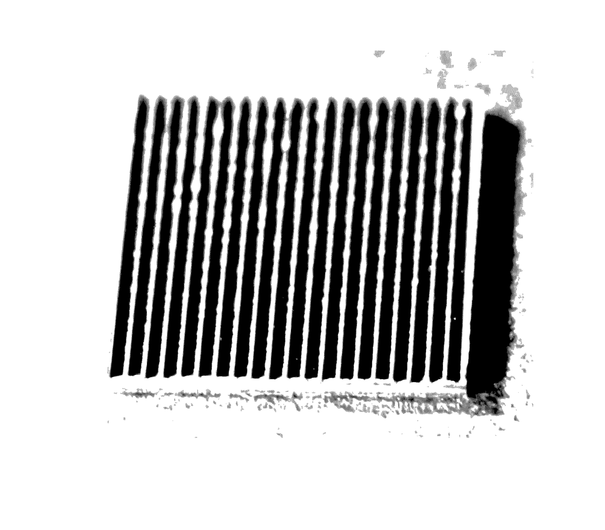
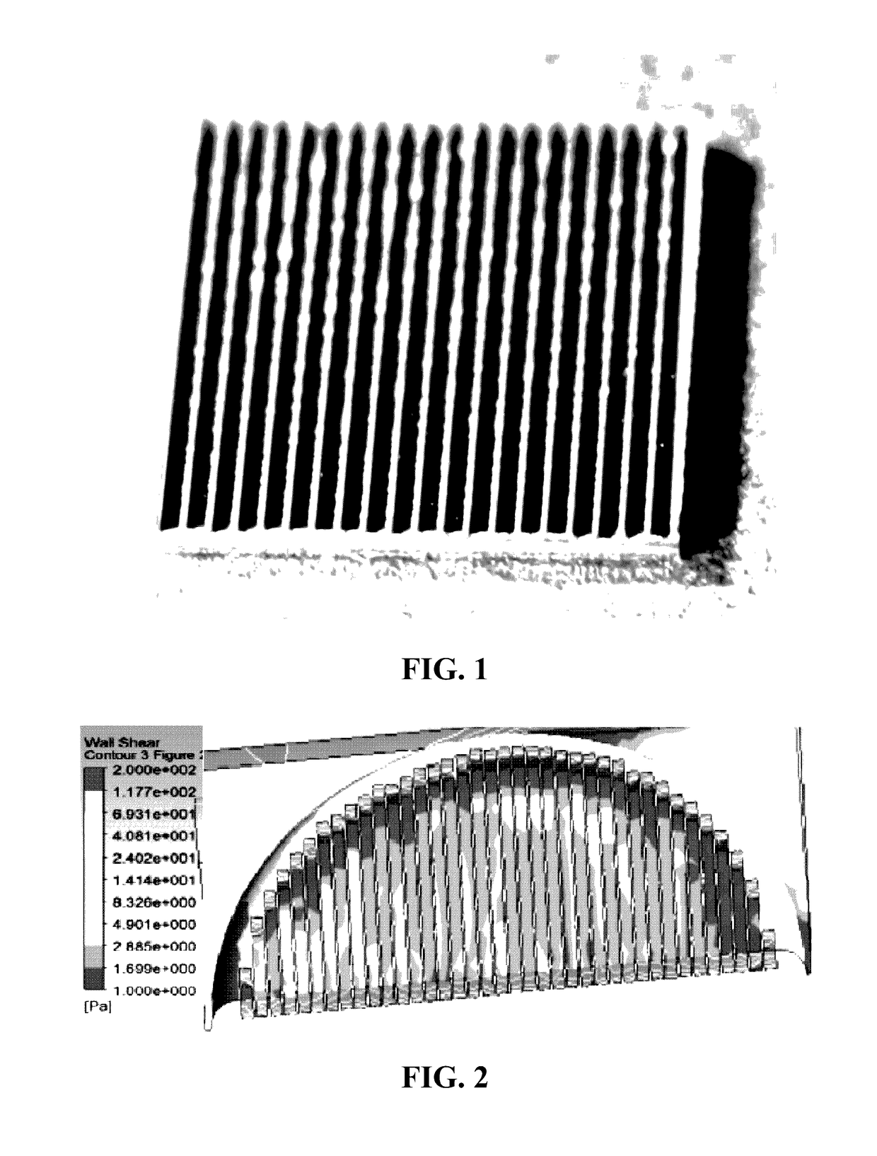
[0084]The material used to generate the sample was a 4″ Silicon wafer that was 1000 um thick; 1 cm×1 cm chips were made for testing out of the 4″ wafer (FIG. 1). The pattern to be etched into the silicon was developed following CAD analysis of blood flow over etched surfaces. This geometry was chosen in particular because of its ability to decrease the shear stress caused by blood to levels within a range where endothelial cells can function normally.

[0085]Optimization of the etched g...

example 2

Mechanical Heart Valve Surface Optimization for Endothelial Cell Adhesion

[0090]Endothelial cells line the vascular system of the human body. Current heart transplant options do not incorporate this vital vascular lining; endothelialization. Biological valves pose little foreign body response from the immune system and can be implanted with little drug assistance, but their drawback is that they require replacement within ten years due to wear. Mechanical heart valves are the alternative as they last forever. The drawback in this case is intense foreign body response from the host; thus, harsh anticoagulants such as warfarin are required for patients with these types of implants. This example was conducted to directly address the problem of endothelialization by seeding endothelial cells onto the surfaces of aortic heart valve implants. The findings show that it is possible to adhere endothelial cells to the surfaces of implanted heart valves, and that with proper topographical adjus...


PUM

PropertyMeasurementUnit
widthaaaaaaaaaa
widthaaaaaaaaaa
widthaaaaaaaaaa

Abstract

The present invention relates to a biomedical implant for use in a fluid shear stress environment of a subject. The biomedical implant of the present invention includes a patterned surface having a plurality of cellular niches. The cellular niches of the patterned surface are effective to maintain at least one localized layer of living cells within the plurality of cellular niches by decreasing fluid shear stress within the cellular niches as compared to fluid shear stress measured outside of the cellular niches, with the fluid shear stress measured outside of the cellular niches having a peak fluid shear stress of at least about 50 dynes per square centimeter (dynes / cm2). The present invention also relates to methods of making and using the biomedical implant. The present invention further relates to a biomedical implant system.

Description

CROSS-REFERENCE TO RELATED APPLICATIONS[0001]This application is a U.S. National Phase filing under 35 U.S.C. §371 of International Application No. PCT / US2012 / 055732, filed Sep. 17, 2012, and published as WO 2013 / 040544-A2 on Mar. 21, 2013, which claims benefit of priority from U.S. Provisional Patent Application Ser. No. 61 / 535,260, filed Sep. 15, 2011. The entire contents of each of the prior applications are incorporated herein by reference in their entirety.FIELD OF THE INVENTION[0002]The present invention relates to a biomedical implant and a biomedical implant system for use in a fluid shear stress environment of a subject. The present invention also relates to a method for maintaining at least one localized layer of living cells on a biomedical implant introduced into a fluid shear stress environment of a subject.BACKGROUND OF THE INVENTION[0003]Various technologies have been described in the art that involve promoting cell growth on the surface of biomedical implants. Exampl...

Claims


Application Information

Patent Timeline
Patent Type & Authority Patents(United States)
IPC IPC(8): A61F2/06A61F2/24A61F2/82A61L31/14A61L27/50A61L27/38
CPCA61F2/06A61F2/82A61F2/24A61L27/3804A61L27/50A61L31/14A61F2240/001
Inventor FRENDL, CHRISTOPHER M.BUTCHER, JONATHAN T.
Owner CORNELL UNIVERSITY
Why Eureka
Try Eureka