
Virion-derived nanospheres for selective delivery of therapeutic and diagnostic agents to cancer cells

a nanosphere and cancer cell technology, applied in the field of molecular biology and medicine, can solve the problems of inability to effectively deliver therapeutic agents to treat cancerous cells by conventional means, adverse side effects, and agents that target actively dividing cells are not able to distinguish between actively dividing cancer cells and actively dividing healthy cells. , to achieve the effect of reducing the immune response and antibody cross-reactivity of a subj

Active Publication Date: 2017-07-11
AURA BIOSCI
View PDF61 Cites 10 Cited by
  • Summary
  • Abstract
  • Description
  • Claims
  • Application Information

AI Technical Summary

Benefits of technology

[0019]According to aspects of the invention, NSPs derived from proteins of certain viruses described herein exhibit broad tumor tropism also referred to herein as universal tumor tropism. Accordingly, NSPs described herein can be used for targeting one or more tumors with a therapeutic agent, for example in a subject diagnosed as having cancer. In some embodiments, the term “subject” includes animals, such as warm-blooded mammals, for example, humans and primates; avians; domestic household or farm animals such as cats, dogs, sheep, goats, cattle, horses and pigs; laboratory animals such as mice, rats and guinea pigs; fish; reptiles; zoo and wild animals; and the like. Also, in some embodiments, NSPs described herein can be used to detect cancer in a subject by selectively delivering a diagnostic agent (for example a detectable agent, a contrast agent, or other diagnostic agent) to cancer tissue. In some embodiments, this can increase the detectability of cancer tissue or cells and / or enhance the contrast between cancer tissue or cells and surrounding healthy tissue, thereby assisting in the detection or diagnosis of cancer. It should be appreciated that diagnostic reagents described herein can be used alone or in combination with other diagnostic procedures to help detect or diagnose cancer.
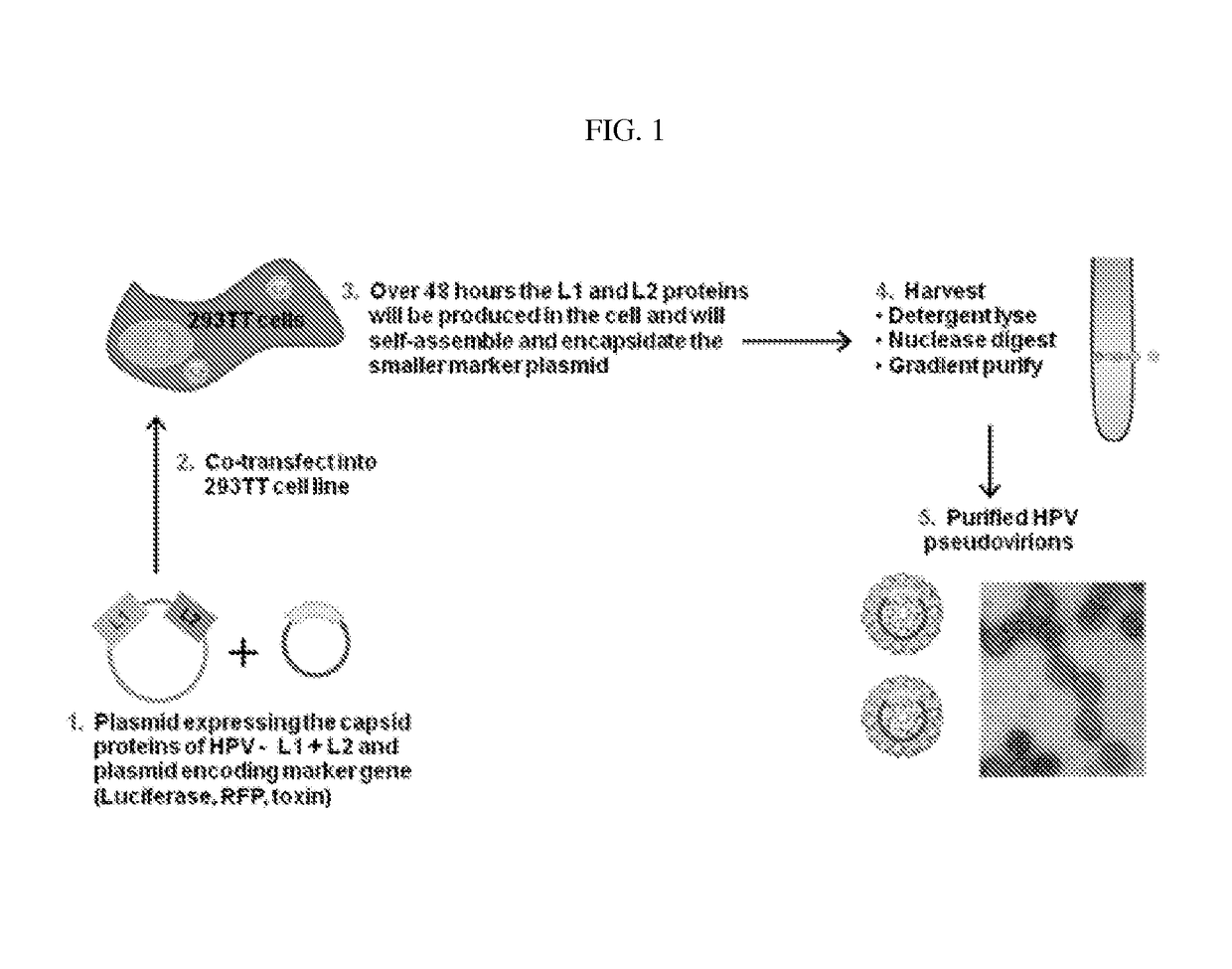
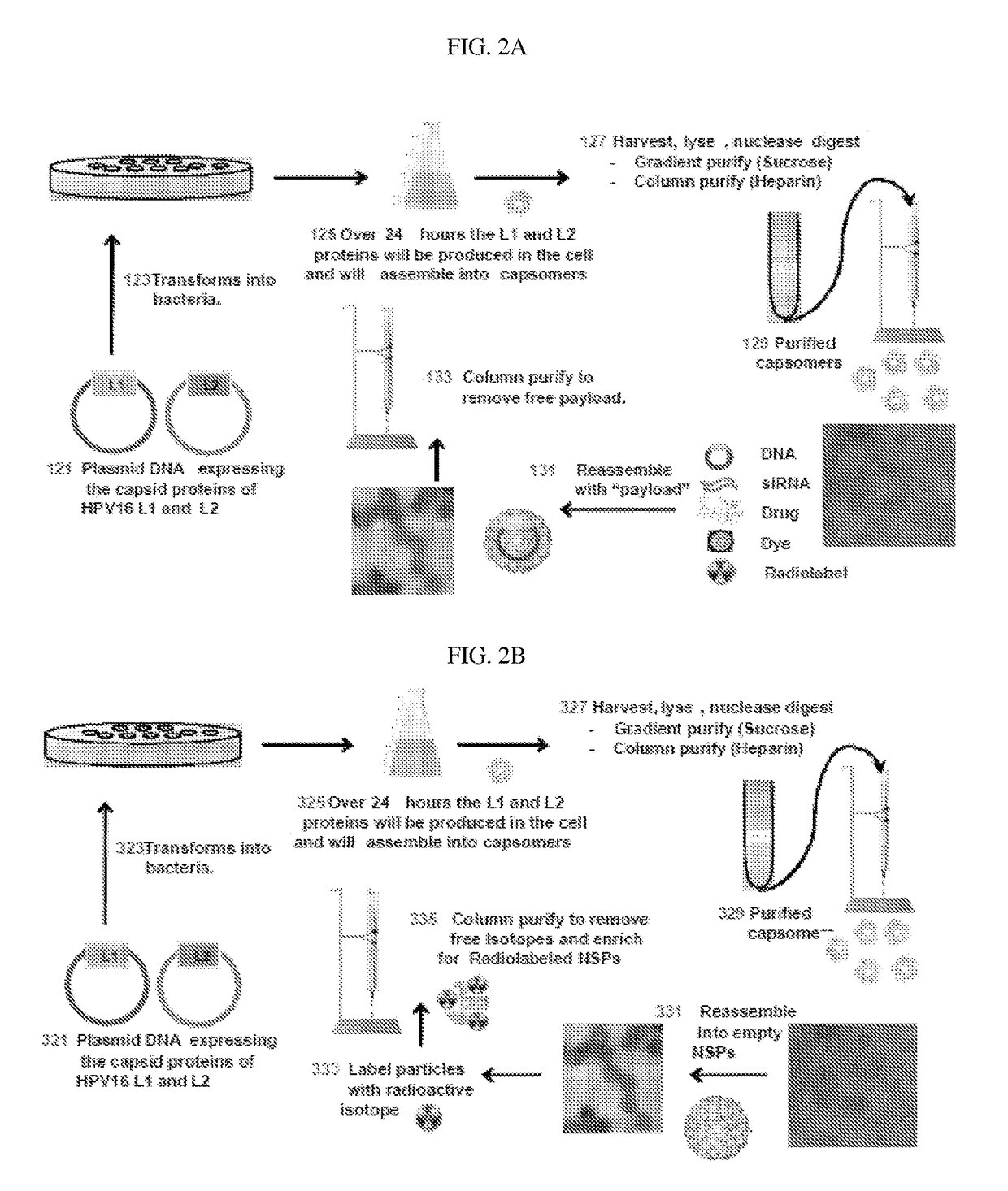
[0032]In some embodiments, provided herein are intracellular methods of encapsidating an agent within a human papillomavirus-derived nanosphere particle comprising: providing a host cell that expresses an L1 capsid protein encoding by a first nucleic acid and an L2 capsid protein encoded by a second nucleic acid different from the first nucleic acid; and incubating the host cell in the presence of an agent within the cell under conditions that promote intracellular nanosphere particle formation, thereby encapsidating the agent within the NSP intracellularly. In some embodiments, the L1 capsid protein comprises a mutation that reduces a subject's immune response and antibody cross-reactivity to the protein.

Problems solved by technology

Many therapeutic agents cannot be delivered effectively to treat cancerous cells by conventional means such as ingestion, injection, inhalation, and topical application because many of these agents are subject to rapid degradation.
Further, direct systemic administration of many therapeutic agents often causes detrimental side-effects.
For example, those agents that target actively dividing cells are not able to discriminate between actively dividing cancer cells and actively dividing healthy cells.
Thus, in the process of destroying or inhibiting the rapidly dividing cancer cells, many of the healthy cells are also damaged.

Method used


Image

  • Virion-derived nanospheres for selective delivery of therapeutic and diagnostic agents to cancer cells
  • Virion-derived nanospheres for selective delivery of therapeutic and diagnostic agents to cancer cells
  • Virion-derived nanospheres for selective delivery of therapeutic and diagnostic agents to cancer cells

Examples


example 1

Production of Mutant L1* and L2 Capsid Proteins in E. coli Cell System (FIG. 2A)

[0148]Purification of VLPs by Sucrose Gradient Centrifugation

[0149]Make a stock solution of 65% sucrose by dissolving 32.5 g of crystalline sucrose (Fisher cat. #57-50-1) to a final volume of 50 ml sample buffer. Sample buffer used for VLP purification is 0.5M NaCl (American Bioanalytical cat. # AB01915) in sterile 1×PBS (Boston BioProducts cat. # BM 220S).

[0150]Make different concentrations of sucrose solution as described in Table 1 by mixing appropriate volumes of 65% sucrose stock solution (Step 1) in sample buffer.

[0151]

TABLE 1mlFinal65%mlsucrose %stockbuffer507.692.31406.153.85304.625.38203.086.92101.548.46

[0152]Gently overlay decreasing concentrations of sucrose (highest concentration at the bottom) in a Beckman Polyallomer centrifuge tube (Cat. #326819). The volumes of different sucrose concentrations in the tube are as follows:

[0153]

65% 0.5 ml50% 0.5 ml40%0.75 ml30%0.75 ml20%0.75 ml10%0.75 ml-1 ...

example 2

Production of Mutant L1* and L2 Capsid Proteins in Mammalian Cell System (FIGS. 4A& B)

[0185]Plasmids containing human-optimized codon sequences were used to produce HPV16 / 31L1 mutant (L1*) and a HPV16L2 capsid proteins using a mammalian cell culture system, as follows.

[0186]HPV L1 / L2 nanosphere particles and L1 only nanosphere particles were added to disassembly buffer (0.5 M NaCl, 10 mM DTT (Dithiothreitol), 20 mM EDTA (Ethylenediaminetetraacetic acid), 50 mM Tris-HCl (pH 8.0)) to reach a final concentration of 0.05 mg / ml. The solution was incubated overnight at 37° C.

[0187]The HPV nanosphere particles were then concentrated to between 0.1-0.2 mg / ml using 100 kD spin columns. DNA or siRNA was added at a ratio of 1 μg of nucleic acid for every 5 μg of nanosphere particle. The resultant mixture was dialyzed using 100 kD cut-off dialysis tubing against one of two reassembly buffers: (1) 0.5M NaCl, 5 mM CaCl2, 40 mM HEPES (pH 6.8); or (2) 0.5M NaCl, 5 mM CaCl2, 40 mM Histidine-HCl (pH ...

example 3

Biodistribution Study in Parental SKOV3 Orthotopic Tumor Model with Spontaneous Metastasis (FIGS. 5A&B)

[0193]An orthotopic murine model for ovarian cancer with spontaneous metastases was used to compare the specificity and efficiency of the nanosphere particle of the present invention with a human papilloma virus (HPV) virus-like particle (VLP) (also referred to herein as PsV particles) for measuring biodistribution. Three groups of severe combined immunodeficiency (SCID) female mice received implantation of SKOV3 (ATCC) parental tumor cell line by unilateral ovarian graft. The experiment was designed to compare HPV pseudovirus (PsV) particles to the nanosphere particles of the present invention. Subjects in group 3 received a single intraperitoneal injection of nanosphere particles (0.65 ml) when tumor sizes reached medium to large by palpitation on day 77 post-tumor implantation.

[0194]FIG. 5A shows the results of in vivo bioluminescent signals as 48 hours after dosing. FIG. 5B sho...


PUM

PropertyMeasurementUnit
pHaaaaaaaaaa
pHaaaaaaaaaa
volumeaaaaaaaaaa

Abstract

The invention relates to methods for producing papilloma-derived nanosphere particles that contain therapeutic, diagnostic, or other agents. The invention also provides nanosphere particle preparations that are useful for selectively delivering therapeutic, diagnostic, and / or other agents to cancer cells of subjects without eliciting a serotype-specific immunogenic response in the subjects.

Description

RELATED APPLICATIONS[0001]This application is a national stage filing under 35 U.S.C. §371 of international PCT application PCT / US2013 / 025230, filed Feb. 7, 2013, which was published under PCT Article 21(2) in English and claims the benefit under 35 U.S.C. §119(e) of U.S. provisional application Ser. No. 61 / 596,042, filed Feb. 7, 2012, each of which is herein incorporated by reference in its entirety.FIELD OF INVENTION[0002]Embodiments described herein relate to the fields of molecular biology and medicine.BACKGROUND OF THE INVENTION[0003]Many therapeutic agents cannot be delivered effectively to treat cancerous cells by conventional means such as ingestion, injection, inhalation, and topical application because many of these agents are subject to rapid degradation. Further, direct systemic administration of many therapeutic agents often causes detrimental side-effects. For example, those agents that target actively dividing cells are not able to discriminate between actively dividi...

Claims


Application Information

Patent Timeline
Patent Type & Authority Patents(United States)
IPC IPC(8): A61K9/16A61K51/08A61K9/51A61K38/16A61K49/00B82Y5/00A61K45/06A61K9/00
CPCA61K49/0056A61K9/16A61K9/5184A61K38/162A61K45/06A61K47/48884A61K51/088A61K9/0019B82Y5/00A61K47/6929A61P35/00A61K47/6901
Inventor DE LOS PINOS, ELISABETKINES, RHONDA C.
Owner AURA BIOSCI
Learn More