Antibody and antibody mimetic for visualization and ablation of endogenous proteins

a technology of endogenous proteins and antibodies, applied in the field of compositions and methods for visualization and ablation of endogenous proteins, can solve the problems of non-physiologic over-expression of protein that can alter, labor- and time-consuming knockin mouse construction, and no opportunity to label the target protein, etc., to achieve the effect of removing background nois

Active Publication Date: 2017-08-29
UNIV OF SOUTHERN CALIFORNIA
View PDF13 Cites 1 Cited by
  • Summary
  • Abstract
  • Description
  • Claims
  • Application Information

AI Technical Summary

Benefits of technology

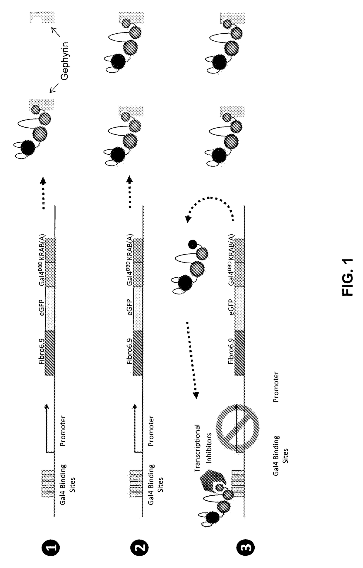
[0011]The present disclosure provides, in one embodiment, compositions and methods for labeling an endogenous protein in a living cell. The composition can be in the form of a polynucleotide encoding a fusion protein that includes a first portion or binding moiety for recognizing the endogenous protein, an optional second portion for emitting a signal, and a third portion that regulates the expression of the fusion protein itself, by virtue of binding to a transcription regulatory element that is operatively linked to the coding sequence. As illustrated in FIG. 1, when the amount of expressed fusion protein exceeds what is required for binding all of the endogenous protein in the cell, the fusion protein binds to the transcription regulatory element and shuts down the expression of itself. As such, no extra fusion protein is expressed in the cell other than those bound to the target protein, eliminating background noise. It does not require integration into the genome—therefore no health-related position effect. The compositions also allow for cell-specific expression in any animal. Transcriptional control can be used for any protein that binds at high affinity to a membrane protein or membrane associated protein.
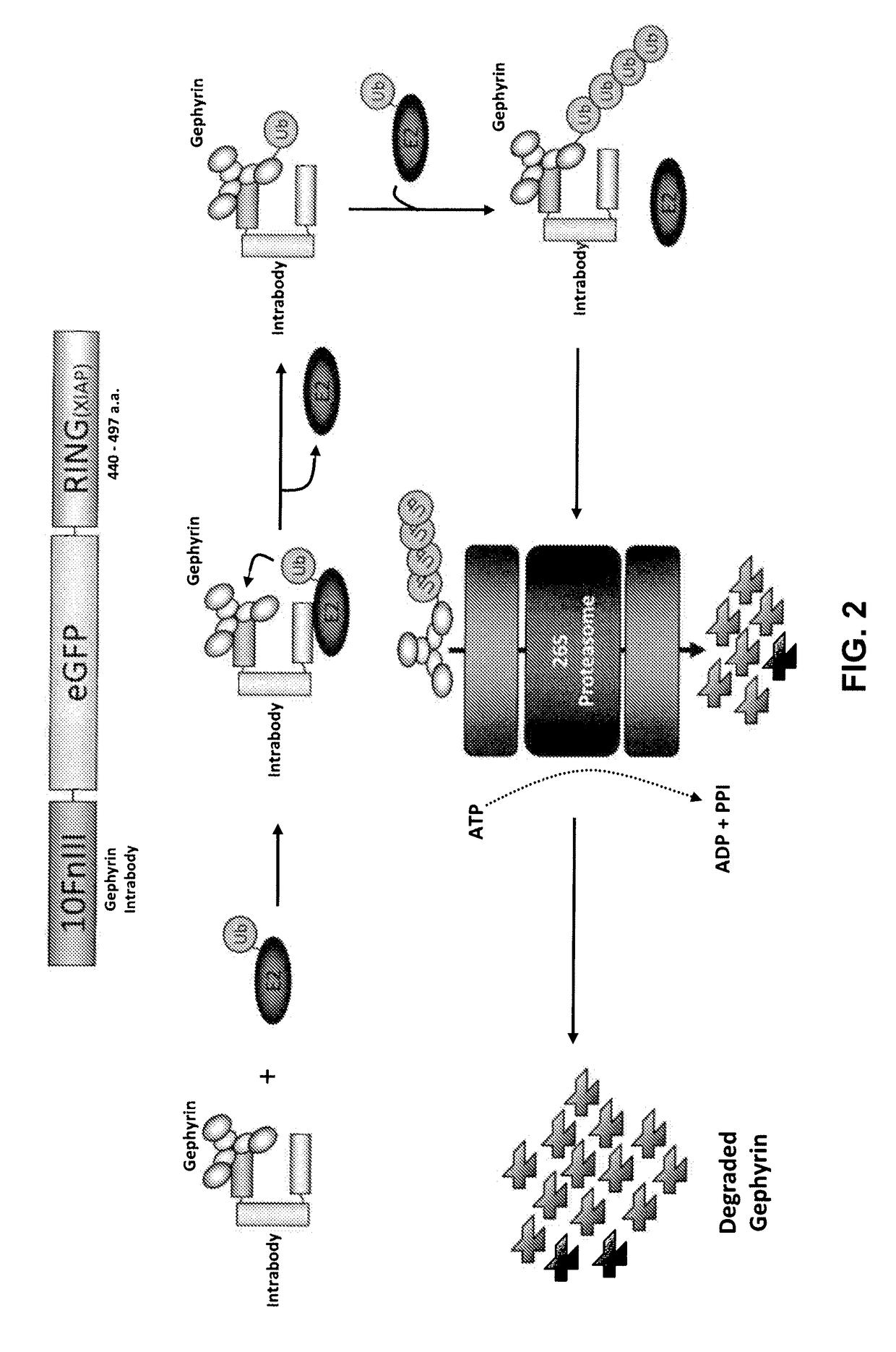
[0012]Also provided is a fusion protein that includes a first portion or binding moiety for recognizing an endogenous protein and a second portion that directs the fusion protein, along with the bound endogenous protein, to a protein degradation pathways, such as through ubiquitination, lysomosal degradation or autophagy (see illustration in FIG. 2). By introducing the fusion protein, or a polynucleotide encoding the fusion protein to a cell, the fusion protein can then effectively recognize the target endogenous protein and effect its removal in the cell.
[0015]Further provided are methods for testing the binding specificity and efficiency of a binding protein, such as an antibody, an antigen binding fragment of an antibody, or an antibody mimetic, to an endogenous protein, in particular intracellularly. Such methods provide a fusion protein that includes the domain of the target protein that is recognized by the binding protein and a peptide that localizes the fusion protein to a specific location, such as an organelle membrane. In the event the binding protein, upon binding to the target protein, is located properly, it is shown that the binding protein works specifically and efficiently.

Problems solved by technology

Direct insertion into cells by transfection often results in non-physiologic over-expression of protein that can alter both the target proteins subcellular localization and cellular morphology.
Construction of knockin mice is labor- and time-intensive and offers no opportunity to label the target protein in a conformation, complex, or modification-selective fashion.
However, this approach is suboptimal as it requires injection of every cell to be visualized, is transient, has limited dosage control over the injection, and because the antibodies themselves will gradually be inactivated / degraded by the reducing environment inside the cell and cellular protein turnover.
Furthermore, the amount of antibody in the cell must match exactly the amount of target, something that is not currently possible, either the target is not sufficiently labeled, or there is a high level of background label.
Currently there is no method for directly degrading specific endogenous proteins.
The second is that in siRNA, there can be “off target” effects that result in blocking protein production from unintended RNA templates.
A third limitation is that siRNA targets all proteins encoded by particular mRNAs.
This limitation makes it impossible to target proteins with specific conformations (e.g., activated or inactivated forms of the protein) or specific post-translational modifications that have the same mRNA sequence.
This limitation also may make it difficult to target / avoid various splice variants from a particular gene.
With regards to oncogenes and oncogeneic mutations, siRNA methods are also limited in targeting specific sites that differ from the wildtype gene sequence by one or a small number of nucleotides.

Method used


Image

  • Antibody and antibody mimetic for visualization and ablation of endogenous proteins
  • Antibody and antibody mimetic for visualization and ablation of endogenous proteins
  • Antibody and antibody mimetic for visualization and ablation of endogenous proteins

Examples


experimental examples

Example 1—Selection of Targeting Intrabodies

[0206]Intrabodies were selected from a double-stranded DNA library encoding a protein based on the 10fnIII scaffold with random substitutions in 17 residues in the BC and FG loops (Koide A, Bailey C W, Huang X, Koide S (1998) The fibronectin type III domain as a scaffold for novel binding proteins. J Mol Biol 284:1141-1151). See illustrations in FIGS. 7 and 8. This library had a diversity of ˜1013 and copy number of ˜5.

[0207]For each round of selection the library was PCR amplified to a concentration of ˜10 ng / μl, as assayed by agarose gel electrophoresis using a small mass analytical DNA ladder. 100 nM of library DNA was then transcribed in vitro by T7 RNA polymerase for 2-4 hours at 37° C. The transcription reaction was stopped by addition of 50 mM EDTA and then phenol / chloroform extracted, desalted using a CENTRI-SEP™ (Applied Biosystems) column, and finally ethanol precipitated.

[0208]Afterward library RNA was resuspended in ddH2O and i...

example 2

tion of Specificity and Efficiency of Intrabodies

[0214]This examples shows how an intrabody, or any other antibody, antibody fragment or antibody mimetic, is tested for its capability, in an intracellular environment, to (1) bind to the target protein, (2) fold correctly intracellulary, (3) not aggregate, and (4) not bind to non-target proteins.

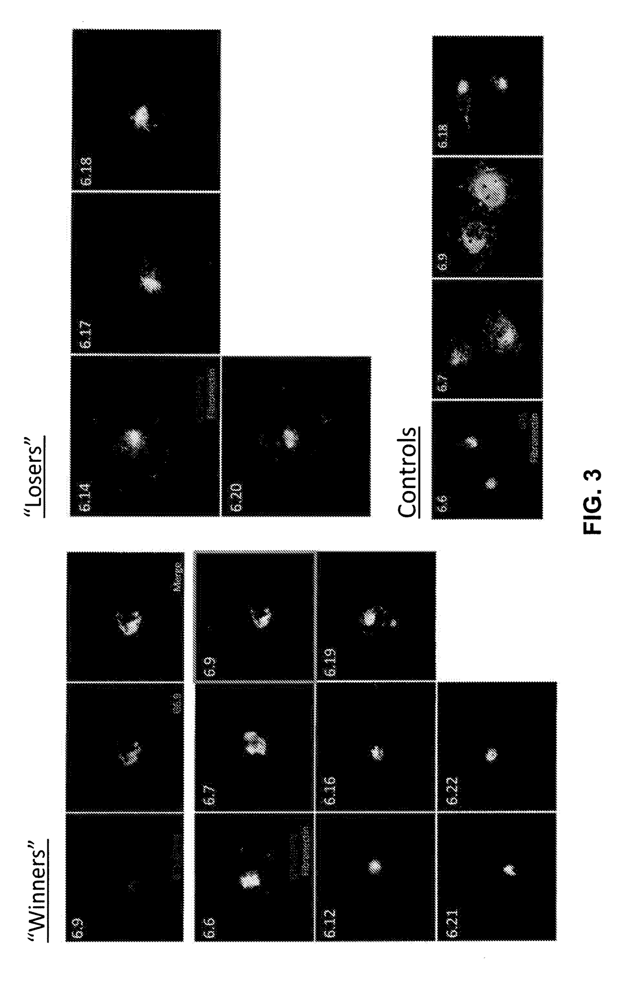
[0215]Following mRNA display selection, Fibronectin clones from late rounds that had been selected to bind with high affinity to a target molecule were cloned into an expression plasmid that has been engineered so that a GFP tag was fused to the fibronectin upon expression. A second construct was made encoding the target domain fused with a short peptide that mediates localization to the Golgi apparatus (Andersson, A. M., et al. (1997) A retention signal necessary and sufficient for Golgi localization maps to the cytoplasmic tail of a Bunyaviridae (Uukuniemi virus) membrane glycoprotein, J Virol 71: 4717-4727).

[0216]The two plasmids above wer...

example 3

Endogenous Protein with an Intrabody

[0222]To generate intrabodies, mRNA display, an in vitro protein selection method previously to produce peptides and proteins that bind targets with nanomolar to picomolar affinity (Roberts R W, Szostak J W (1997) RNA-peptide fusions for the in vitro selection of peptides and proteins. Proc Natl Acad Sci USA 94:12297-12302), was used (see Example 1).

[0223]Gephyrin and PSD-95, scaffolding proteins that are localized to inhibitory and excitatory postsynaptic sites, respectively, were used as targets in two separate mRNA selection procedures (see procedure in Example 2). In both cases intrabodies that bind very tightly and specifically to the target proteins (FIG. 15). Using biochemical methods it was estimated that the Kds for both intrabodies are ˜50 μM, indicating that they bind with better affinities than the best monoclonal antibodies.

[0224]When expressed in cortical neurons in culture, each intrabody was almost perfectly colocalized with its ta...


PUM

PropertyMeasurementUnit
timeaaaaaaaaaa
concentrationaaaaaaaaaa
fluorescentaaaaaaaaaa

Abstract

Provided are compositions and methods for labeling an endogenous protein, in particular, in a live cell, or for ablating an endogenous or target protein. The compositions relate to a fusion protein having a binding moiety such as an antibody, an antigen binding fragment of an antibody or an antibody mimetic that recognizes the endogenous or target protein.

Description

CROSS-REFERENCE TO RELATED APPLICATIONS[0001]This application is a national stage application under 35 U.S.C. §371 of International Application No. PCT / US2012 / 028947 filed Mar. 13, 2012 which in turn claims priority under 35 U.S.C. §119(e) of U.S. Provisional Application Nos. 61 / 452,535 and 61 / 532,065, filed Mar. 14, 2011 and Sep. 7, 2011, respectively, the contents of each of which are incorporated by reference into the present disclosure.STATEMENT OF GOVERNMENT SUPPORT[0002]This invention was made with government support under Contract No. GM 083898 awarded by the National Institutes of Health and National Institute of General Medical Sciences. The government has certain rights to the invention.FIELD OF INVENTION[0003]The present disclosure generally relates to compositions and methods for visualization and ablation of endogenous proteins.BACKGROUND OF THE INVENTION[0004]Throughout and within this application various technical and patent literature are referenced either explicitly...

Claims


Application Information

Patent Timeline
Patent Type & Authority Patents(United States)
IPC IPC(8): C07H21/04C12N15/62G01N33/58C07K14/78C07K16/18C07K16/40C07H21/02C12N15/09
CPCG01N33/581C07K14/78C07K16/18C07K16/40C12N15/62G01N33/582C07K2317/80C07K2317/81C07K2317/92C07K2318/20C07K2319/60C07K2319/70C07K2319/81
Inventor ARNOLD, DON B.ROBERTS, RICHARD W.GROSS, GARRETT G.MORA, RUDYJUNGE, JASON
Owner UNIV OF SOUTHERN CALIFORNIA
Why Eureka
Try Eureka