Autonomous gamma, X-ray, and particle detector

a detector array and gamma technology, applied in the field of scintillator arrays, can solve the problems of icu bound patients, difficult to evaluate mri, and inability to perform mri, etc., to improve image reconstruction, improve scintillator, and screen quickly and reliably tbi

Active Publication Date: 2017-12-05
UNIVERSITY OF SOUTH CAROLINA +3
View PDF653 Cites 5 Cited by
  • Summary
  • Abstract
  • Description
  • Claims
  • Application Information

AI Technical Summary

Benefits of technology

[0042]A positron emission tomography (PET) imager is provided for example to acquire Brain PET (BET) to rapidly and reliably screen for TBI at the theater or field levels. BET is based on hardware utilizing solid-state thin film electronics and improved scintillators, and software algorithms that improve image reconstruction. BET provides brain-imaging capabilities not possible with conventional technologies, and are lightweight, small, and less expensive to build, operate, and maintain. BET's γ-detector may be composed of massively parallel thin-film photodetectors (TFPD) and electronics integrated with voxel based scintillators into multiple layers resulting in improved solid angle of collection and depth of interaction resolution. The detector may be helmet shaped, resulting in significantly improved sensitivity and image resolution. BET preferably has one of two configurations: 1) a wearable helmet shaped imager supported by a backpack frame allowing subject mobility, and 2) a fixed but portable version with adjustable tilt (upright to horizontal) and higher sensitivity and image resolution. Both configurations may be low mass, rugged, low-power, low-voltage, ultra-high spatial resolution (˜1 mm) and high sensitivity. BET's improved sensitivity translates into smaller amounts of label and less radiation being administered to the subject, reducing operational costs and radiation burden. These systems are easily operated in theater or field level hospitals, emergency departments, and intensive care units (ICU).
[0043]A preliminary investigation of the effects of geometry and scintillator material on the performance of a dedicated spherically shaped brain PET (BET) imaging camera shows that a spherical cap provides improved NEC while preserving spatial resolution when compared to an equivalent dedicated cylindrical PET brain camera, and greatly improved PET performance relative to a conventional whole body PET / CT. In addition, the simulations show that LSO will generally outperform LaBr3 for NEC unless the timing resolution is considerably smaller than 300 ps. Furthermore, LaBr3 has a larger Compton scatter fraction that can degrade the system point spread function relative to LSO and contribute to count loss. [173]. The three basic detector geometries investigated were spherical cap, dedicated brain cylinder, and whole body cylinder. The spherical cap BET device extended 120 degrees from the vertical axis and had a radius of 12.5 cm. The cylindrical brain PET (CYL) device had a 25 cm diameter and the same total volume of scintillator for a 10 mm thick scintillator resulting in a height of 22 cm. The whole body (WB) PET system had ring geometry with a 41 cm radius, a 15.8 cm axial field-of-view (FOV), and, because this configuration represented a real PET / CT scanner (GE D690 / 710), a fixed scintillator thickness of 25 mm. Assuming a 10 and 25 mm thick scintillator for BET and CYL geometry, and 25 mm for WB system the scintillator volumes were ˜1,800 ml and ˜10,500 ml, respectively.

Problems solved by technology

Furthermore, LaBr3 has a larger Compton scatter fraction that can degrade the system point spread function relative to LSO and contribute to count loss.
This is a critical issue because presently only 6% of them receives TPA [1]; (2) Patients with severe traumatic brain injury (TBI) who are intensive care unit (ICU) bound are very difficult to evaluate [2] due to the issues associated with moving life-support services to an imaging suite.
Typically, MRI is all but impossible [3].
PET has been successfully used to evaluate these patients [4-6] but is logistically complex and resource intensive.
Presently, the extent of damage for mild to moderate TBI is difficult to ascertain using CT; (4) Brain PET scans while people move and / or interact naturally with their environment (rather than lying prone) in studies of obesity, gambling, chronic fatigue syndrome, etc.
; (5) Brain PET scans of people for whom traditional PET scans may not be feasible (e.g. mania, dementia); (6) Rapid confirmation of brain death—presently very difficult in some cases; (6) 1-mm in vivo spatial resolution vs.

Method used


Image

  • Autonomous gamma, X-ray, and particle detector
  • Autonomous gamma, X-ray, and particle detector
  • Autonomous gamma, X-ray, and particle detector

Examples


Embodiment Construction

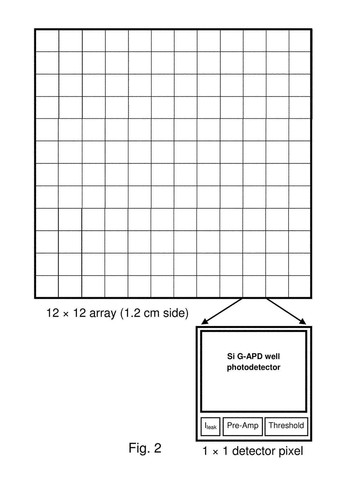
[0086]The system level technology includes both fabrication of autonomous detector voxels (ADVs) containing thin-film scintillation photon detector (e.g. Geiger mode avalanche photodiode, G-APD) with suitable electronics integrated with appropriate scintillator volume and the integration of these ADVs into autonomous detector arrays (ADAs) or autonomous detector blocks (ADBs) and assembling them into top-system level PET scanner with desired shape and providing the full functionality with desired spatial, temporal and energy resolution and uniformity.

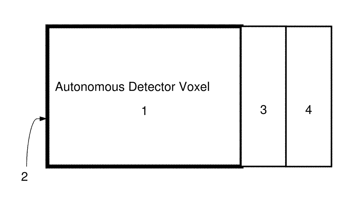
[0087]Autonomous Detector Voxel (ADV):

[0088]The ADV is comprised of a scintillator, one or more coupled electronics module(s), optically reflecting and isolating surface layers, and electrically isolating coatings. The detection and processing electronics are coupled to or fabricated onto one or more of the scintillator surfaces. The detection electronics contain everything necessary to detect and transmit the data of a scintillation ev...


PUM


Abstract

A system and method for imaging gamma- and x-ray, and charged particles sources employing a three dimensional array of scintillation elements arranged surrounding an emission source. According to a preferred embodiment, each element of the array comprises a scintillator element, a solid-state photon detector, and processing electronics to output an electronic signal. The elements may be efficiently packed in both the X-Y plane and stacked in the Z-axis, to provide depth of interaction information. The elements of the array are preferably hierarchically arranged with control electronics provided together for subarray modules (e.g., an n×m×1 module), and synchronization electronics provided at a larger scale. The modules preferably communicate with a control system through a shared addressable packet switched digital communication network with a control and imaging system, and receive control information from that system through the network.

Description

CROSS REFERENCE TO RELATED APPLICATIONS[0001]The present application is a Continuation under 35 U.S.C. §120 of U.S. patent application Ser. No. 15 / 080,073, filed Mar. 24, 2016, now U.S. Pat. No. 9,606,245, issued Mar. 28, 2017, which is a non-provisional of, and claims benefit of priority under 35 U.S.C. §119(e) from, U.S. Provisional Patent Application No. 62 / 137,276, filed Mar. 24, 2015, the entirety of which are expressly incorporated herein by reference.FIELD OF THE INVENTION[0002]The present invention relate to the field of scintillation detector arrays.BACKGROUND OF THE INVENTION[0003]Positron Emission Tomography (PET) is a proven and powerful research and clinical tool that uses molecular biomarkers for imaging the function and metabolism of the human body and brain. These biomarkers can be tailored to a high degree of specificity to highlight abnormal metabolism and uptake and are often used to diagnose diseases such as brain tumor, strokes, neuron-damaging disease (dementia...

Claims


Application Information

Patent Timeline
Patent Type & Authority Patents(United States)
IPC IPC(8): G01T1/20A61B6/03G01T1/208G01T1/24A61B6/00
CPCG01T1/208G01T1/245G01T1/2018A61B6/037A61B6/501G01T1/1642G01T1/20183G01T1/20185G01T1/20181G01T1/20182G01T1/2006
Inventor CZARNECKI, STEVEKROL, ANDRZEJMANDAL, KRISHNAPOLIKS, MARK D.SCHMIDTLEIN, C. ROSSTHOMPSON, MICHAELTURNER, JAMES
Owner UNIVERSITY OF SOUTH CAROLINA
Why Eureka
Try Eureka