Method and apparatus for automated measurement of chiral analyte concentration

a technology of chiral analytes and automated measurement, which is applied in the field of physiological analytes measurement, can solve the problems of insufficient accuracy of procedures, limited space and personnel available for monitoring patients, and inability to accurately measure concentration, etc., and achieves the effects of convenient and reliable measurement, accurate measurement, and convenient monitoring

Active Publication Date: 2018-04-10
LESZINSKE ALAN J
View PDF3 Cites 0 Cited by
  • Summary
  • Abstract
  • Description
  • Claims
  • Application Information

AI Technical Summary

Benefits of technology

[0030]Accordingly, it is an object of the invention to provide a practical, reliable, affordable, and automated apparatus capable of accurately measuring the concentration of an optically active ingredient such as glucose in a sample.
[0031]It is another object of the invention to provide an automated device capable of accurately measuring the glucose concentration in a blood product acquired from patient blood before, during, and after major surgical procedures (perioperatively) and in critical care settings. During surgery, the device can monitor directly from ultrafiltrate effluent discharged from the hemoconcentrator without additional invasive measures or attachments to the patient (subject).
[0037]The monochromatic light source in one embodiment is a laser diode or light emitting diode assembly with a known wavelength in the visual and near IR range. Applicant's system is presently designed for 635 nm. This specific wavelength was utilized to permit “off-the-shelf” optics; however, the system has successfully been operated at 780 nm although this range requires safety devices such as interlocks to protect operators. It was found that 780 nm wavelength provided much better transmission, with less rotation, but negligible interference. The light source assembly is equipped with feedback temperature controller to stabilize the light source. The required stability of the monochromatic source received by the detector allows for fluctuations totaling not more than four percent of the range of response for glucose concentrations from 0 mg to 400 mg / dL (dynamic range), and preferably an order of magnitude below. As an example for operating parameters with a dynamic range of 240 mV the tolerance for baseline fluctuations would be no greater than + / −5 mV.
[0038]The first polarizer is incorporated in the optical platform in front of the light source and is oriented to provide a specific state of polarized light. As in most polarimetry and ellipsometry the optical components are selected to match the wavelength of the light source, and the optics have anti-reflective coatings of the same wavelength mounted on their surfaces. The resolution of the degree to which the optical signal can be managed is highly dependent on the extinction ratios of the polarizers. In the preferred embodiment extinction ratios range between 1:10,000 and 1:100,000 are obtained. This is described as an optical component through which one part of light in 10,000 to 100,000 of the light is incident on the detector; for a very carefully oriented polarizer and analyzer system the transmission is about 1 part in 100,000. The utilization of a retarder further increases the precision by providing greater resolution of the signal of the basic polarimeter.

Problems solved by technology

In modern surgical suites there are limited space and personnel available for monitoring patient blood analytes, though studies have shown the importance for tightly controlled glucose during cardiothoracic and other major surgical procedures.
These procedures lack sufficient accuracy, take too long, and require that limited staff take time away from critical functions to draw a blood sample, analyze it, or send it off to the hospital's lab for analysis (and wait for results).
The time lag between sampling and delivery of results is detrimental to optimum blood glucose management.
The “Portland Protocol” (Furnary 2004), where insulin is continuously provided to the patient during open heart surgical procedures, was developed to keep blood glucose from rising above 180 mg / dL, but does not address hypoglycemic (low blood glucose) conditions that lead to other postoperative complications.
These complications include mediastinitis or deep sternal wound infection, loss of mental acuity, respiratory infections, and death.
There are currently no automated blood glucose monitoring systems utilized to monitor critically ill patients or perioperatively.
Some companies have developed implantable monitors for use in critical care settings, but none for intraoperative monitoring.
Little is known about how this might interact with the hemoconcentrator or heart-lung bypass process, in addition to a lack of measurement precision and sensitivity.
It has been designed for use in clinical settings by healthcare professionals, but requires calibration with inaccurate and labor intensive fingerstick or laboratory analysis.
While continuous monitors based on microdialysis technologies for diabetic patients have been released to market in the United States, they are not stand-alone monitors, and require that the patient calibrate and make adjustments to treatment (insulin injection or medication) based on readings from old inaccurate existing finger-stick method and monitor, or confirmation readings from clinical laboratory devices.
Time between testing and receipt of results can be more than an hour, and presumably because of this, patient sampling is done at least on an hourly basis during cardiothoracic surgeries that last on average about six hours, which does not provide sufficiently frequent measurement to permit timely adjustment of insulin and / or glucose.
The incidence of diabetes, and therefore the number of diabetic patients undergoing surgery has increased, pushing mortality, morbidity, and hospitals costs upwards.
A variety of clinical procedures have been implemented that have helped slow the increase in the incidence of deep sternal wound infections, but none has addressed it as sufficiently as shown in the clinical studies over the past four years.
Microdialysis based continuous monitors remain targeted at the much larger consumer monitor market, and have not yet been applied successfully to perioperative monitoring, and continue to be used as ancillary products to track trends as opposed to adjusting or directing treatment, due to their lack of sufficient accuracy and necessary precision.
The prior art includes an injectable product that glows in the presence of glucose, however the results obtained are general in nature and not sufficiently specific to provide guidance in therapeutic treatment.
Most utilize RFID or Bluetooth technology to transmit measurement data to the monitors, again with high costs and insufficient measurement precision.
Most require too much hands-on effort, frequent calibration, implantables, transmission radio frequencies, or other issues that would preclude their providing the required accurate, safe, convenient, and automated real-time measurement system that displays results on-demand.
This approach suffers in sensitivity of measurement (according to the authors) which is probably due to noise problems associated with the motor driven phase modulator as well as other unidentified problems.
Again, noise levels in the system represent the major problem in achieving the required sensitivity.
The reported data show a scatter that is unacceptable for a working blood glucose sensor.
The methods therein described suffer from a common problem of cost and complexity that reduce their commercial utility from a practical standpoint.

Method used


Image

  • Method and apparatus for automated measurement of chiral analyte concentration
  • Method and apparatus for automated measurement of chiral analyte concentration
  • Method and apparatus for automated measurement of chiral analyte concentration

Examples


Embodiment Construction

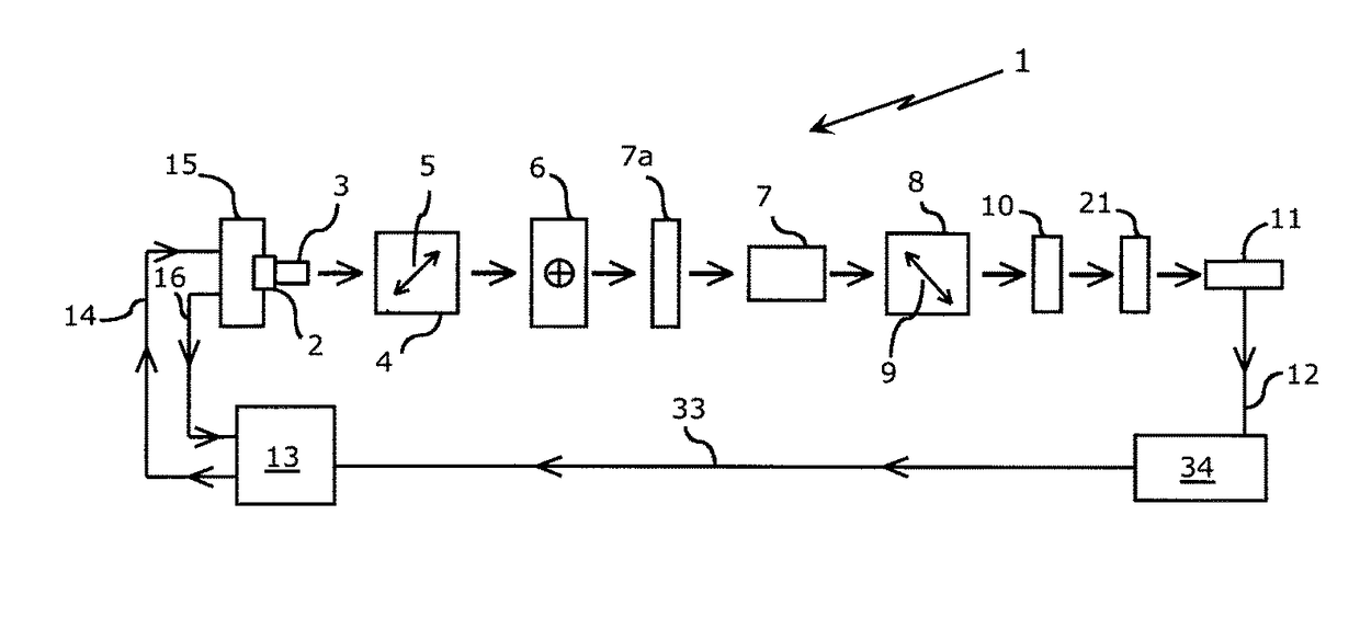
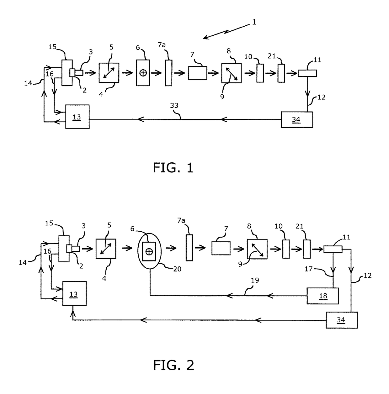
[0051]In one embodiment of the proposed device, light from a monochromatic source, such as an LED or laser diode, is collimated and polarized. This light beam is passed through a retarder and subsequently through a high quality optical quartz glass flow cell through which the sample ultrafiltrate, calibration standard or flush solvent are drawn by the device's pump and manifold system. The light emanating from the measurement flow cell is routed through the analyzer, and optionally through a focusing lens and / or bandpass filter of known wavelength, before it is directed onto the detector.

[0052]Referring to FIG. 1, a monochromatic light source (laser diode or light emitting diode) 2 is shown, the output of which is collimated by an optical component 3 (if needed) and transmitted to a polarizer 4. Applicant has found that the utilization of a 10 mW, 635 nm laser diode having an integrated, internal beam corrected optic is suitable. Double ended arrow 5 indicates the direction of polar...


PUM

PropertyMeasurementUnit
concentrationsaaaaaaaaaa
wavelengthaaaaaaaaaa

Abstract

Perioperative patient blood glucose concentrations are determined by imposing patient effluent ultrafiltrate through a sample cell incorporated in an automated polarimeter. The device includes an optical platform, fluid handling subassembly, controlling electronics, and integration software. A stable collimated light source of known intensity and distinct specified wavelength is passed through an optical platform including a polarizer, retarder, bandpass filters, sample flow cell, analyzer and detector. The angular rotation of the transmitted light resulting from the glucose contained in patient ultrafiltrate collected in the sample flow cell is recorded and provides a measure of the glucose concentration.

Description

RELATED APPLICATIONS[0001]This application is related to and claims priority to a provisional application entitled “METHOD AND APPARATUS FOR AUTOMATED MEASUREMENT OF CHIRAL ANALYTE CONCENTRATION” filed Jun. 5, 2012, and assigned Ser. No. 61 / 655,806.FIELD OF THE INVENTION[0002]The present invention relates to the measurement of physiological analytes and particularly to automated systems for the measurement of such analytes throughout major surgical procedures and the measurement of such analytes in the care of critically ill patients.BACKGROUND OF THE INVENTION[0003]The detrimental effects of elevated perioperative blood glucose have been reported in a large and growing body of peer-reviewed medical publications. Pre-operative and intraoperative hypoglycemia and hyperglycemia have been reported as independent risk factors for postoperative complications, including infection and death. In modern surgical suites there are limited space and personnel available for monitoring patient bl...

Claims


Application Information

Patent Timeline
Patent Type & Authority Patents(United States)
IPC IPC(8): A61B5/1455G01N21/21A61B5/153G01J4/04A61B5/1495A61B5/155A61B5/15A61B5/145A61B5/00A61B5/1459
CPCA61B5/14532A61B5/1459A61B5/1495A61B5/14558A61B5/153A61B5/15003A61B5/155A61B5/150229A61B5/150992A61B5/4836A61B5/6866A61B5/7246G01J4/04G01N21/21A61B5/1455G01N2201/127A61B2505/05A61B2560/0233G01N2201/062G01N2201/0612
Inventor LESZINSKE, ALAN J.
Owner LESZINSKE ALAN J
Why Eureka
Try Eureka