Method for detecting protein or protein complex by identifying a plurality of epitopes synchronously

A protein and epitope technology, applied in the field of proteomics research, can solve the problems of cumbersome, high GC content of oligonucleotide aptamers, extension of auxiliary fragments, etc.

Inactive Publication Date: 2009-06-17
EAST CHINA NORMAL UNIV
View PDF0 Cites 9 Cited by
  • Summary
  • Abstract
  • Description
  • Claims
  • Application Information

AI Technical Summary

Problems solved by technology

[0003] In protein detection, the method of proximity dependent DNA ligation can detect trace amounts of protein, but because the final quantitative method it adopts is the method of fluorescent quantitative PCR, it is often caused by the non-specificity of PCR amplification or the adaptation of oligonucleotides. The GC content of the daughter is too high to make the PCR reaction impossible, so the auxiliary fragment has to be extended
In addition, this method can only detect the same epitope in binary complexes of the same protein
Of course, the main disadvantage of this method is the presence of background ligation reactions, which require tedious optimization of reaction conditions to minimize interference
This method can only detect two epitopes at most at the same time, so more flexible methods are needed for proteomics research

Method used


Image

  • Method for detecting protein or protein complex by identifying a plurality of epitopes synchronously
  • Method for detecting protein or protein complex by identifying a plurality of epitopes synchronously
  • Method for detecting protein or protein complex by identifying a plurality of epitopes synchronously

Examples


Embodiment 1

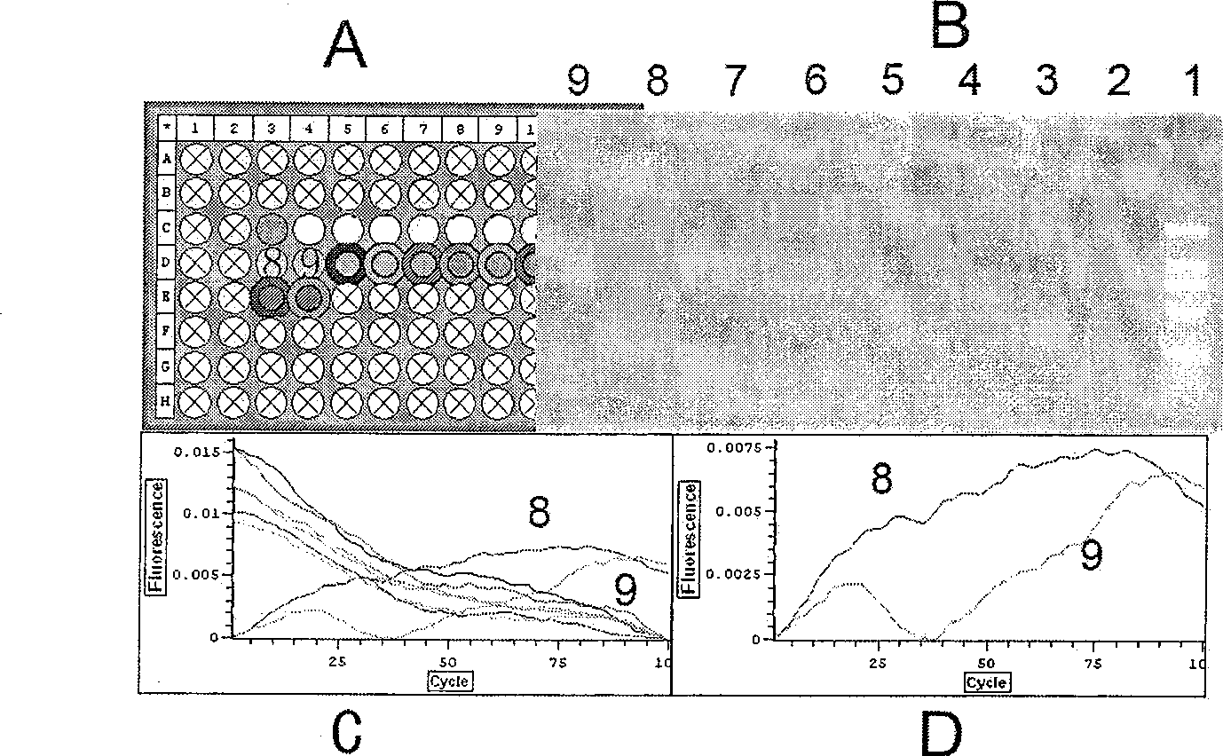
[0049] Four oligonucleotide aptamers detect CatD protein

[0050] 1. Add 1.0uL 100pM four kinds of oligonucleotide aptamer mixture into the PCR tube

[0051] 2. Dilute the CatD protein with 1% BSA as the diluent, then add the CatD protein with a concentration of 1.0aM, 100.0aM and 10.0fM and 1% BSA as a control in different tubes, and place it at room temperature for 1 hour

[0052] 3. Add 0.1uL 25uM four kinds of linker mixture to the buffer containing 1.0U T4 DNA Ligase, add this mixture to the reaction tube and place it at room temperature for 5 hours for ligation reaction

[0053] 4. Add 1.0uL 10X phi29 buffer, 0.5uL 10mM dNTP, 0.2uL 100X BSA, 0.3uL phi DNA polymerase, 7.5uL ddH to each tube 2 O, 0.5 uL Eva Green, 0.1 uL primer1 and 0.1 uL primer2. Amplify at 30°C for 8-12 hours

[0054] 5. Start to use OPTICON2 continuous fluorescence detector (MJ Research) to record data for four hours at the 8th hour, read the value every 2.5 minutes, and last for about four hours, mai...

Embodiment 2

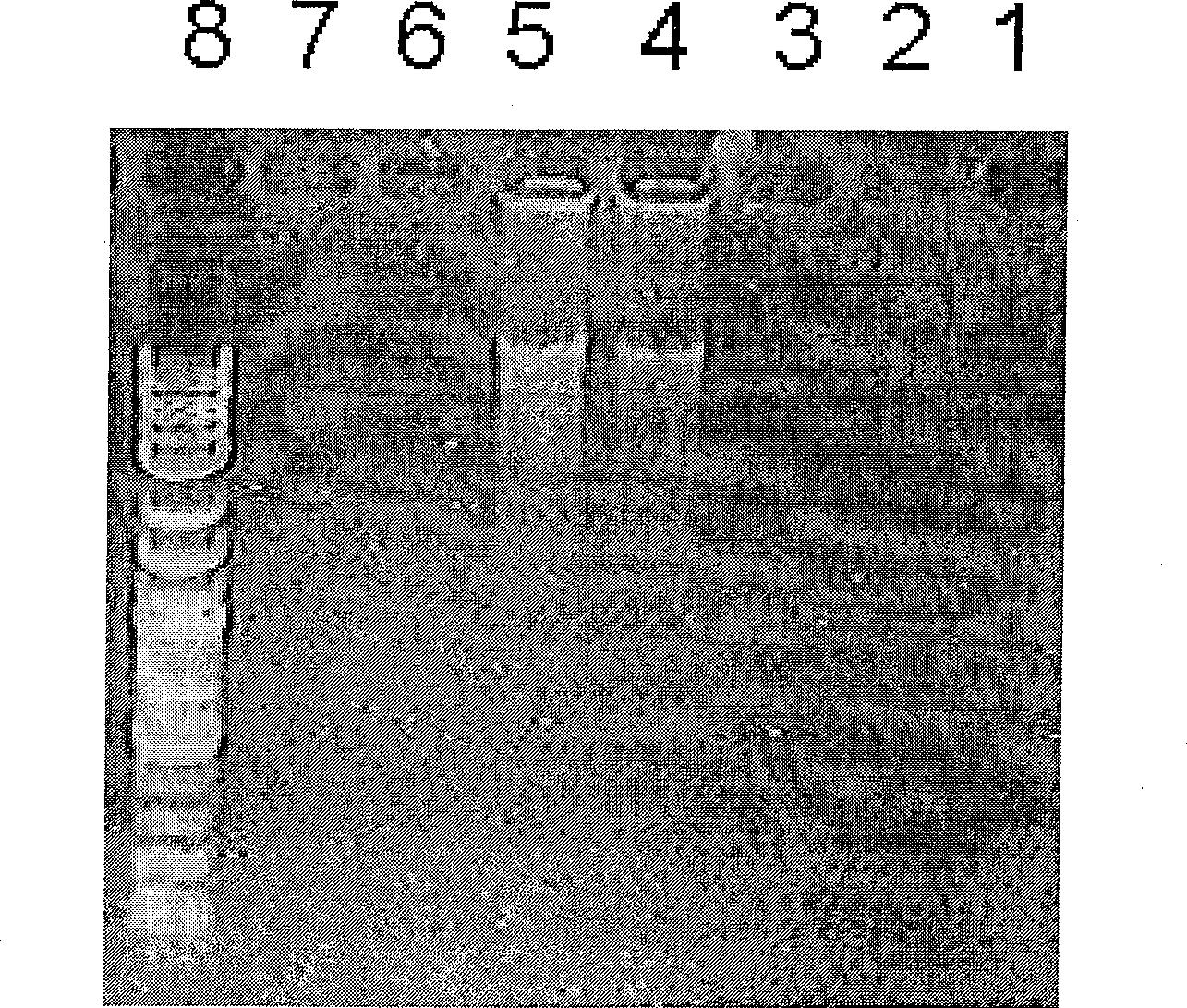
[0057] Four oligonucleotide aptamers detect CatD protein

[0058] 1. Add 1.0uL 100pM four kinds of oligonucleotide aptamer mixture into the PCR tube

[0059] 2. Dilute the CatD protein with 1% BSA as the diluent, and then add CatD protein with a concentration of 1.0aM, 100.0aM and 10.0fM and 1% BSA as a control in different tubes; CatD protein with three epitopes was used as a control, that is, 10.0 nM and 1.0 nM proteins were added to the tube, and left at room temperature for 1 hour

[0060] 3. Add 0.1uL of 25uM four kinds of linker mixture to the buffer containing 1.0U of T4 DNA Ligase, add this mixture to the reaction tube and place it at room temperature for 7 hours to carry out the ligation reaction

[0061] 4. Add 1.0uL 10X phi29 buffer, 0.5uL 10mM dNTP, 0.2uL 100X BSA, 0.3uL phi DNA polymerase, 8.0uL ddH to each tube 2 O, 0.1uL primer1 and 0.1uL primer2 were amplified at 30°C for 36 hours

[0062] 5. Use 2% agarose electrophoresis for detection

[0063] 6. The resu...


PUM


Abstract

The invention adopts oligonucleotide aptamers for identifying special tripeptide sequence KAI, GEL, DGI and LAS, extanding oligonucleotide aptamers by Structure-switch and phosphorylation decorating 5' end thereof, synthetising four connexons; connecting oligonucleotide aptamers combinated with target albumen through proximity dependent DNA Ligation technique as chain ringlike DNA; amplifying detecting signals via rolling-circle DNA duplicate method; the technique can detect target albumen sensitively, the least detecting concentration can get nM level; this method with flexibility can adjust according needed numbers of ligand; can detect albumen composite, provide sensitive detecting method for chip technique using oligonucleotide aptamers as ligand.

Description

technical field [0001] The invention relates to proteomics research technology, in particular to a method in which an oligonucleotide aptamer (Nucleic Acid Aptamer) that recognizes protein epitopes is used to simultaneously recognize multiple epitopes to detect proteins or protein complexes. Background technique [0002] Protein expression can exist as a monomer or a dimer, and there are more complex complexes formed by multiple proteins. The detection of these proteins and even complexes is very important, especially in proteomics research , we not only need to know what kind of protein is expressed, but we also need to know the state of the protein, including modifications and complexes. Therefore, the detection of proteins, especially the detection of various states of proteins after translation is very important. The study of proteomics requires high-throughput technology and sensitive detection methods. Recently, two-dimensional electrophoresis technology and protein c...

Claims


Application Information

Patent Timeline
Patent Type & Authority Patents(China)
IPC IPC(8): C12Q1/68G01N33/68
CPCG01N33/6842
Inventor 牛文泽江楠胡应和
Owner EAST CHINA NORMAL UNIV
Why Eureka
Try Eureka