DNA binding protein detection method based on moving endonuclease

A technology that combines proteins and detection methods, applied in the field of non-labeled detection in biomedicine, can solve the problems of low-abundance protein loss, improve detection efficiency and sensitivity, time-consuming and labor-intensive problems, achieve comprehensive biological significance, avoid radioactive pollution, and operate Safe and fast effect

Inactive Publication Date: 2007-10-24
SOUTHEAST UNIV
View PDF0 Cites 3 Cited by
  • Summary
  • Abstract
  • Description
  • Claims
  • Application Information

AI Technical Summary

Problems solved by technology

Although these conventional methods have played an important role in the study of sequence-specific DNA-binding proteins, they are labor-intensive, time-consuming and labor-intensive to operate, and are likely to cause contamination by radioactive substances, and cannot be used for high-throughput detection.
There are currently some methods for detecting DNA-binding proteins using non-radioactive labels, but most of these methods require fluorescent molecular labeling of proteins before detection. This process may lead to the loss of lo

Method used


Image

  • DNA binding protein detection method based on moving endonuclease
  • DNA binding protein detection method based on moving endonuclease
  • DNA binding protein detection method based on moving endonuclease

Examples


Embodiment 1

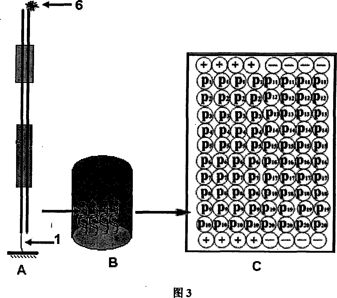
[0030] Example 1. Non-labeled detection of DNA-binding proteins based on streptavidin-coated 96-well plates (this method is applicable to all commercial microplate reader detection systems, and the detection instrument recommended in this embodiment is Tecan, Switzerland Infinite TM F200 automatic microplate reader. )

[0031] 1. Probe synthesis and immobilization

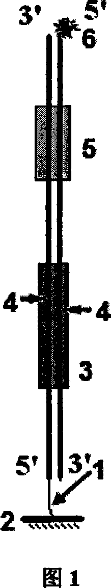
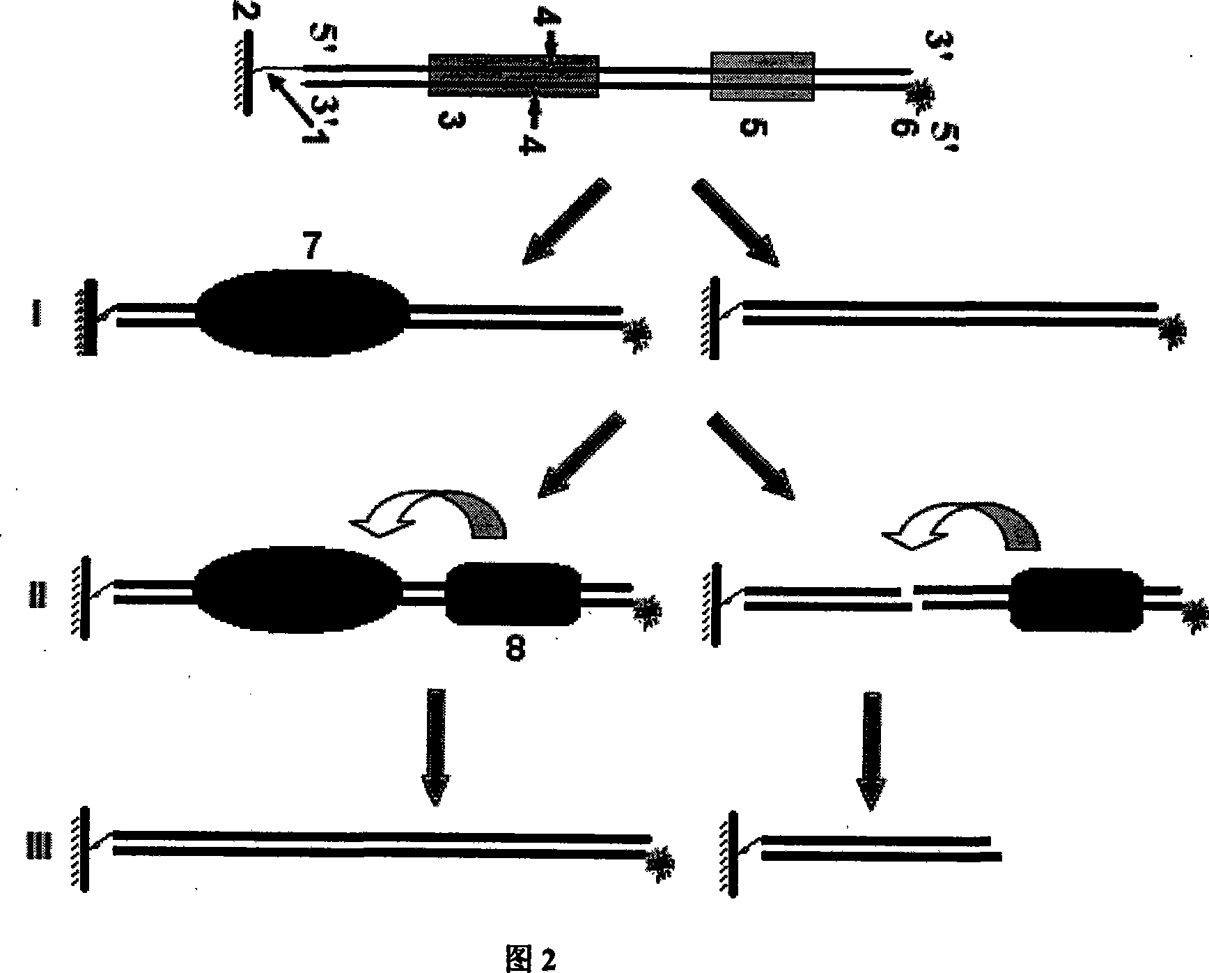
[0032] 1.1 According to the above description of the probe design method and accompanying drawing 3, double-stranded oligonucleotide detection probe P n The linking group 1 on the biotin molecule, CP n The signal molecule 6 above is digoxin molecule. As shown in Table 1, the 20 detection probes have the same sequence except the binding site sequence of the DNA-binding protein according to different proteins to be detected. In addition, two pairs of complementary sequences were separately synthesized as negative (-) and Positive control (+).

[0033] serial number

P n

transcription factor...

Embodiment 2

[0042] Example 2. Non-labeled detection of DNA-binding proteins based on DNA microarray chips (this method is applicable to the general operation of all DNA microarray chips)

[0043] 1. Synthesis of detection probes

[0044] The design and synthesis of all probes were the same as in Example 1. The difference is that the P n Linking group 1 in is labeled with an amino group, and the CP n The signal molecule 6 in is labeled with Cy3 fluorescent molecule.

[0045] 2. Microarray preparation

[0046] According to the probe prepared in step 1, as shown in Figure 4, the probe P n and its corresponding complementary probe CP n All were mixed and dissolved in double-distilled water at a concentration of 100 μM, incubated at 95°C for 5 minutes, and then naturally cooled to room temperature overnight to make P n and CP n Fully annealed to a double-stranded structure. Take the above refolding solution and dissolve it into an equal volume of 2×carbonate buffer (0.2M Na 2 CO 3 / 0...


PUM


Abstract

The invention relates to a DNA combine protein check method based on mobile cutting tangent enzyme, which designs a section of double stranded probe for the recognize sequence of an object DAN combine protein, wherein one single strand 5' end is marked with a connecting group 1 to be fixed on the surface of a solid carrier 2, and another single strand 5' end is marked with a signal molecule 6, the 3' end of the recognize point 3 with the object protein is arranged with a IIS type tangent enzyme recognize point 5, adjusts the distance between two recognize points to arrange the enzyme tangent pint 4 in the combine point 3. And the operation comprises that I, cultivating the solution with object DNA combine protein with the fixed double stranded probe, to process enzyme tangent reaction in a tangent enzyme system, II, when the object protein exists, the signal molecule is held on the surface of the carrier, and when the object protein does not exist, the probe is cut off by the tangent enzyme in the idle protein combine point, III, based on the signal strength, realizing the non-mark check on the object protein.

Description

technical field [0001] The present invention relates to a method for non-marker detection of deoxyribonucleic acid (DNA) binding protein, where the DNA binding protein mainly refers to a regulatory factor with a specific DNA binding sequence, and the mobile cutting restriction endonuclease here mainly refers to Type IIS restriction endonuclease that cleaves double-stranded DNA outside of its recognition site. The invention belongs to the technical field of label-free detection in biomedicine. Background technique [0002] The sequence-specific recognition and interaction of DNA and proteins play an important role in the regulation of biological gene expression, and the occurrence and development of almost all diseases are related to the abnormality of DNA-binding proteins. The study of DNA-binding proteins has attracted more and more attention, and has become one of the important contents of genome and proteome research. It is very important to detect the amount of one or ...

Claims


Application Information

Patent Timeline
IPC IPC(8): G01N33/68C12Q1/68
Inventor 白云飞王进科张定东孙蓓丽葛芹玉陆祖宏
Owner SOUTHEAST UNIV
Why Eureka
Try Eureka