
Colloidal gold method for fast quantitative determination of C-reaction protein and its application

A quantitative detection and reactive protein technology, applied in the field of clinical immunology detection, can solve the problems of long detection time, poor reaction homogeneity, unsuitable for single test, etc., to shorten the cure time and reduce medical expenses.

Active Publication Date: 2008-12-10
SHANGHAI UPPER BIO TECH PHARMA
View PDF0 Cites 16 Cited by
  • Summary
  • Abstract
  • Description
  • Claims
  • Application Information

AI Technical Summary

Problems solved by technology

[0007] One of the technical problems to be solved by the present invention is to overcome the shortcomings of conventional C-reactive protein detection methods that are not suitable for single-person testing, long detection time, and poor reaction homogeneity, and provide a fast, simple and accurate rapid quantitative detection Colloidal gold method for C-reactive protein

Method used


Image

  • Colloidal gold method for fast quantitative determination of C-reaction protein and its application
  • Colloidal gold method for fast quantitative determination of C-reaction protein and its application
  • Colloidal gold method for fast quantitative determination of C-reaction protein and its application

Examples

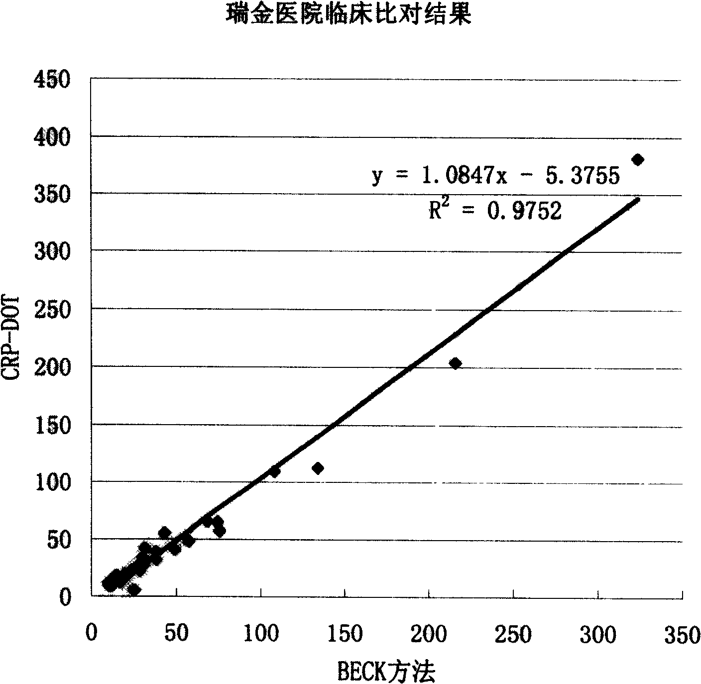

example 1

[0031] Example 1 uses the preparation of the CRP gold standard method rapid quantitative detection reagent of two monoclonal antibodies

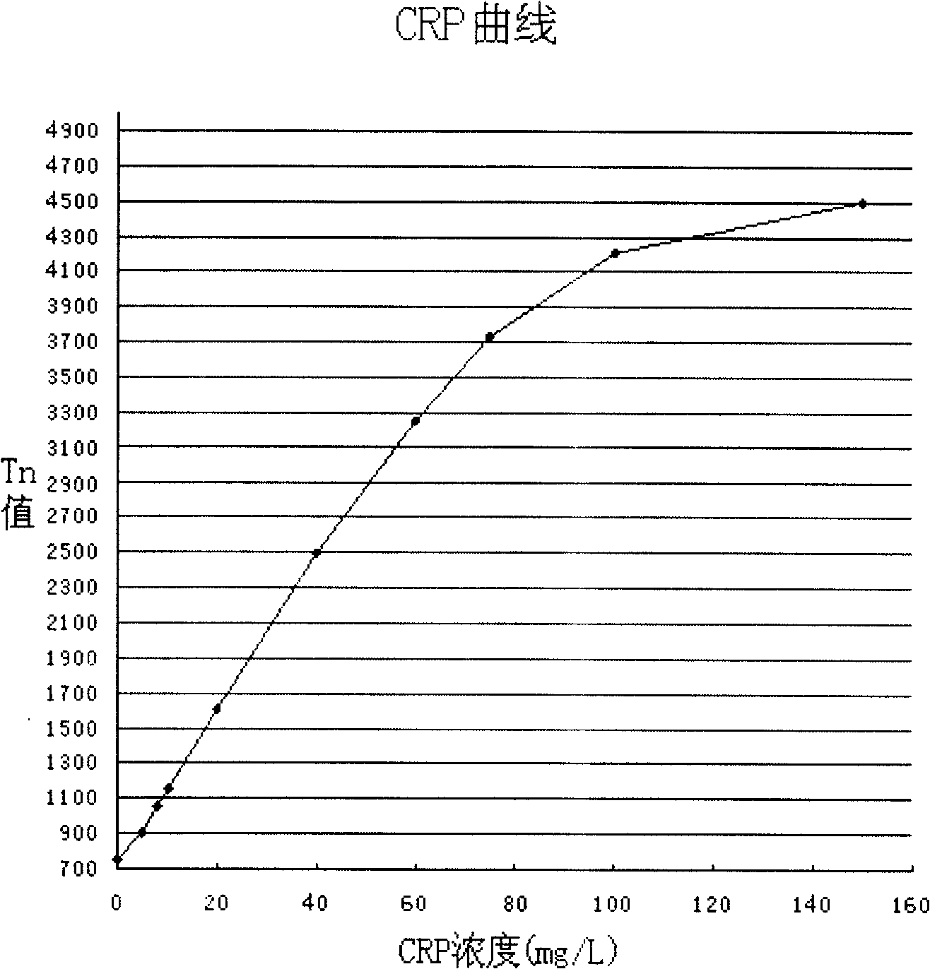
[0032]1. Preparation of freeze-dried anti-CRP immune colloidal gold and reaction plate: according to the method introduced by Lu Shengkai et al. (Chinese Journal of Microbiology and Immunology, 13(2):125, 1993; Shanghai Journal of Medical Laboratory, 5(1):62 , 1990) prepared colloidal gold and anti-CRP immune colloidal gold conjugates. However, the colloidal gold is coated with purified CRP monoclonal antibody, 0.15mg of CRP monoclonal antibody is used to coat 10ml of colloidal gold, and 2500ml of colloidal gold is finally recovered to 1500ml anti-CRP immune colloidal gold application solution, buffer containing 1% BSA, 0.5mg / ml PEG 20M, 0.1% sodium azide. The pre-cooling temperature for freeze-drying is -35°C, the maximum temperature rise is 30°C, and the vacuuming time is 20 hours. The filling volume of the bottle can be set as required, ...

example 2

[0041] Example 2 CRP colloidal gold method rapid quantitative detection method

[0042] 1. Take out the reagent from the cold storage place, equilibrate at room temperature for at least half an hour, and accurately add 1.1ml of CRP freeze-dried product reconstituted solution to the anti-CRP gold standard solution freeze-dried product and shake well.

[0043] 2. Use a clean tip to absorb 100 μl of the reconstituted CRP gold standard solution and add it to an empty reaction tube. The tip only absorbs the CRP gold standard solution to prevent any contamination.

[0044] 3. Place the CRP reaction plate flat on the test bench, add 2 drops of CRP blocking solution to the reaction well, and wait until it is completely infiltrated

[0045] 4. Fresh serum samples do not need to be pretreated. Serum that has been frozen or stored at 4-8°C for several days and centrifuged can be used as the supernatant; accurately pipette 10 μl of serum and add it to the CRP dilution tube (see the bottle...


PUM

PropertyMeasurementUnit
Ionic strengthaaaaaaaaaa

Abstract

The invention overcomes the defects that the routine C-reactive protein (CRP) quantitative detection method is not suitable to single detection and takes long time, and provides a quick, convenient and accurate method for detecting the colloidal gold of C-reactive protein quickly and quantitatively. The reaction principles of the method is the gold-standard dot immune filtration assay of the solid phase double antibody sandwich method; a sample to-be-detected is diluted 40-480 times and then mixed evenly with immune colloidal gold in a liquid phase homogeneous medium and finally put on a reaction board; wherein, the contained sample to-be-detected which is mixed with the immune colloidal gold can be specifically captured by the monoclonal antibodies or the polyclonal antibodies of another determinant of anti sample to-be-detected, which is fixed on a piece of membrane; and the to-be-detected sample is shown like red spots, and the brightness of the color of spots is in direct proportion to the CRP concentration in the sample.

Description

technical field [0001] The invention belongs to the field of clinical immunology detection, in particular to a colloidal gold method for rapid quantitative detection of C-reactive protein and its application in the preparation of diagnostic kits. Background technique [0002] C-reactive protein (CRP) is a capsular C polysaccharide that can bind to Streptococcus pneumoniae. It was discovered by Tillett and Francis (1930) and studied by Abernethy and Avery (1941). CRP is composed of five identical subunits ( 23KD) is a ring-shaped pentameric protein formed by non-covalent chain aggregation, with a molecular weight of 115KD. It is an acute phase protein synthesized by liver cells when the body is subjected to inflammatory stimuli such as microbial invasion or tissue damage. CRP increases within a few hours of inflammation, and its content returns to normal with the recovery of tissue structure and function. Therefore, the determination of CRP is of great significance for disea...

Claims


Application Information

Patent Timeline
IPC IPC(8): G01N33/53G01N33/68G01N33/577
Inventor 徐建新
Owner SHANGHAI UPPER BIO TECH PHARMA
Learn More