
Recombinant canine adenovirus type 2 transfer vector, construction method and application thereof

A transfer vector, adenovirus technology, applied in the field of genetic engineering, to achieve the effect of reducing workload and stabilizing biological characteristics

Inactive Publication Date: 2009-02-04
HARBIN VETERINARY RES INST CHINESE ACADEMY OF AGRI SCI
View PDF0 Cites 7 Cited by
  • Summary
  • Abstract
  • Description
  • Claims
  • Application Information

AI Technical Summary

Problems solved by technology

Different from the above research results, Reddy et al. (2002)[ps} for the first time inserted the GFP gene without any regulatory sequence into the Sac I and Snag I sites of the E3 region of PAV3, and made it consistent with the direction of E3 transcription. However, only the virus in which the GFP gene is inserted into the SnagI site can be obtained, which indicates that the E3ORF where the SacI site is located may be required for virus replication

Method used


Image

  • Recombinant canine adenovirus type 2 transfer vector, construction method and application thereof
  • Recombinant canine adenovirus type 2 transfer vector, construction method and application thereof
  • Recombinant canine adenovirus type 2 transfer vector, construction method and application thereof

Examples


Embodiment 1

[0058] Example 1 Construction of recombinant plasmid pMD-E3

[0059] 1 Materials and methods

[0060] Cells, Viruses and Strains

[0061] MDCK cells are subcultured by this laboratory, and the culture medium is DMEM containing 10% fetal bovine serum; It was isolated by the method disclosed in the following documents: Li Liujin et al., Isolation, Identification and Screening of Canine Adenovirus Type II Attenuated Vaccine Strain. Qinghai Science and Technology, 2000, June, Volume 7, Supplement), and propagated on MDCK cells; The recipient strain DH5a was preserved by our laboratory.

[0062] main reagent

[0063] PrimeSTAR HS DNA polymerase, rTaq DNA polymerase, pMD18-T vector, dNTP, IPTG, X-Gal were purchased from TaKaRa Company. DMEM was purchased from GIBCO-BRL Company, and fetal bovine serum was purchased from Tianjin Haoyang Biological Company. The gel extraction kit (Gel Extraction Mini Kit) was purchased from Shanghai Huashun Biological Engineering Co., Ltd., and ot...

Embodiment 2

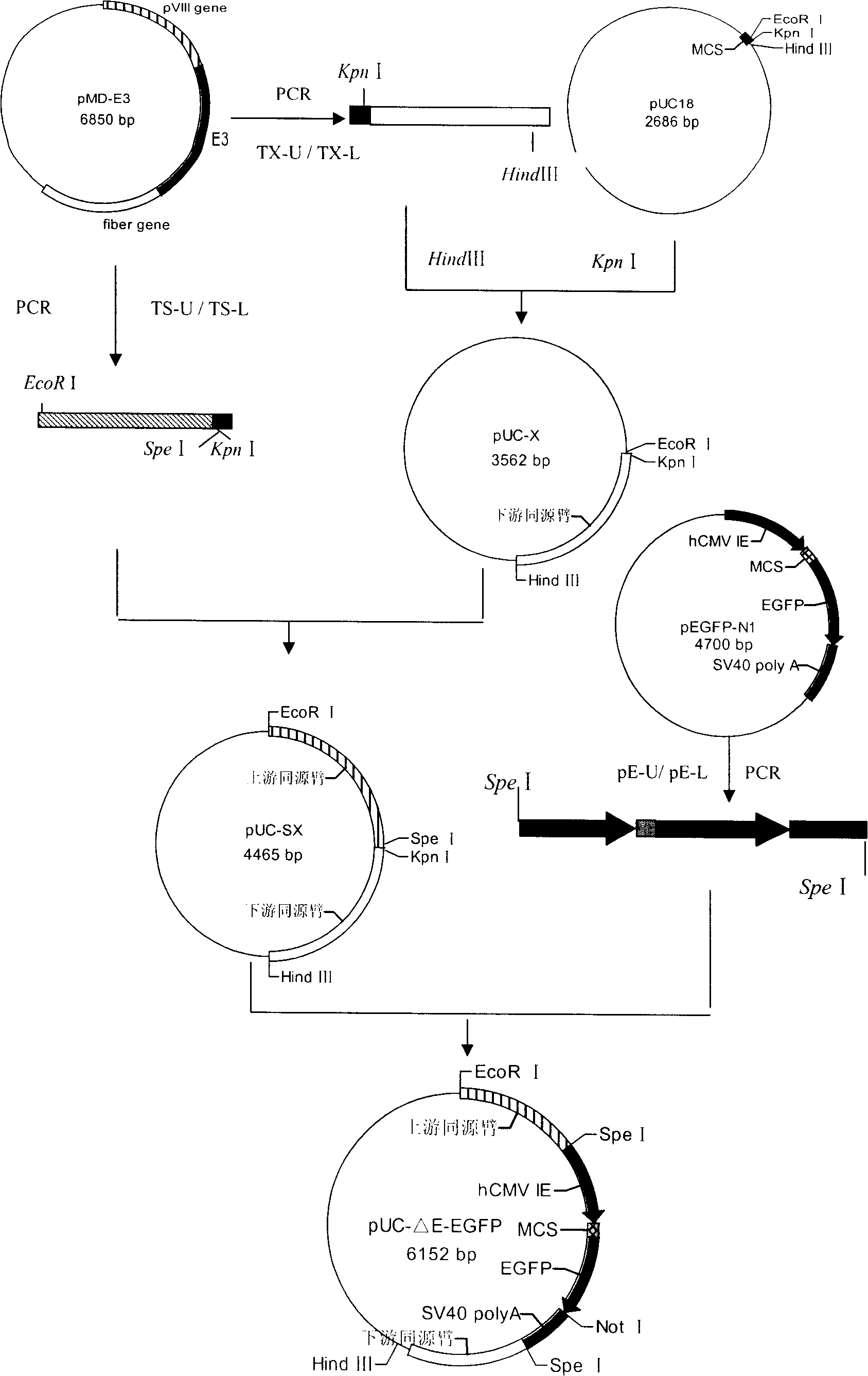
[0087] Example 2 Construction of recombinant canine adenovirus type 2 with E3 deletion and analysis of its biological characteristics

[0088] 1 Materials and methods

[0089] 1.1 Viruses, vectors and cells

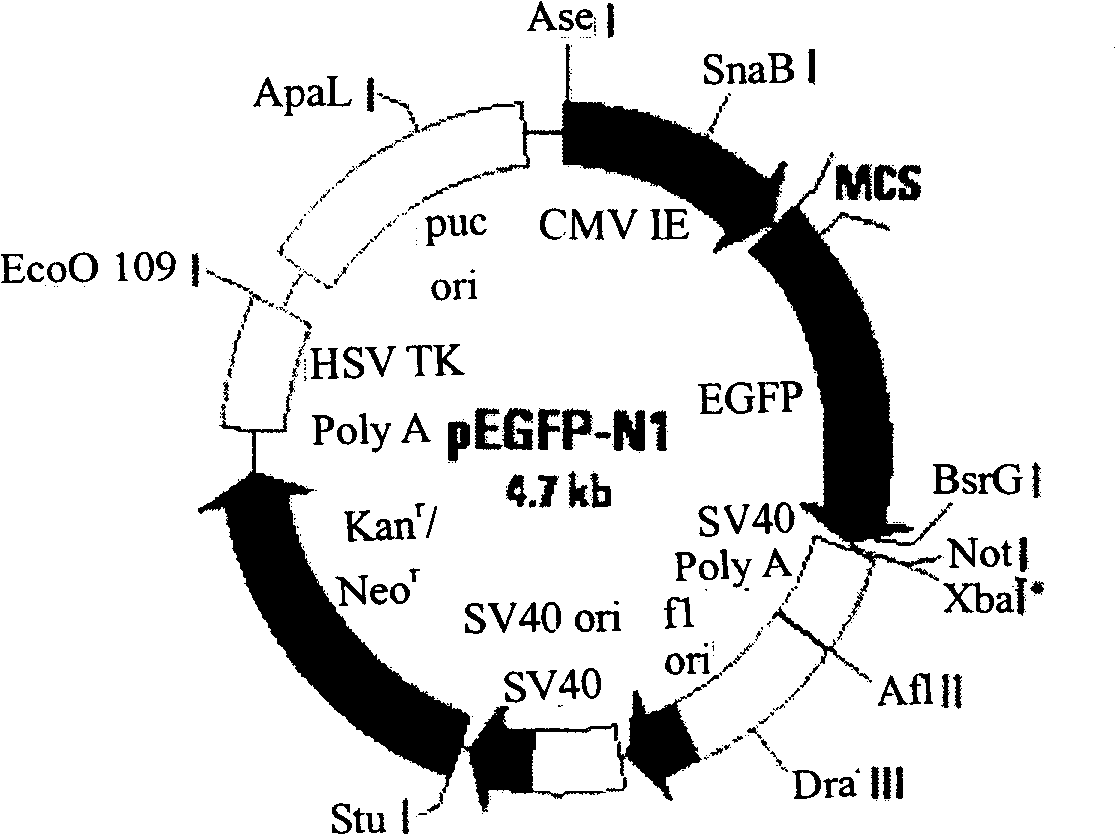
[0090] The sources of CAV-2 vaccine strain and competent cell DH5a are the same as in Example 1; the pUC18 vector was purchased from Dalian Bao Biological Engineering Company; the plasmid pEGFP-N1 (see Figure 4 ) is a product of Clontech Company, with hCMV immediate early promoter, MCS, EGFP and SV40 polyA signal.

[0091] 1.2 Main reagents

[0092]DMEM was purchased from GIBCO-BRL; fetal bovine serum was purchased from Tianjin Haoyang Biological Company; PrimeSTAR HS DNA Polymerase, dNTP, DNA Marker, T4 DNA ligase, restriction enzymes EcoRI, Kpn I and Spe I were purchased from Dalian Bao Biological Engineering Company; Calcium Phosphate Ttansfection Kit was purchased from Invitrogen Company; small amount of gel recovery kit was purchased from Shanghai Huashun Bioengi...


PUM


Abstract

The present invention discloses a recombinant canine adenovirus-2 transfer vector, a method for constructing the recombinant canine adenovirus-2 transfer vector and an applications thereof. The recombinant canine adenovirus-2 transfer vector loses the 1412bp fragment of the E3 region, a large fragment of exogenous gene can be inserted, and moreover, a hCMV IE promoter, a multiple cloning site, an enhanced green fluorescent protein gene and a SV40 early transcribed Poly A signal sequence can be inserted in the position of the lost fragment of the E3 region. The method successfully constructs the lost recombinant CAV-2 transfer vector of the E3 region, obtains a purified canine adenovirus-2 strain containing EGFP reporter gene and optimizes the cloning and purification method thereof, an evaluation indicates that the biological property of the recombinant virus is stable, and therefore the present invention provides a technical platform for the further development of CAV-2 live vector vaccine and related fundamental researches.

Description

technical field [0001] The invention relates to a recombinant virus transfer vector, in particular to a recombinant canine type 2 adenovirus transfer vector, its construction method and application, and belongs to the field of genetic engineering. Background technique [0002] The E3 region is a non-essential region for adenovirus replication in vitro. Inserting foreign genes in the E3 region does not affect the growth characteristics of the virus, and is often used to clone foreign gene regions. Among human and animal adenoviruses, except for sheep adenovirus (Vrati et al., 1995), the E3 region is located between the pVIII protein gene in the L4 region of the adenovirus genome and the fibrin gene in the L5 region, but the genome sizes of various adenoviruses are different. The start site of the E3 region is also different. EThe complete adenovirus E3 region contains 9 to 12 open reading frames (ORFs), of which 7 encoded products have been identified in infected cells, name...

Claims


Application Information

Patent Timeline
IPC IPC(8): C12N15/861C12N15/47C12N7/01
Inventor 曲连东姜骞周洁陈洪岩刘家森李昌文张洪英司昌德刘立奎韩凌霞孟庆文
Owner HARBIN VETERINARY RES INST CHINESE ACADEMY OF AGRI SCI
Learn More